VIP in brief
Vasoactive intestinal peptide is an endogenous 28-residue signalling peptide of the secretin and glucagon superfamily, linear, with a C-terminal asparagine amide and no disulfide, cut from a precursor that also yields PHM-27. It was isolated from hog small intestine in 1970 and sequenced in 1974, and it is made in neurons throughout the nervous system and in the gut; the best-known pathology of it is the VIP-secreting tumour. It binds the class B1 receptors VPAC1 and VPAC2 with high affinity and PAC1 far more weakly, and a 2.7 Å cryo-EM structure shows it bound to VPAC1. It is cleared from plasma within minutes, which is what most of the analogue chemistry on it is answering. The amidated peptide, C147H238N44O42S, is the substance registered under the name aviptadil; several public records, including the one a name search reaches, describe the free acid instead, and no VIP-bound VPAC2 structure was found.[1,2,3,4,5,6,7,8,9,10,11]
VIP regulatory status
No FDA, EMA or Health Canada marketing authorisation for synthetic VIP (INN aviptadil) was found in Drugs@FDA (queried through openFDA), the EMA medicines data report or the Health Canada Drug Product Database (searched November 2026). Aviptadil is investigational: the highest phase reached is phase 3, in a completed randomised, blinded trial in hospitalised adults with acute respiratory distress syndrome associated with COVID-19.[12,13,14,15,7]
VIP at a glance
- A 28-residue linear peptide, all L-amino acids, with a C-terminal asparagine amide and no disulfide bond. It is cut from a 170-residue precursor that also yields the separate peptide PHM-27.[1,3]
- Isolated from hog small intestine in 1970 and sequenced in 1974; a synthetic peptide matching the proposed sequence reproduced the activity of the natural material in the assays of the day, which is how the sequence was confirmed chemically.[2,3,16]
- Three class B1 G protein-coupled receptors respond to it. VPAC1 and VPAC2 bind VIP with high affinity; the PAC1 receptor bound PACAP with about 1000-fold higher affinity than VIP when first cloned and expressed.[17,18,19,4,20]
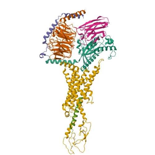
- A cryo-EM structure at 2.7 A resolution shows the peptide bound to VPAC1 with the stimulatory G protein attached, and molecular dynamics run alongside it found fewer stable peptide-receptor contacts for VIP than for PACAP-27 at the same receptor.[21,5]
- It is an endogenous signal rather than a designed molecule, made in neurons throughout the central and peripheral nervous system and in the gut, and it does not last in circulation: a radioimmunoassay study in conscious dogs measured a half-life of about 1.8 minutes and a transhepatic loss of nearly three quarters in a single pass through the liver. Synthetic VIP has its own International Nonproprietary Name, aviptadil.[1,9,10,7]
What is VIP?
Vasoactive intestinal peptide is an endogenous signalling peptide, not a designed analogue. It was purified from the small intestine of the hog and reported in 1970 as a 28-residue polypeptide chemically distinct from the kinins, substance P, glucagon and secretin; the name records the vasodilator activity seen in the assays used for its isolation, which identified no receptor. Its amino acid sequence was determined in 1974 using kallikrein cleavage, and a synthetic octacosapeptide built to the proposed sequence, together with a set of C-terminal fragments of increasing length, was reported in 1973 to carry the expected activity, with the shorter fragments weaker than the longer ones.[2,3,16]
Chemically it belongs to the superfamily that also contains secretin, glucagon, the glucagon-like peptides, glucose-dependent insulinotropic polypeptide and growth-hormone-releasing hormone. The family relationship is practical as well as taxonomic: the first VIP receptor cDNA was isolated from a rat lung library by cross-hybridisation with the secretin receptor cDNA, and that receptor turned out to be a seven-transmembrane, Gs-coupled protein related to the secretin, calcitonin and parathyroid hormone receptors. A second VIP-responsive receptor was cloned from rat tissue in 1993 and its human counterpart from a placenta library in 1995. Current nomenclature calls them VPAC1 and VPAC2, with PAC1 as the third, PACAP-preferring member of the group.[17,18,19,20]
Where it is made and what it does are the reasons it is studied. UniProt describes it as a neuropeptide abundantly expressed throughout the central and peripheral nervous systems, where the annotation records neuroprotective and immune-modulating roles, and the RefSeq gene summary lists the classical physiology the name was given for: vasodilation, lower arterial blood pressure, stimulation of myocardial contractility, increased glycogenolysis and relaxation of the smooth muscle of the trachea, stomach and gall bladder, with antibacterial and antifungal activity recorded as well. In the brain it is also an anatomical marker: the central circadian clock of the suprachiasmatic nucleus is a network of GABAergic neuron types in which the VIP-producing cells occupy the core and arginine vasopressin cells the shell, and work on the clock is written in those terms.[1,9,22]
The best-known pathology is a disease of too much of it. A VIPoma is a neuroendocrine tumour, mostly pancreatic, that secretes VIP in quantity; a 2022 review notes that most are already metastatic when found and that the condition is rare enough that no prospective study of it exists. That tumour is also why VIP is measured in plasma at all, and the measurement is an immunoassay rather than a mass measurement, which matters: in the 1981 dog study the peptide was altered during hepatic transit so completely that it escaped recognition by the assay, so an immunoassay figure reports what the antibody still binds rather than how much peptide passed.[11,10]
In the human precursor, recorded as UniProt P01282, the mature peptide occupies residues 125 to 152 and the asparagine at 152 is annotated as an amide. The same chain yields PHM-27 at residues 81 to 107, with its own C-terminal methionine amide, so two different peptides come out of one precursor. The mature sequence is conserved: a purification from rabbit small intestine confirmed that rabbit VIP is identical to porcine VIP, and the human sequence read off the precursor is the same 28 residues.[1,23]
The name is easy to confuse with PACAP, a separate gene product sharing close to 70 per cent sequence identity with VIP over the overlapping region, and with the glycine-extended 29-residue chain that precedes amidation. Neither is VIP. The supplier lists this entry by the bare abbreviation, which identifies the peptide by name only; the sequence, the amide and the receptor evidence below come from public records.[24,21,25]
The peptide is also short-lived, which is the single property that explains most of the analogue chemistry done on it. In five conscious dogs the measured half-life was 1.80 plus or minus 0.1 minutes with a metabolic clearance rate of 39.3 plus or minus 5.2 mL/kg/min and a distribution volume of 103.7 plus or minus 14.8 mL/kg, and 72.9 plus or minus 2.1% of the immunoreactive peptide was lost in one pass through the liver, against 27.5 plus or minus 12.5% for somatostatin measured the same way. Chemistry consistent with that is on record: a membrane-bound endoprotease purified from porcine antral mucosa cleaves VIP at three peptide bonds containing no arginine, with a Michaelis constant of 7.7 micromolar and a catalytic efficiency of 7.4 x 10^6 per molar per second. No figure for the human peptide in humans was located in the sources read here.[10,26]
Not to be confused with
- PACAP-27 and PACAP-38
- Pituitary adenylate cyclase-activating polypeptide is a separate gene product, isolated from ovine hypothalamus in 1989, whose 38-residue form shares roughly 70 per cent sequence identity with VIP across the overlapping region. The two peptides are not interchangeable at the receptor level: the cloned PAC1 receptor bound PACAP with about 1000-fold higher affinity than VIP, while VPAC1 and VPAC2 bind both with high affinity. Position 4 differs between them (Ala in VIP, Gly in PACAP), and chimeric peptides swapping that residue were used to test its contribution.[24,4,21,20]
- PHM-27 and PHI
- PHM-27 (peptide histidine methionine) comes from the same human precursor protein as VIP, at residues 81 to 107, and carries its own C-terminal methionine amide. It is a different peptide cut from the same chain, not a form of VIP; the porcine and rabbit counterpart is called PHI.[1,23]
- VIP-Gly (glycine-extended VIP)
- The 29-residue glycine-extended chain is the amidation precursor and is the molecule used in the deposited solution NMR structures, whose entity sequence runs HSDAVFTDNYTRLRKQMAVKKYLNSILNG. It is one residue longer than mature VIP and carries no C-terminal amide, so those coordinates are not coordinates of the mature peptide.[25,27]
- Secretin, glucagon, GLP-1, GIP and GHRH
- All belong to the same structurally related peptide superfamily and act on class B1 receptors, which is why cross-hybridisation with the secretin receptor cDNA is how the first VIP receptor was cloned. They are separate hormones with separate receptors; family membership is not identity.[20,17]
VIP structure and sequence
The chain is HSDAVFTDNYTRLRKQMAVKKYLNSILN, read from the amino terminus. It is linear and unbridged: with no cysteine there is no disulfide, and there is no cyclisation, lipid arm, linker or sugar anywhere in the molecule. Its only chemical decoration is at the far end, where the C-terminal asparagine is an amide rather than a free acid. That single difference matters for identity, because the computed public record most easily reached by searching the name describes the free acid, with a formula of C147H237N43O43S and an average mass of 3326.8 g/mol, while the mature peptide annotated in the precursor record is the amide. The two are different substances, and the figures above are reported here for the form they actually describe.[1,6]
Free in solution the peptide has no single shape. Solution NMR of the glycine-extended chain in two different membrane-mimicking environments gave a disordered N-terminal region followed by a long alpha-helix, straight in one environment and curved when bound to micelles. In the micelle-bound form the side chains of Phe6, Tyr10, Leu13 and Met17 lie on the concave face and form a hydrophobic patch that sits at the water-micelle boundary, the same residue positions that form the micelle-binding face of PACAP-38. Those coordinates are deposited for the 29-residue precursor rather than for mature VIP, which is why they are described here as a related molecule and not as a structure of this entry.[27,25]
The three-dimensional view on this page is the receptor-bound state: a cryo-EM reconstruction at 2.7 A of the 28-residue peptide (chain P) in the VPAC1 receptor, with the Gs heterotrimer and a stabilising nanobody also in the file. In that model the peptide's N-terminal residues reach into the transmembrane bundle while the helical remainder runs up past the extracellular loops toward the receptor's extracellular domain. Hydrogen bonds assigned in the deposited complex include His1 to a transmembrane glutamine (3.16 A), Asp3 to a transmembrane arginine side chain (2.87 A), Thr7 to a lysine (2.46 A), Thr11 to an aspartate in extracellular loop 2 (3.01 A) and the Leu27 backbone to a tyrosine of the extracellular domain (2.35 A). The view does not show the amide group, which the deposited entity does not model, and it does not show the ensemble: extracellular loop 3 is poorly resolved in this complex and the peptide's C-terminal half moves with the mobile extracellular domain.[5,21]
C-terminus: NH2
- His1
- Ser2
- Asp3
- Ala4
- Val5
- Phe6
- Thr7
- Asp8
- Asn9
- Tyr10
- Thr11
- Arg12
- Leu13
- Arg14
- Lys15
- Gln16
- Met17
- Ala18
- Val19
- Lys20
- Lys21
- Tyr22
- Leu23
- Asn24
- Ser25
- Ile26
- Leu27
- Asn28
Modifications
All 28 residues are L-amino acids and the chain is linear. There is no cysteine, so there is no disulfide bond; there is no cyclisation, no lipidation, no PEG, no glycosylation and no metal coordination. The only post-translational change is the C-terminal amide. Methionine at position 17 is the one oxidation-labile side chain in the sequence, and it is also one of the residues that forms the hydrophobic face of the helix in the micelle-bound NMR structure.[1,27,5]
What studies on VIP show
Each card names the experimental system and what was measured. A result in one system is not evidence about another.
Cryo-EM, molecular dynamics simulation and cell-based cAMP and binding assays · Purified human VPAC1 and PAC1 receptor complexes with the Gs heterotrimer and nanobody 35; cAMP accumulation and NanoBRET competition binding in transfected cells
The VIP-VPAC1-Gs complex was resolved at 2.7 A and the peptide N-terminus bound in the transmembrane bundle was the stable part of the structure, while its C-terminus moved with the extracellular domain. Fewer stable interactions were assigned between VPAC1 and VIP than between VPAC1 and PACAP-27, VIP showed more extensive dynamics with extracellular loop 3, and simulations of VIP modelled into PAC1 predicted more transient core contacts than at VPAC1.[21]
- Material
- Synthetic human VIP (28 residues) and PACAP-27, plus the chimeric peptides Gly4-VIP and Ala4-PACAP27
- Measured
- Map resolution and modelled receptor-peptide hydrogen bonds; persistence of contacts in simulation; concentration-response for cAMP production; competition binding at PAC1
- Compared with
- PACAP-27 at the same receptors, and VIP modelled into PAC1 from the PACAP-27-bound template
- Limitation
- The PAC1 comparison for VIP is a simulation built on a PACAP-27-bound template rather than an experimental VIP-PAC1 structure, and the engineered receptor plus nanobody is a purified preparation, not a membrane.
Piper SJ et al., 2022, Nature Communications
Cryo-EM with molecular dynamics and multi-endpoint signalling assays · Gs-coupled complexes of the PAC1 receptor short splice variant and the null variant; a panel of functional endpoints in cells
VIP, but not PACAP, showed globally greater activity across the endpoints tested at the short variant than at the null variant, and the two structures with simulations were used to attribute that difference to splicing within the receptor extracellular domain.[28]
- Material
- Human VIP bound to PAC1sR and to PAC1nR
- Measured
- Structures of VIP-bound PAC1sR and PAC1nR; activity of VIP and of PACAP across the endpoint panel at each variant
- Compared with
- PACAP at the same two receptor variants
- Limitation
- This characterises an alternatively spliced receptor variant rather than the peptide, and the endpoint panel is cellular; the structure of VIP bound to PAC1sR carries the same preparation caveats as other Gs-coupled cryo-EM complexes.
Lu JJ et al., 2025, Proceedings of the National Academy of Sciences of the United States of America
Peptide alanine and glycine scanning with binding and adenylyl cyclase assays, plus ab initio modelling · Membranes from cell clones stably expressing recombinant human VPAC1 or VPAC2
Substitution at 14 of the 28 positions raised the inhibition constant or the half-maximal concentration at VPAC1 more than tenfold. Changes at His1, Val5, Arg14, Lys15, Lys21, Leu23 and Ile26 lowered activity without altering the predicted peptide conformation, which the authors read as direct receptor contact. Behaviour at VPAC2 was similar except at Thr11, Tyr22 and Asn28, and combining substitutions at 11, 22 and 28 gave an analogue reported as more than 1000-fold selective for VPAC1.[29]
- Material
- Synthetic VIP analogues with a single substitution at each of the 28 positions
- Measured
- Inhibition constant against radioiodinated VIP binding and half-maximal concentration for adenylyl cyclase stimulation
- Compared with
- Unmodified VIP
- Limitation
- The conformational part of the argument rests on modelling rather than on measured structures of the analogues, and the assays are on membranes from a recombinant cell line.
Nicole P et al., 2000, Journal of Biological Chemistry
Alanine and D-amino acid scanning with binding and activation assays across three species · Transfected CHO and PANC1 cells expressing human, rat or guinea pig VPAC1, and T47D cells carrying a native receptor
His1, Asp3, Phe6, Arg12, Arg14 and Leu23 were the positions most important for affinity, while Ser2, Asp8, Asn9, Thr11, Val19, Asn24, Ser25, Leu27 and Asn28 were not required for high-affinity interaction. An analogue substituting alanine at nine positions (2, 8, 9, 11, 19, 24, 25, 27 and 28, giving eleven alanines in the chain) was equipotent with VIP at VPAC1 in all three species. Human VPAC1 expressed in CHO cells behaved markedly differently from the native receptor in T47D cells.[30]
- Material
- Single-substitution VIP analogues, and an analogue carrying alanine at nine substituted positions
- Measured
- Receptor affinity and activation per analogue and per species
- Compared with
- VIP at each receptor and in each cell background
- Limitation
- The cell-background discrepancy is itself the finding: a pharmacophore measured in one transfected system does not transfer to a native receptor, so these position assignments are system-dependent.
Igarashi H et al., 2002, Journal of Pharmacology and Experimental Therapeutics
Photoaffinity labelling with receptor cleavage and gel analysis · Human VPAC1 receptor expressed in cells
The VIP probe labelled a fragment corresponding to residues 130 to 137 of the receptor N-terminal ectodomain, placing the peptide's own N-terminus against that domain in continuity with the previously mapped 6 to 28 segment. The antagonist probe labelled a different fragment, residues 43 to 66.[31]
- Material
- Bpa0-VIP, a VIP probe carrying a photoreactive benzoylphenylalanine ahead of residue 1, alongside a probe built on the peptide antagonist PG97-269
- Measured
- Which receptor fragment carried the covalent label after photolysis and cleavage
- Compared with
- The antagonist probe labelled with the same method
- Limitation
- A crosslink reports proximity of a modified probe, not the geometry of the unmodified peptide, and the added photoreactive residue changes the molecule being mapped.
Ceraudo E et al., 2008, Journal of Molecular Neuroscience
Receptor cloning, expression and radioligand binding · COS cells expressing the cloned rat PACAP receptor; in situ hybridisation in rat brain
The cloned receptor bound PACAP with about 1000-fold higher affinity than VIP, which is the measurement that separates this receptor, now called PAC1, from the two that respond to both peptides.[4]
- Material
- Rat PACAP receptor cDNA isolated by cross-hybridisation with the rat VIP receptor cDNA
- Measured
- Binding affinity and adenylate cyclase stimulation
- Compared with
- VIP against PACAP at the same receptor
- Limitation
- A single cloned rat receptor in a heterologous cell line; the figure is an affinity ratio in that system and not a statement about any tissue.
Hashimoto H et al., 1993, Neuron
Receptor cloning with radioligand binding and cAMP measurement · COS-7 cells transfected with the human VIP-2 receptor clone
Radioiodinated VIP bound with high affinity, reported at a half-maximal inhibitory concentration of 0.93 nM, displaced by unlabelled PACAP-38 at 6.2 nM, and VIP and PACAP-38 were equipotent in raising cAMP in these cells.[19]
- Material
- Full-length human VPAC2 cDNA from a placenta library
- Measured
- Half-maximal inhibitory concentration against radioiodinated VIP, and cAMP accumulation
- Compared with
- PACAP-38 against VIP at the same receptor
- Limitation
- Transient expression in a non-native cell line, with figures that depend on the radioligand and assay conditions used.
Adamou JE et al., 1995, Biochemical and Biophysical Research Communications
Enzyme purification and substrate kinetics · Porcine antral mucosal membranes
A membrane-bound endoprotease purified to homogeneity cleaved VIP at three peptide bonds containing no arginine, with a Michaelis constant of 7.7 micromolar and a catalytic efficiency of 7.4 x 10^6 per molar per second at pH 7.6, and hydrolysed most other peptides tested far more slowly.[26]
- Material
- Natural VIP as substrate for the purified endoprotease
- Measured
- Cleavage sites, Michaelis constant and catalytic efficiency, inhibitor sensitivity
- Compared with
- Secretin, substance P and other neuropeptides as alternative substrates
- Limitation
- A purified enzyme acting on a peptide in a detergent-containing assay, from porcine tissue; it establishes chemistry of the cleavage, not what happens in any intact animal.
Jeohn GH et al., 1995, Journal of Biological Chemistry
How VIP is studied at the three class B1 receptors that recognise it
The receptor-level account of this peptide follows the two-domain pattern of class B1 G protein-coupled receptors. The receptor's large N-terminal extracellular domain, folded as a sushi-type module, captures the helical middle and C-terminal part of the peptide, while the peptide's first few residues insert into the pocket formed by the transmembrane helices and trigger the conformational change that opens the intracellular face to the stimulatory G protein. Photoaffinity mapping placed both the 6 to 28 segment and the peptide's own N-terminus against that ectodomain; mutating a conserved glutamate in the ectodomain of either VPAC receptor to alanine left no detectable radioiodinated-VIP binding and only very weak cAMP stimulation. The deposited VPAC1 complex shows the N-terminal residues contacting transmembrane positions while the Gs alpha carboxy-terminal helix is bound in the receptor's intracellular cavity.[32,31,33,5,21]
Selectivity among the three receptors is measured rather than assumed, and it is asymmetric. VPAC1 and VPAC2 both respond to VIP and to PACAP with high affinity, and at the cloned human VPAC2 the two peptides were equipotent for cAMP accumulation. PAC1 is the selective one: it bound PACAP with about 1000-fold higher affinity than VIP. Structural and simulation work has tried to explain that gap at the level of individual contacts, reporting fewer persistent VIP contacts at VPAC1 than PACAP-27 makes there, and more transient VIP contacts in the PAC1 core; alternative splicing of the PAC1 extracellular domain shifts the balance, with VIP showing greater activity at the short variant than at the full-length one. Every one of those statements is a measurement in a defined assay or a simulation of a defined model, and none of them describes what happens in a person.[19,4,21,28,20]
What is still unknown about VIP
The identity question a reader should know about is the C-terminal amide. The mature peptide is amidated: the precursor record annotates the final asparagine as an amide, and the 29-residue glycine-extended chain is its precursor. Yet the computed chemical record that a name search reaches, and the FDA substance record titled vasoactive intestinal peptide with CAS 37221-79-7, describe the free acid (C147H237N43O43S, 3326.8 g/mol), while the FDA record for aviptadil and PubChem CID 16140438 describe the amide (C147H238N44O42S, 3325.8 g/mol). The deposited cryo-EM entity for the peptide lists 28 standard residues with no non-standard monomer, so the amide is absent from the model as well. Nothing read here established the functional contribution of the amide for this peptide. Treating the free-acid figures as figures for the amide is wrong by one chemical group, about 1 Da.[1,6,5,27,7,34,8]
Structural coverage is uneven across the three receptors. VIP has been resolved bound to VPAC1 with Gs, and to the PAC1 short and null variants. The first structure of VPAC1 itself, reported in 2020, carried PACAP-27 rather than VIP. No VIP-bound VPAC2 structure was found: the VPAC2 complexes located carry PACAP-27, and the only VPAC2-specific structure of the receptor alone is a crystal structure of its isolated extracellular domain. So the receptor at which VIP and PACAP were reported equipotent for cAMP is the one with no deposited picture of VIP in it.[5,35,36,37,38,39,19]
The free peptide's conformation is also not settled in the way a single rendered structure suggests. The deposited solution NMR coordinates describe the glycine-extended precursor rather than mature VIP, and they differ between two membrane-mimicking environments, one giving a straight helix and the other a curved one. Within the receptor complex the C-terminal half of the peptide and the receptor's extracellular loop 3 are the mobile, least well resolved parts of the map. A flexible peptide that orders on binding is the working picture, and the parts of it that matter most for selectivity are the parts measured least firmly.[27,25,21]
Residue-level pharmacophore maps disagree with each other for a reason that is itself a finding. One alanine scan assigned more than a tenfold penalty to substitution at 14 of 28 positions at VPAC1; another, scanning across human, rat and guinea pig receptors, found several of those same positions dispensable for high-affinity interaction and reported that human VPAC1 expressed in CHO cells behaved markedly differently from the native receptor in a human cell line. Species and expression background therefore change which residues look essential, which limits how far any single pharmacophore list can be carried.[29,30]
Key papers on VIP
- Polypeptide with broad biological activity: isolation from small intestine.
Said SI, Mutt V (1970), Science · Primary isolation
The isolation paper: where the 28-residue peptide and its name come from.
- Structure of the porcine vasoactive intestinal octacosapeptide. The amino-acid sequence. Use of kallikrein in its determination.
Mutt V, Said SI (1974), European Journal of Biochemistry · Primary structure determination
The sequence determination, which is the chemical identity everything else rests on.
- Understanding VPAC receptor family peptide binding and selectivity.
Piper SJ, Deganutti G, Lu J, Zhao P, Liang YL, Lu Y, Fletcher MM, Hossain MA, Christopoulos A, Reynolds CA, Danev R, Sexton PM, Wootten D (2022), Nature Communications · Structure paper with pharmacology
The cryo-EM structure of VIP bound to VPAC1 with Gs, read alongside simulations and cAMP data; the single most informative molecular description available.
- Identification of key residues for interaction of vasoactive intestinal peptide with human VPAC1 and VPAC2 receptors and development of a highly selective VPAC1 receptor agonist. Alanine scanning and molecular modeling of the peptide.
Nicole P, Lins L, Rouyer-Fessard C, Drouot C, Fulcrand P, Thomas A, Couvineau A, Martinez J, Brasseur R, Laburthe M (2000), Journal of Biological Chemistry · Peptide structure-activity study
Position-by-position mapping of which residues the two VPAC receptors need, and the VPAC1-selective analogue that came out of it.
- Molecular cloning and tissue distribution of a receptor for pituitary adenylate cyclase-activating polypeptide.
Hashimoto H, Ishihara T, Shigemoto R, Mori K, Nagata S (1993), Neuron · Receptor cloning and binding
The measurement that defines PAC1 as PACAP-preferring, which is what makes VIP's receptor profile specific rather than generic.
- Pharmacology and functions of receptors for vasoactive intestinal peptide and pituitary adenylate cyclase-activating polypeptide: IUPHAR review 1.
Harmar AJ, Fahrenkrug J, Gozes I, Laburthe M, May V, Pisegna JR, Vaudry D, Vaudry H, Waschek JA, Said SI (2012), British Journal of Pharmacology · Review
The IUPHAR nomenclature review: the authoritative map of the three receptors and the peptide superfamily around them.
- Structural difference of vasoactive intestinal peptide in two distinct membrane-mimicking environments.
Umetsu Y, Tenno T, Goda N, Shirakawa M, Ikegami T, Hiroaki H (2011), Biochimica et Biophysica Acta · Solution NMR
What the free peptide looks like in membrane-mimicking solution, and why the deposited NMR coordinates are of the glycine-extended precursor.
- Structural basis of modified ligand selectivity from N-terminal PAC1R alternative splicing.
Lu JJ, Deganutti G, Li M, Humphrys LJ, Li Y, Nettleton TJ, Venugopal H, Julita V, Christopoulos G, Reynolds CA, Sexton PM, Wootten D, Zhao P, Piper SJ (2025), Proceedings of the National Academy of Sciences of the United States of America · Structure paper with pharmacology
The most recent structural work involving VIP, at a spliced PAC1 variant.
- Radioimmunoassay study of hepatic clearance and disappearance half-time of somatostatin and vasoactive intestinal peptide in dogs.
Chayvialle JA, Rayford PL, Thompson JC (1981), Gut · Pharmacokinetics in dogs
The source of the half-life and hepatic-clearance figures, and of the warning that an immunoassay stops recognising the peptide after it has passed through the liver.
References
- 1.UniProt Consortium. UniProtKB entry P01282 (VIP_HUMAN): VIP peptides precursor. UniProtKB. source · database record
- 2.Said SI, Mutt V. (1970). Polypeptide with broad biological activity: isolation from small intestine. Science. 169:1217-1218. doi:10.1126/science.169.3951.1217 · abstract only
- 3.Mutt V, Said SI. (1974). Structure of the porcine vasoactive intestinal octacosapeptide. The amino-acid sequence. Use of kallikrein in its determination. European Journal of Biochemistry. 42:581-589. doi:10.1111/j.1432-1033.1974.tb03373.x · abstract only
- 4.Hashimoto H, Ishihara T, Shigemoto R, Mori K, Nagata S. (1993). Molecular cloning and tissue distribution of a receptor for pituitary adenylate cyclase-activating polypeptide. Neuron. 11:333-342. doi:10.1016/0896-6273(93)90188-w · abstract only
- 5.Piper SJ, Deganutti G, Lu J, Zhao P, Liang YL, Lu Y, Fletcher MM, Hossain MA, Christopoulos A, Reynolds CA, Danev R, Sexton PM, Wootten D. (2022). Cryo-EM structure of the VPAC1R-VIP-Gs complex. Protein Data Bank entry 8E3Z. RCSB Protein Data Bank. source · database record
- 6.PubChem. PubChem Compound Summary for CID 53314964. PubChem, US National Library of Medicine. source · database record
- 7.U.S. Food and Drug Administration. Global Substance Registration System record A67JUW790C: AVIPTADIL. FDA Global Substance Registration System (GSRS). source · database record
- 8.PubChem. PubChem Compound Summary for CID 16140438, VIP (Human, Porcine). PubChem, US National Library of Medicine. source · database record
- 9.National Center for Biotechnology Information. (2026). Gene record 7432, VIP, vasoactive intestinal peptide (Homo sapiens). NCBI Gene. source · database record
- 10.Chayvialle JA, Rayford PL, Thompson JC. (1981). Radioimmunoassay study of hepatic clearance and disappearance half-time of somatostatin and vasoactive intestinal peptide in dogs. Gut. 22:732-7. doi:10.1136/gut.22.9.732 · abstract only
- 11.Azizian A, König A, Ghadimi M. (2022). Treatment options of metastatic and nonmetastatic VIPoma: a review. Langenbeck's archives of surgery. 407:2629-2636. doi:10.1007/s00423-022-02620-7 · abstract only
- 12.U.S. Food and Drug Administration. (2026). Drugs@FDA data, openFDA drugsfda endpoint: active-ingredient search for aviptadil (no matches). openFDA. source · database record
- 13.European Medicines Agency. (2026). Medicines for human use: full EPAR data report (JSON), 2,754 records, retrieved 2026-11-02. European Medicines Agency. source · database record
- 14.Health Canada. (2026). Drug Product Database API: active-ingredient search for aviptadil (no records). Health Canada Drug Product Database. source · database record
- 15.ClinicalTrials.gov. NCT06729606: A Multicenter, Adaptive, Randomized, Blinded Controlled Trial of the Safety and Efficacy of Investigational Therapeutics for Hospitalized Patients With Acute Respiratory Distress Syndrome Associated With COVID-19 (Trial H1: Aviptadil). ClinicalTrials.gov, US National Library of Medicine. source · database record
- 16.Bodanszky M, Klausner YS, Said SI. (1973). Biological activities of synthetic peptides corresponding to fragments of and to the entire sequence of the vasoactive intestinal peptide. Proceedings of the National Academy of Sciences of the United States of America. 70:382-384. doi:10.1073/pnas.70.2.382 · abstract only
- 17.Ishihara T, Shigemoto R, Mori K, Takahashi K, Nagata S. (1992). Functional expression and tissue distribution of a novel receptor for vasoactive intestinal polypeptide. Neuron. 8:811-819. doi:10.1016/0896-6273(92)90101-i · abstract only
- 18.Lutz EM, Sheward WJ, West KM, Morrow JA, Fink G, Harmar AJ. (1993). The VIP2 receptor: molecular characterisation of a cDNA encoding a novel receptor for vasoactive intestinal peptide. FEBS Letters. 334:3-8. doi:10.1016/0014-5793(93)81668-p · abstract only
- 19.Adamou JE, Aiyar N, Van Horn S, Elshourbagy NA. (1995). Cloning and functional characterization of the human vasoactive intestinal peptide (VIP)-2 receptor. Biochemical and Biophysical Research Communications. 209:385-392. doi:10.1006/bbrc.1995.1515 · abstract only
- 20.Harmar AJ, Fahrenkrug J, Gozes I, Laburthe M, May V, Pisegna JR, Vaudry D, Vaudry H, Waschek JA, Said SI. (2012). Pharmacology and functions of receptors for vasoactive intestinal peptide and pituitary adenylate cyclase-activating polypeptide: IUPHAR review 1. British Journal of Pharmacology. 166:4-17. doi:10.1111/j.1476-5381.2012.01871.x · abstract only
- 21.Piper SJ, Deganutti G, Lu J, Zhao P, Liang YL, Lu Y, Fletcher MM, Hossain MA, Christopoulos A, Reynolds CA, Danev R, Sexton PM, Wootten D. (2022). Understanding VPAC receptor family peptide binding and selectivity. Nature Communications. 13:7013. doi:10.1038/s41467-022-34629-3 · full text read
- 22.Peng Y, Tsuno Y, Maejima T, Wang M, Jung J, Matsui A, et al. (2026). A GABAergic network from AVP- to VIP-neurons in the suprachiasmatic nucleus sets the timing of circadian behavior rhythms. PLoS biology. 24:e3003706. doi:10.1371/journal.pbio.3003706 · abstract only
- 23.Gossen D, Buscail L, Cauvin A, Gourlet P, De Neef P, Rathe J, Robberecht P, Vandermeers-Piret MC, Vandermeers A, Christophe J. (1990). Amino acid sequence of VIP, PHI and secretin from the rabbit small intestine. Peptides. 11:123-128. doi:10.1016/0196-9781(90)90120-t · abstract only
- 24.Miyata A, Arimura A, Dahl RR, Minamino N, Uehara A, Jiang L, Culler MD, Coy DH. (1989). Isolation of a novel 38 residue-hypothalamic polypeptide which stimulates adenylate cyclase in pituitary cells. Biochemical and Biophysical Research Communications. 164:567-574. doi:10.1016/0006-291x(89)91757-9 · abstract only
- 25.Umetsu Y, Tenno T, Goda N, Shirakawa M, Ikegami T, Hiroaki H. (2011). NMR structure of vasoactive intestinal peptide in Methanol. Protein Data Bank entry 2RRH (companion entry 2RRI, micelle-bound). RCSB Protein Data Bank. source · database record
- 26.Jeohn GH, Takahashi K. (1995). Purification and characterization of a vasoactive intestinal polypeptide-degrading endoprotease from porcine antral mucosal membranes. Journal of Biological Chemistry. 270:7809-7815. doi:10.1074/jbc.270.14.7809 · abstract only
- 27.Umetsu Y, Tenno T, Goda N, Shirakawa M, Ikegami T, Hiroaki H. (2011). Structural difference of vasoactive intestinal peptide in two distinct membrane-mimicking environments. Biochimica et Biophysica Acta. 1814:724-730. doi:10.1016/j.bbapap.2011.03.009 · abstract only
- 28.Lu JJ, Deganutti G, Li M, Humphrys LJ, Li Y, Nettleton TJ, Venugopal H, Julita V, Christopoulos G, Reynolds CA, Sexton PM, Wootten D, Zhao P, Piper SJ. (2025). Structural basis of modified ligand selectivity from N-terminal PAC1R alternative splicing. Proceedings of the National Academy of Sciences of the United States of America. 122:e2521157122. doi:10.1073/pnas.2521157122 · abstract only
- 29.Nicole P, Lins L, Rouyer-Fessard C, Drouot C, Fulcrand P, Thomas A, Couvineau A, Martinez J, Brasseur R, Laburthe M. (2000). Identification of key residues for interaction of vasoactive intestinal peptide with human VPAC1 and VPAC2 receptors and development of a highly selective VPAC1 receptor agonist. Alanine scanning and molecular modeling of the peptide. Journal of Biological Chemistry. 275:24003-24012. doi:10.1074/jbc.M002325200 · abstract only
- 30.Igarashi H, Ito T, Hou W, Mantey SA, Pradhan TK, Ulrich CD 2nd, Hocart SJ, Coy DH, Jensen RT. (2002). Elucidation of vasoactive intestinal peptide pharmacophore for VPAC(1) receptors in human, rat, and guinea pig. Journal of Pharmacology and Experimental Therapeutics. 301:37-50. doi:10.1124/jpet.301.1.37 · abstract only
- 31.Ceraudo E, Tan YV, Nicole P, Couvineau A, Laburthe M. (2008). The N-terminal parts of VIP and antagonist PG97-269 physically interact with different regions of the human VPAC1 receptor. Journal of Molecular Neuroscience. 36:245-248. doi:10.1007/s12031-008-9073-7 · abstract only
- 32.Couvineau A, Laburthe M. (2012). VPAC receptors: structure, molecular pharmacology and interaction with accessory proteins. British Journal of Pharmacology. 166:42-50. doi:10.1111/j.1476-5381.2011.01676.x · abstract only
- 33.Nicole P, Du K, Couvineau A, Laburthe M. (1997). [Directed mutagenesis of human recombinant receptors of vasoactive intestinal peptide of VIP1 and VIP2 type. Difference in structure-activity relationship]. Gastroenterologie Clinique et Biologique. 21:365-369. PMID 9208011 · abstract only
- 34.U.S. Food and Drug Administration. Global Substance Registration System record 6J2WVD66KR: VASOACTIVE INTESTINAL PEPTIDE. FDA Global Substance Registration System (GSRS). source · database record
- 35.Lu JJ, Deganutti G, Li M, Humphrys LJ, Li Y, Nettleton TJ, Venugopal H, Julita V, Christopoulos G, Reynolds CA, Sexton PM, Wootten D, Zhao P, Piper SJ. (2025). Cryo-EM structure of the PAC1sR-VIP-Gs complex. Protein Data Bank entry 9P93. RCSB Protein Data Bank. source · database record
- 36.Duan J, Shen DD, Zhou XE, Bi P, Liu QF, Tan YX, Zhuang YW, Zhang HB, Xu PY, Huang SJ, Ma SS, He XH, Melcher K, Zhang Y, Xu HE, Jiang Y. (2020). Cryo-EM structure of an activated VIP1 receptor-G protein complex revealed by a NanoBiT tethering strategy. Nature Communications. 11:4121. doi:10.1038/s41467-020-17933-8 · abstract only
- 37.Duan J, Shen DD, Zhou XE, Bi P, Liu QF, Tan YX, Zhuang YW, Zhang HB, Xu PY, Huang SJ, Ma SS, He XH, Melcher K, Zhang Y, Xu HE, Jiang Y. (2020). Cryo-EM structure of an activated VIP1 receptor-G protein complex. Protein Data Bank entry 6VN7. RCSB Protein Data Bank. source · database record
- 38.Xu Y, Feng W, Zhou Q, Liang A, Li J, Dai A, Zhao F, Yan J, Chen CW, Li H, Zhao LH, Xia T, Jiang Y, Xu HE, Yang D, Wang MW. (2022). A distinctive ligand recognition mechanism by the human vasoactive intestinal polypeptide receptor 2. Nature Communications. 13:2272. doi:10.1038/s41467-022-30041-z · abstract only
- 39.RCSB Protein Data Bank depositors. (2010). Crystal structure of the extracellular domain of human Vasoactive intestinal polypeptide receptor 2. Protein Data Bank entry 2X57. RCSB Protein Data Bank. source · database record
Related entries
- SermorelinSermorelin is a synthetic 29-residue fragment of growth hormone-releasing hormone, the hypothalamic peptide that signals the pituitary, ending in an arginine amide.
- TesamorelinTesamorelin is the full 44-residue human growth hormone-releasing hormone sequence, C-terminally amidated as the natural hormone is. It carries one added group: a trans-3-hexenoyl acyl chain on the amine of the N-terminal tyrosine.
- GLP-1 SMGLP-1 SM is a long-acting synthetic GLP-1 receptor agonist, a 31-residue analogue of human glucagon-like peptide 1 with a fatty-acid side chain. That side chain binds serum albumin, and two backbone substitutions stiffen the cleavage site and leave the side chain only one place to attach.
- GLP-3 RTGLP-3 RT is an investigational synthetic 39-residue peptide from Eli Lilly, carrying a fatty-diacid side chain on Lys17. Within one molecule it activates three human class B1 G protein-coupled receptors, the GIP, GLP-1 and glucagon receptors.
