GLP-1 SM in brief
GLP-1 SM is a synthetic 31-residue analogue of human glucagon-like peptide 1, reported by Novo Nordisk in 2015, with three changes: 2-aminoisobutyric acid at position 2, arginine replacing a lysine, and a C18 fatty diacid on the one remaining lysine through a gamma-glutamate and two AEEA spacers. It is an approved medicine in the United States, the European Union and Canada. That side chain binds serum albumin reversibly, the route the design took to keep the peptide circulating far longer than the native hormone. In the receptor measurements reported, it behaves close to native GLP-1. A cryo-EM structure shows the bound backbone, but the fatty diacid has never been resolved in atomic detail. The one published receptor affinity figure comes from the originating laboratory.[1,2,3,4,5,6]
GLP-1 SM regulatory status
GLP-1 SM is an approved medicine. The US FDA has approved it for type 2 diabetes (glycaemic control, and reduction of cardiovascular and kidney-outcome risk), chronic weight management, cardiovascular risk reduction in adults with established cardiovascular disease and obesity or overweight, and noncirrhotic metabolic dysfunction-associated steatohepatitis (MASH) with moderate to advanced liver fibrosis under accelerated approval; the European Union, on EMA assessment, has authorised it for type 2 diabetes, weight management and noncirrhotic MASH, and Health Canada for type 2 diabetes, chronic weight management, cardiovascular risk reduction and noncirrhotic MASH under a conditional authorisation. In Drugs@FDA the original approvals of the three US applications naming this substance were granted on 5 December 2017 (NDA 209637), 20 September 2019 (NDA 213051) and 4 June 2021 (NDA 215256), and the later indications were added as efficacy supplements to those applications rather than as new applications; the European and Canadian authorisations are recorded in the agency databases cited here. Markers of GLP-1 SM are monitored in and out of competition under the WADA 2026 Monitoring Program. The approval carries a boxed warning, the strongest warning a US label can hold, on the risk of thyroid C-cell tumours: in rodents the substance causes dose-dependent and treatment-duration-dependent thyroid C-cell tumours at clinically relevant exposures, and whether it does the same in humans, including medullary thyroid carcinoma, is undetermined, so the label contraindicates it in anyone with a personal or family history of that carcinoma or with multiple endocrine neoplasia syndrome type 2. The same label lists acute pancreatitis, diabetic retinopathy complications, hypoglycaemia alongside insulin or an insulin secretagogue, acute kidney injury from volume depletion, severe gastrointestinal reactions, hypersensitivity reactions, acute gallbladder disease and pulmonary aspiration under general anaesthesia or deep sedation among its serious adverse reactions, and gives the adverse reactions reported at an incidence of 5 per cent or more as nausea, abdominal pain, diarrhoea, decreased appetite, vomiting and constipation.[7,8,9,10,11,12,6,13]
GLP-1 SM at a glance
- This is an approved medicine, not an investigational one: regulators in the United States, the European Union and Canada have authorised it, the earliest of those approvals dating from December 2017, and the registry identity behind it is settled across several independent records (CAS 910463-68-2, UNII 53AXN4NNHX, PubChem CID 56843331, ChEBI:167574, KEGG D10025). Markers of the molecule are monitored in and out of competition under the WADA 2026 Monitoring Program.[6,7,9,10,14,15,16,17,12]
- It is a single-receptor agonist and that is what separates it from the peptides it is shelved beside. GLP-2 TZ reaches the GIP receptor as well and GLP-3 RT adds a glucagon arm, both on their own backbones; GLP-1 LR is the earlier molecule from the same programme with a shorter mono-acid and no constrained second residue; taspoglutide carries no lipid at all. In one signalling panel the two multi-receptor peptides internalised the receptor and recruited arrestin far less than this molecule did.[4,1,2,3,18]
- One cryo-EM structure shows the peptide bound to the human GLP-1 receptor with heterotrimeric Gs at 2.5 Å, and an earlier crystal structure shows the unacylated chain with the receptor's extracellular domain at 1.8 Å. Neither contains the fatty diacid, and there is no structure of the molecule alone.[5,19,3,2]
- The side chain is the design: the discovery report describes a search for the fatty acid and linker combination giving the highest albumin affinity while keeping receptor potency, and gives the receptor affinity of the selected molecule as 0.38 plus or minus 0.06 nM, about three-fold lower than GLP-1 LR's with albumin affinity higher. That figure comes from the originating laboratory and was read here as an abstract only.[1,2]
- In solution the molecule does not stay a monomer. Separate groups report dimers and trimers at low concentration, oligomers, micelles and short fibrils as concentration and time increase, and beta-sheet fibrils at low pH, with albumin binding reported to pull oligomers apart.[20,21,22,23]
What is GLP-1 SM?
GLP-1 SM is a laboratory-designed analogue of glucagon-like peptide 1, reported in 2015 by a group at Novo Nordisk. Glucagon-like peptide 1 is a short-lived gut hormone; its receptor, UniProtKB P43220, is a class B1 G protein-coupled receptor that couples to the stimulatory G protein Gs. The design problem the molecule answers is chemical rather than biological: how to keep a peptide of this sequence intact and circulating much longer than the native hormone without losing its grip on the receptor.[1,2,24]
The route taken was reversible binding to serum albumin. A fatty acid hung off a lysine side chain lets the peptide ride on albumin, which both shields it from proteases and slows renal clearance. The published account of the programme describes an alanine scan that identified which positions tolerate modification, a series of mono-acids and diacids of varying length, and the conclusion that a C18 diacid with a gamma-glutamate and two polyethylene-glycol spacers gave the best combination of albumin affinity and receptor potency, with potency falling off again for diacids longer than C18.[2,1]
Two numbering conventions for the same chain are in general use and they are a standing source of confusion. The registry records, and the sequence index of the deposited peptide entities, number the 31 residues from 1, which puts the constrained residue at position 2, the acylated lysine at 20 and the substituted arginine at 28. Most of the chemistry literature, and the author residue ids inside the deposited coordinate files, keep GLP-1 numbering, where the same three are Aib8, Lys26 and Arg34. A position number copied between the two conventions without saying which is meant is wrong by six. One further distinction worth keeping straight is between the molecule and the products it has been formulated into: one approved formulation is co-formulated with the permeation enhancer sodium N-[8-(2-hydroxybenzoyl)amino]caprylate, and the published work on that formulation, including simulation and spectroscopy of enhancer-filled membrane defects, is about the excipient and the membrane rather than about the peptide's own chemistry.[14,15,5,2,25,26]
It is routinely grouped with peptides that are not chemically the same thing. GLP-1 LR is the earlier molecule from the same programme with a shorter mono-acid and no constrained second residue; taspoglutide carries no lipid at all; GLP-2 TZ and GLP-3 RT are separate chains engineered to reach more than one incretin receptor. Analytical work on preparations labelled as this peptide is therefore an identity question before it is anything else: comparisons of follow-on and compounded material against reference product report distinct impurity patterns, amino acid deletions and additions, high molecular weight species and, in some products, less peptide than the label claimed. Those categories map onto the chemistry rather than onto a brand: a deletion or an addition in the chain is an assembly error in the peptide backbone, and a high molecular weight species is the self-association this molecule is already documented to undergo.[3,4,27,28]
Not to be confused with
- Human GLP-1(7-37)
- The endogenous 31-residue peptide the molecule was derived from. GLP-1 SM differs by two backbone substitutions, 2-aminoisobutyric acid for Ala8 and arginine for Lys34 in GLP-1 numbering, and by the side-chain conjugate on the remaining lysine. The substitution at the second position is the one reported to resist dipeptidyl peptidase IV cleavage, and the arginine was introduced so that acylation goes to one lysine only.[1,2]
- GLP-1 LR
- The earlier acylated GLP-1 analogue from the same programme. GLP-1 LR keeps the native alanine at the second position and carries a C16 mono-acid on Lys26 through a gamma-glutamate alone. GLP-1 SM replaces that with a C18 diacid on a gamma-glutamate plus two polyethylene-glycol spacers, and the reported receptor affinity of GLP-1 SM is about three-fold lower than GLP-1 LR's while its albumin affinity is higher.[1,2]
- Taspoglutide
- A different GLP-1 analogue that carries 2-aminoisobutyric acid at two positions and no lipid side chain at all. It was solved in the same cryo-EM study, in its own deposited entry, so the two are separate molecules compared side by side rather than variants of one.[3,18]
- GLP-2 TZ, GLP-3 RT and the other multi-receptor incretin peptides
- GLP-1 SM engages one receptor, the GLP-1 receptor. GLP-2 TZ is a dual GIP and GLP-1 receptor agonist and GLP-3 RT adds a glucagon receptor arm; both are chemically distinct chains with their own backbones and their own lipid attachment points. Mass-spectrometric methods separate and identify them individually.[4,38,40,41]
GLP-1 SM structure and sequence
Read along the chain, the sequence is His-Aib-Glu-Gly-Thr-Phe-Thr-Ser-Asp-Val-Ser-Ser-Tyr-Leu-Glu-Gly-Gln-Ala-Ala-Lys-Glu-Phe-Ile-Ala-Trp-Leu-Val-Arg-Gly-Arg-Gly, ending in a free carboxylate. Aib is 2-aminoisobutyric acid, an alanine with a second methyl group on the alpha carbon, which removes the stereocentre and stiffens the backbone at the position where the native hormone is cleaved by dipeptidyl peptidase IV. The single lysine at position 20 carries the arm: its side-chain nitrogen is acylated by two AEEA spacers in series, then a gamma-linked L-glutamate, then octadecanedioic acid, whose far carboxylate stays free. The arginine at position 28 is there so that the arm has only one place to go.[14,15,1,2]
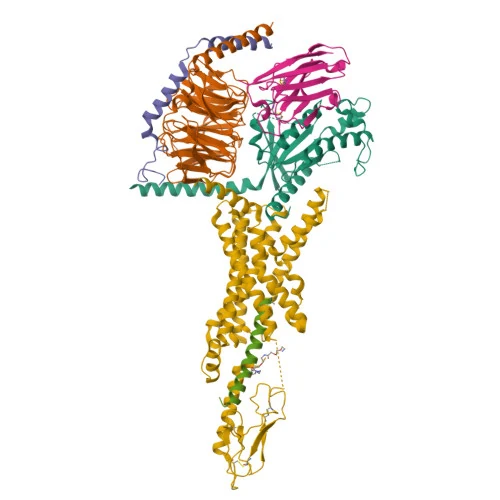
What the 3D view shows is the peptide inside its receptor, not the molecule by itself. In PDB 7KI0 the chain is entity 5, label chain E, auth chain P, modelled over residues 1 to 30 of 31 against the receptor's transmembrane and extracellular domains, with the Gs heterotrimer and a nanobody below. The only part of the side chain with coordinates is the pair of AEEA spacers, deposited as the separate chemical component WF1 and bonded to the lysine side-chain nitrogen; the gamma-glutamate and the entire eighteen-carbon diacid are missing. So the picture settles how the backbone sits in the pocket and says nothing about the feature the molecule was built for.[5,3]
The earlier crystal structure makes the same point from the other side. The programme's own review states that it was not possible to obtain a crystal structure of the molecule, so the unacylated chain was solved with the receptor's extracellular domain, and the overall arrangement was reported as the same as native GLP-1(7-37). In that structure the substituted arginine takes a more defined orientation than the lysine it replaced, with a water molecule coordinated between it and a glutamate of the peptide, which is an observation about the backbone substitution rather than about the lipid.[2,19]
Set against its nearest relatives, the design reads as a short list of decisions. Native GLP-1(7-37) is the same 31-residue chain with alanine at the second position, no constrained residue and no lipid, and it is cleaved at that second position by dipeptidyl peptidase IV. GLP-1 LR, the earlier molecule from the same programme, keeps the native alanine there and hangs a sixteen-carbon mono-acid on the lysine through a gamma-glutamate alone. GLP-1 SM changes three things at once: 2-aminoisobutyric acid at the second position, so the cleavage site is stiffened and the residue loses its stereocentre; arginine for a lysine near the C-terminus, so one lysine is left and the acylation has a single site; and an eighteen-carbon diacid reached through a gamma-glutamate and two AEEA spacers, which is the longest arm the published series found before receptor potency began to fall again. Taspoglutide, solved in the same cryo-EM study, takes the other road entirely: two constrained positions and no lipid. The sources describe this as a trade, not a free gain, since the chemistry that raised albumin affinity lowered receptor affinity about three-fold against GLP-1 LR.[1,2,3,18]
- His1
- Aib2
- Glu3
- Gly4
- Thr5
- Phe6
- Thr7
- Ser8
- Asp9
- Val10
- Ser11
- Ser12
- Tyr13
- Leu14
- Glu15
- Gly16
- Gln17
- Ala18
- Ala19
- Lys(acyl)20
- Glu21
- Phe22
- Ile23
- Ala24
- Trp25
- Leu26
- Val27
- Arg28
- Gly29
- Arg30
- Gly31
Modifications
- 2 (GLP-1 numbering 8) 2-aminoisobutyric acid (Aib, alpha-methylalanine) in place of alanine. The second methyl group on the alpha carbon makes the residue achiral and constrains the local backbone. Modelled in the deposited cryo-EM entry as chemical component AIB.[1,15,5]
- 20 (GLP-1 numbering 26) The lysine side-chain nitrogen is acylated by two consecutive 2-[2-(2-aminoethoxy)ethoxy]acetyl (AEEA, also written OEG) spacers, then a gamma-linked L-glutamate, then octadecanedioic acid, an eighteen-carbon chain with a carboxylic acid at each end. The distal carboxylate is left free. The registry systematic name gives the whole arm as N6-[N-(17-carboxy-1-oxoheptadecyl)-L-gamma-glutamyl-AEEA-AEEA].[14,15,1,2]
- 28 (GLP-1 numbering 34) Arginine in place of the lysine present in human GLP-1. The point of the substitution is that it leaves one lysine in the chain, so acylation is site specific.[1,2,15]
The backbone is all-L apart from the four glycines and the single Aib residue, which are achiral. The linker glutamate is joined through its gamma carboxyl, and the FDA substance record names it as the L enantiomer (N6-[N-(17-carboxy-1-oxoheptadecyl)-L-gamma-glutamyl-...]); the stereo-specific structure PubChem holds for CID 56843331 encodes that one centre the other way, which is recorded under gaps. There is no cysteine in the chain, so no disulfide; the peptide is linear and uncyclised, with no glycosylation and no metal coordination in the molecule as defined. The only branch point is the Lys20 side-chain nitrogen bearing the two AEEA spacers, the gamma-glutamate and the C18 diacid.[15,14,1]
What studies on GLP-1 SM show
Each card names the experimental system and what was measured. A result in one system is not evidence about another.
Cryo-electron microscopy with 3D variability analysis · Purified complexes of the human GLP-1 receptor with heterotrimeric Gs and the nanobody Nb35
The peptide makes interactions with the receptor reported as similar to those of native GLP-1, while the two analogues differ from each other and from the native hormone in the motions seen within the receptor and the bound peptide. The deposited entry resolves the complex at 2.5 Å.[3]
- Material
- GLP-1 SM, modelled over residues 1 to 30 with only the two AEEA spacers of the side chain present
- Measured
- Structure of the agonist-bound receptor and G protein complex, and the distribution of motions within receptor and peptide extracted from the particle set
- Compared with
- Taspoglutide, solved in the same study and deposited separately as 7KI1, and published complexes of the same receptor with native GLP-1
- Limitation
- Read here as an abstract plus the deposited entries; the full text was behind a paywall, so the contact-by-contact description and any numerical comparison of the motions were not verified at source. One laboratory, one deposition pair, and the lipid arm is almost entirely unmodelled.
Zhang X et al., 2021, Cell Reports
Receptor binding, albumin affinity and pharmacokinetic screening · Cloned human GLP-1 receptor; serum albumin binding assays; minipigs
Receptor affinity of the selected molecule was 0.38 plus or minus 0.06 nM, about three-fold lower than GLP-1 LR's, with albumin affinity increased; plasma half-life in minipigs was reported as 46.1 h. The design conclusion recorded is that the fatty acid and the linking chemistry together carry both properties.[1]
- Material
- GLP-1 SM and a series of analogues of the same backbone with fatty acids and linkers varied
- Measured
- Receptor affinity, albumin affinity, and plasma half-life in minipigs
- Compared with
- GLP-1 LR and native GLP-1
- Limitation
- Abstract only, and the work is the originating laboratory's own. Assay formats, albumin concentrations and the species of receptor construct were not verified at source, and no independent curator of these affinity values was reachable.
Lau J et al., 2015, Journal of Medicinal Chemistry
Structure-activity review reproducing the programme's own assay tables · Baby hamster kidney cells expressing the cloned human GLP-1 receptor, with and without added human serum albumin; analytical ultracentrifugation; rats and minipigs
Receptor potency fell sharply when the fatty acid was placed near the N-terminus and when two acids were attached; among diacids, potency rose with chain length up to C18 and reversed beyond it, and the C18 diacid showed the largest affinity shift on adding albumin. Reported half-lives in rats ranged from 1.2 h for the C12 diacid to about 7 h for the selected molecule, with longer values in minipigs.[2]
- Material
- Aib8,Arg34-GLP-1(7-37) acylated at Lys26 with diacids from C12 to C22 through gamma-glutamate and OEG-based linkers
- Measured
- Half-maximal effective concentration at the receptor, relative albumin affinity, and half-life in rats and minipigs across the series
- Compared with
- Native GLP-1(7-37), GLP-1 LR, and acylation at other lysine positions
- Limitation
- A review restating one sponsor's data rather than an independent replication, and the figures are spread across adapted tables and figures from earlier papers. Potencies are cell-based readouts in a recombinant system and the albumin affinities are relative rather than absolute.
Knudsen LB et al., 2019, Frontiers in Endocrinology
Bioluminescence resonance energy transfer signalling and trafficking assays with live-cell microscopy · HEK293T cells transiently transfected with tagged human GLP-1 or GIP receptors and reporter constructs, plus a Min6 beta cell model
GLP-1 SM recruited Gs comparably to native GLP-1 at every concentration tested and produced similar receptor internalisation dynamics, while recruiting 67 percent and 78 percent of the native peptide's maximal beta-arrestin 1 and beta-arrestin 2 response. It did not differ from the native peptide in co-localisation of the receptor with early or recycling endosome markers. In the same panel the two dual agonists showed markedly reduced internalisation and arrestin recruitment.[4]
- Material
- GLP-1 SM supplied by the originator, beside synthesised comparator peptides
- Measured
- Gs and Gq recruitment, cAMP accumulation, receptor internalisation, beta-arrestin 1 and 2 recruitment, and co-localisation of the receptor with Rab5, Rab7 and Rab11 endosome markers
- Compared with
- Native GLP-1(7-36) amide, native GIP(1-42), a pharmacokinetically matched acylated GLP-1 analogue, and the dual agonists GLP-2 TZ and MAR709
- Limitation
- Transient over-expression in a cell line, with the authors noting that Rab marker over-expression can itself distort trafficking and that transfer to physiologically relevant beta cells was limited to one model. The comparison that the study was designed to make is about the dual agonists; GLP-1 SM is the mono-agonist control.
Novikoff A et al., 2021, Molecular Metabolism
Nuclear magnetic resonance, surface plasmon resonance and an in vitro tissue catabolism assay with liquid chromatography and high-resolution mass spectrometry · Solutions of the peptides, immobilised albumin, hypodermal tissue in vitro, and rat plasma
Oligomerisation had a shielding effect against catabolism, while albumin binding gave only limited protection in the hypodermal tissue compartment because the unbound fraction there is higher than in plasma. Catabolites recovered from rat plasma agreed with the in vitro set, with the major cleavage site reported as Tyr19-Leu20.[29]
- Material
- GLP-1 SM and GLP-1 LR and analogues of both
- Measured
- Oligomerisation propensity, albumin interaction, and the identity of catabolites
- Compared with
- The two marketed analogues against each other and against their variants
- Limitation
- Abstract only, so the numbering convention behind the reported cleavage site was not confirmed against the paper's own sequence figure. An in vitro tissue assay is a model of the hypodermal tissue compartment and not a measurement in an animal.
Gallo M et al., 2022, Journal of Pharmaceutical and Biomedical Analysis
Structural mass spectrometry, several methods combined · Aqueous solutions of the peptide at varying concentration, with and without added metal ions, and with human serum albumin
Oligomers up to ten subunits formed within 24 hours. Low micromolar copper and zinc produced higher-order and more abundant oligomers, attributed to metal binding stabilising the small oligomers. Oligomers associated through their hydrophobic C-terminal ends in a parallel orientation, leaving the N-terminal region and the lipid chain loosely engaged and available to bind albumin, an interaction reported to dissociate the oligomers and to do so more strongly when the albumin was glycated.[20]
- Material
- GLP-1 SM
- Measured
- Oligomer stoichiometry and heterogeneity, thermal and structural stability, the interface used for self-assembly, and relative monomer orientation
- Compared with
- Solutions without metal ions, and albumin in glycated versus non-glycated form
- Limitation
- Abstract only. Gas-phase and solution measurements on a purified peptide under chosen buffer conditions; the concentrations are assay conditions and say nothing about any other setting.
Huang S et al., 2026, Journal of the American Chemical Society
Cryogenic transmission electron microscopy, small-angle X-ray scattering and molecular dynamics · Aqueous solutions across a concentration range, plus a Caco-2 epithelial cell monolayer
Slow aggregation into spherical micelles at sufficiently high concentration, with a small population of needle-shaped fibrils, and dimers and trimers at lower concentration; micelles once formed were stable to further ageing. The aggregation state altered the peptide's effect on the permeability of the epithelial model membrane.[23]
- Material
- GLP-1 SM
- Measured
- Aggregate morphology and size, the species present at each concentration, and permeability of the cell monolayer
- Compared with
- Solutions at lower concentration and freshly prepared against aged
- Limitation
- Abstract only, and the companion study from the same group reports beta-sheet fibrils at pH 2.4 as a separate regime, so morphology here is a function of the chosen conditions rather than a property of the molecule on its own.
Hamley IW et al., 2025, Biomacromolecules
Impurity profiling with an antigen-presentation proteomics assay and a fibrillation assay · Monocyte-derived dendritic cells from healthy donors stimulated with sample impurities; peptide samples held under light exposure
Follow-on and compounded samples carried impurity profiles distinct from reference product, including amino acid deletions and additions and impurities that were not identified, and presented a different set of peptides on the dendritic cells. Compounded material differed significantly from reference product in strength, summed impurities and high molecular weight protein level after light exposure.[28]
- Material
- Follow-on and compounded material labelled as GLP-1 SM or GLP-1 LR, as drug substance and as finished product
- Measured
- Which peptides were presented on the cells, impurity profiles by chromatography and mass spectrometry, strength, total impurity, high molecular weight protein content, and physical stability
- Compared with
- The corresponding reference products
- Limitation
- Abstract only, and the work is by the reference product's manufacturer, which is a clear interest in the result. The assays report what a sample contains and what a cell assay presented; they establish nothing about what any particular preparation elsewhere contains.
Kopp KL et al., 2026, Pharmaceutical Research
Radiolabelled absorption, metabolism and excretion study · Healthy human volunteers, with parallel studies in rats and monkeys; radioactivity measured in blood, plasma, urine and faeces, and in expired air for humans and rats
The intact molecule was the primary circulating component in all three species, accounting for 69 to 83 percent of total compound-related material, and was metabolised before excretion. Recovery of excreted radioactivity was 75.1 percent in humans, 72.1 percent in rats and 58.2 percent in monkeys, with urine the primary route in all three. Breakdown was by proteolytic cleavage of the peptide backbone together with sequential beta-oxidation of the fatty acid side chain, and was not confined to particular organs. Intact molecule recovered in urine accounted for 3.1 percent of the administered radioactivity in humans, under 1 percent in rats, and none was detected in monkeys.[30]
- Material
- Tritium-labelled GLP-1 SM
- Measured
- Blood-to-plasma ratio, the share of circulating compound-related material accounted for by the intact molecule, metabolite profiles in plasma, urine and faeces, and recovery of excreted radioactivity
- Compared with
- The two nonclinical species against the human data
- Limitation
- One study, largely by the originating company, read here as an abstract. Radiolabel accounting says where compound-related material went and does not identify the structure of every metabolite, and the percentages belong to the species and the sampling schedule used.
Jensen L et al., 2017, European Journal of Pharmaceutical Sciences
Bioanalytical method development with a disposition study in rats · Rat plasma and brain tissue; a liquid chromatography tandem mass spectrometry assay built and validated for the purpose
The paper's contribution is the assay rather than a result about the molecule: it reports a method sensitive and specific enough to follow this compound in rat plasma and then applies it to a disposition and brain distribution study in that species. It is cited here as the analytical basis for measuring the compound, which is the kind of work the rest of this page's figures depend on.[31]
- Material
- The compound itself, measured as the intact analyte rather than through a surrogate
- Measured
- Concentrations of the compound over time in plasma and in brain tissue, by the validated assay
- Compared with
- Plasma against brain tissue, as the two compartments sampled
- Limitation
- Abstract read rather than full text, so the assay's validation figures, the sampling schedule and the exposure amounts are not carried here. It is one species and the brain compartment measurement is tissue concentration, which does not by itself establish that the molecule crosses into brain parenchyma rather than remaining in its vasculature.
Lee TS et al., 2023, Journal of Chromatography B
What has been measured about GLP-1 SM at the GLP-1 receptor, and what the lipid arm does instead
At the receptor, the evidence is that this molecule behaves close to the native hormone. The cryo-EM structure reports interactions similar to those of GLP-1, and the signalling panel that put it beside the dual agonists found Gs recruitment, internalisation and endosomal routing indistinguishable from GLP-1(7-36) amide, with arrestin recruitment somewhat lower at 67 and 78 percent of the native maximum for the two arrestins. That is the opposite of the pattern reported for the dual agonists in the same experiment, which recruited far less arrestin and internalised the receptor much less. So the backbone changes, which were chosen to resist an enzyme and to free a single lysine, did not buy a different signalling profile and were not meant to.[3,4]
The side chain carries the property the design was for, and it works away from the receptor. A C18 diacid on a gamma-glutamate and two polyethylene-glycol spacers binds serum albumin reversibly; the published structure-activity series shows the tension directly, since the same chemistry that raises albumin affinity lowers receptor potency once it is too long or placed too near the N-terminus. A molecular dynamics study of the peptide with human serum albumin ranks the FA3 to FA4 fatty acid site as the most favourable of those tested and names two arginine residues of albumin as localising the diacid, which is a computed result consistent with earlier solution spectroscopy rather than an experimental structure of the pair.[2,1,32]
Two further behaviours of the molecule are chemistry rather than pharmacology and are measured as such. It self-associates, through its hydrophobic C-terminal end, into species ranging from dimers to ten-subunit oligomers, micelles and fibrils depending on concentration, time, pH and the presence of copper or zinc, and that association both shields it from catabolism and competes with albumin binding; a separate study reports that hydrophobic surfaces catalyse the formation of a colloidal phase from solutions of the peptide, with the size of the colloids predicted by a classical model. Fate in the body has been traced with a tritium label: the intact molecule was the main circulating species in humans, rats and monkeys, and it was broken down by cleavage of the peptide backbone together with stepwise beta-oxidation of the fatty acid chain, which is degradation of both halves of the design rather than of one. And in rodents the labelled molecule was reported not to cross the blood-brain barrier, reaching the brain instead at circumventricular organs and sites adjacent to the ventricles, with c-Fos activity measured in ten brain areas of treated rats and transcript changes in the area postrema; that is a distribution and activity measurement in named species and it is not a statement about any other organism.[20,21,22,23,33,29,30,34]
What is still unknown about GLP-1 SM
There is no structure of this molecule by itself, and no structure of its lipid arm. The deposited cryo-EM entry models the backbone to residue 30 and only the two AEEA spacers of the side chain; the crystal structure that preceded it used the unacylated chain because, as the programme's own review states, it was not possible to obtain a crystal structure of the molecule. The consequence is that the one feature distinguishing it from the native hormone has never been seen in atomic detail, either free or on albumin. The albumin interaction is described from relative affinity measurements, from solution spectroscopy and from simulation, not from a solved complex.[5,19,2,32]
The receptor affinity figure quoted everywhere traces to one abstract from the originating laboratory. The Guide to Pharmacology record for this ligand exists but its web service refused an unauthenticated request, the discovery paper is not open access, and the review that restates the structure-activity tables is by the same authors. So the single number on this page has no independent source behind it, and that is a gap rather than a doubt about the molecule's identity, which is settled by several mutually consistent registry records.[1,2,14,15]
The registry records do not agree on the formula. Rebuilding the molecule from its sequence and side chain reproduces the PubChem formula and monoisotopic mass exactly, while the FDA substance record prints a different formula and a lower calculated weight. The FDA record does carry the correct structural modifications, so this reads as a computation on an incomplete representation rather than a dispute about what the molecule is, but anyone copying a formula from a registry field should check which one they have taken.[14,15]
What a given preparation contains is an open question that only measurement closes. Comparisons of follow-on and compounded material against reference product report new impurities, unidentified impurities, deletions and additions in the chain, high molecular weight species, trace metals and counterions, and in some products a lower peptide content than the label stated; those studies come from the reference manufacturer, which is an interest to weigh, but the analytical findings are specific and checkable. Independent laboratories have in parallel built and validated methods that identify this peptide and its metabolites in serum, dried blood spots and urine, so the analytical means to answer the question for any particular sample exist and are published.[27,28,35,36,37,38,39]
Key papers on GLP-1 SM
- Discovery of the Once-Weekly Glucagon-Like Peptide-1 (GLP-1) Analogue [GLP-1 SM].
Lau J, Bloch P, Schäffer L, Pettersson I, Spetzler J, Kofoed J, Madsen K, Knudsen LB, McGuire J, Steensgaard DB, Strauss HM, Gram DX, Knudsen SM, Nielsen FS, Thygesen P, Reedtz-Runge S, Kruse T (2015), Journal of Medicinal Chemistry · Discovery report with receptor binding, albumin affinity and animal pharmacokinetics
The discovery report. Every later statement about the backbone substitutions, the choice of a C18 diacid and the receptor affinity traces here.
- The Discovery and Development of [GLP-1 LR] and [GLP-1 SM].
Knudsen LB, Lau J (2019), Frontiers in Endocrinology · Review by the programme's own authors, with reproduced assay tables
Open access, and the only place the programme's structure-activity tables are laid out in full, including the potency cost of moving or lengthening the fatty acid and the reason the molecule itself was not crystallised.
- Structure and dynamics of [GLP-1 SM]- and taspoglutide-bound GLP-1R-Gs complexes.
Zhang X, Belousoff MJ, Liang YL, Danev R, Sexton PM, Wootten D (2021), Cell Reports · Cryo-EM structures with 3D variability analysis
The structural account, and the entry behind the 3D view on this page. Read it together with the deposited file to see how little of the side chain is actually modelled.
- Spatiotemporal GLP-1 and GIP receptor signaling and trafficking/recycling dynamics induced by selected receptor mono- and dual-agonists.
Novikoff A, O'Brien SL, Bernecker M, Grandl G, Kleinert M, Knerr PJ, Stemmer K, Klingenspor M, Zeigerer A, DiMarchi R, Tschöp MH, Finan B, Calebiro D, Müller TD (2021), Molecular Metabolism · Signalling, internalisation and endosomal trafficking assays
The most informative independent receptor-level measurement located: it puts this peptide beside the native hormone and the dual agonists in one panel of signalling and trafficking assays, and it is open access.
- Structural Mass Spectrometry Resolves the Conformational Dynamics of [GLP-1 SM] Oligomerization.
Huang S, Zhou Y, Lui TY, R Benzenberg L, Zenobi R, Wu R (2026), Journal of the American Chemical Society · Structural mass spectrometry of oligomerisation
The current best account of what the molecule does in solution on its own, including the self-assembly interface and the effect of copper and zinc.
- Oligomerization, albumin binding and catabolism of therapeutic peptides in the subcutaneous compartment: An investigation on lipidated GLP-1 analogs.
Gallo M, Vanni D, Esposito S, Alaimo N, Orvieto F, Rulli F, Missineo A, Caretti F, Bonelli F, Veneziano M, Orsatti L, Monteagudo E (2022), Journal of Pharmaceutical and Biomedical Analysis · NMR and SPR with an in vitro tissue catabolism assay
Independent work connecting self-association, albumin binding and proteolysis, and the source of the reported major cleavage site.
- Impurities and Potential Immunogenicity Associated With Follow-on and Compounded Glucagon-like Peptide-1 Receptor Agonists.
Kopp KL, Lamberth K, Schelde O, Øgendahl AK, Wojcieszek M, Mogensen JE, Ramírez-Andersen HS, Schneider CL, Staby A, Hach M (2026), Pharmaceutical Research · Impurity profiling with an antigen-presentation assay
Read with its authorship in mind, it is the most detailed published analysis of how material labelled as this peptide can differ from reference product.
- Novel LC-MS/MS analysis of the GLP-1 analog [GLP-1 SM] with its application to pharmacokinetics and brain distribution studies in rats.
Lee TS, Park EJ, Choi M, Oh HS, An Y, Kim T, Kim TH, Shin BS, Shin S (2023), Journal of Chromatography B · LC-MS/MS method with rat pharmacokinetics and tissue distribution
A worked example of the analytical side: an independent validated method quantifying the peptide in rat plasma and brain tissue, with the partition into brain tissue reported as very low.
References
- 1.Lau J, Bloch P, Schäffer L, Pettersson I, Spetzler J, Kofoed J, Madsen K, Knudsen LB, McGuire J, Steensgaard DB, Strauss HM, Gram DX, Knudsen SM, Nielsen FS, Thygesen P, Reedtz-Runge S, Kruse T. (2015). Discovery of the Once-Weekly Glucagon-Like Peptide-1 (GLP-1) Analogue [GLP-1 SM]. Journal of Medicinal Chemistry. 58:7370-7380. doi:10.1021/acs.jmedchem.5b00726 · abstract only
- 2.Knudsen LB, Lau J. (2019). The Discovery and Development of [GLP-1 LR] and [GLP-1 SM]. Frontiers in Endocrinology. 10:155. doi:10.3389/fendo.2019.00155 · full text read
- 3.Zhang X, Belousoff MJ, Liang YL, Danev R, Sexton PM, Wootten D. (2021). Structure and dynamics of [GLP-1 SM]- and taspoglutide-bound GLP-1R-Gs complexes. Cell Reports. 36:109374. doi:10.1016/j.celrep.2021.109374 · abstract only
- 4.Novikoff A, O'Brien SL, Bernecker M, Grandl G, Kleinert M, Knerr PJ, Stemmer K, Klingenspor M, Zeigerer A, DiMarchi R, Tschöp MH, Finan B, Calebiro D, Müller TD. (2021). Spatiotemporal GLP-1 and GIP receptor signaling and trafficking/recycling dynamics induced by selected receptor mono- and dual-agonists. Molecular Metabolism. 49:101181. doi:10.1016/j.molmet.2021.101181 · full text read
- 5.Zhang X, Belousoff MJ, Liang YL, Danev R, Sexton PM, Wootten D. (2021). PDB 7KI0: [GLP-1 SM]-bound Glucagon-Like Peptide-1 (GLP-1) Receptor in Complex with Gs protein. RCSB Protein Data Bank. doi:10.2210/pdb7KI0/pdb · database record
- 6.U.S. Food and Drug Administration. (2026). Drugs@FDA (openFDA drugsfda endpoint): submission records for applications NDA 209637, NDA 213051 and NDA 215256, original approval dates and approved efficacy supplements. Drugs@FDA. source · database record
- 7.U.S. Food and Drug Administration. (2026). Prescribing information, section 1, Indications and Usage: [GLP-1 SM type 2 diabetes medicine], NDA 209637 (label effective 2026-07-30). openFDA drug label database. source · full text read
- 8.U.S. Food and Drug Administration; DailyMed, U.S. National Library of Medicine. (2026). Prescribing information, section 1, Indications and Usage: [GLP-1 SM chronic weight management medicine], NDA 215256 (revised June 2026). DailyMed. source · full text read
- 9.European Medicines Agency. (2026). Medicines output: European public assessment reports data set, entries for the authorised [GLP-1 SM] medicines (type 2 diabetes, weight management, MASH). European Medicines Agency, medicines data (download). source · database record
- 10.Health Canada. (2026). Product monograph: [GLP-1 SM type 2 diabetes medicine], DIN 02471469, section 1, Indications. Health Canada Drug Product Database. source · full text read
- 11.Health Canada. (2026). Product monograph: [GLP-1 SM chronic weight management medicine], DIN 02522578, section 1, Indications. Health Canada Drug Product Database. source · full text read
- 12.Nationale Anti Doping Agentur Deutschland. (2025). WADA publishes Prohibited List 2026 (including changes to the 2026 Monitoring Program). NADA Germany, news. source · full text read
- 13.U.S. Food and Drug Administration. (2026). Prescribing information, boxed warning and section 6, Adverse Reactions: [GLP-1 SM], risk of thyroid C-cell tumours, serious adverse reactions and reactions at an incidence of 5 per cent or more (label set id 27f15fac-7d98-4114-a2ec-92494a91da98, effective 2026-01-30). openFDA drug label database. source · full text read
- 14.PubChem. [GLP-1 SM], compound summary CID 56843331. PubChem, National Center for Biotechnology Information. source · database record
- 15.FDA Global Substance Registration System. [GLP-1 SM], substance record, UNII 53AXN4NNHX. FDA GSRS. source · database record
- 16.ChEBI. [GLP-1 SM], CHEBI:167574. Chemical Entities of Biological Interest, EMBL-EBI. source · database record
- 17.Kanehisa Laboratories. KEGG DRUG entry D10025: [GLP-1 SM]. KEGG, Kyoto Encyclopedia of Genes and Genomes. source · database record
- 18.Zhang X, Belousoff MJ, Liang YL, Danev R, Sexton PM, Wootten D. (2021). PDB 7KI1: Taspoglutide-bound Glucagon-Like Peptide-1 (GLP-1) Receptor in Complex with Gs Protein. RCSB Protein Data Bank. doi:10.2210/pdb7KI1/pdb · database record
- 19.Lau J, Bloch P, Schäffer L, Pettersson I, Spetzler J, Kofoed J, Madsen K, Knudsen LB, McGuire J, Steensgaard DB, Strauss HM, Gram DX, Knudsen SM, Nielsen FS, Thygesen P, Reedtz-Runge S, Kruse T. (2015). PDB 4ZGM: Crystal structure of [GLP-1 SM] peptide backbone in complex with the GLP-1 receptor extracellular domain. RCSB Protein Data Bank. doi:10.2210/pdb4ZGM/pdb · database record
- 20.Huang S, Zhou Y, Lui TY, R Benzenberg L, Zenobi R, Wu R. (2026). Structural Mass Spectrometry Resolves the Conformational Dynamics of [GLP-1 SM] Oligomerization. Journal of the American Chemical Society. 148:41169-41183. doi:10.1021/jacs.6c12077 · abstract only
- 21.Castelletto V, de Mello LR, Seitsonen J, Hamley IW. (2026). Fibrillar and Micellar Aggregation of [GLP-1 SM] and Formation of a Chiral-Imprinted Glass. Biomacromolecules. 27:2818-2827. doi:10.1021/acs.biomac.5c02669 · abstract only
- 22.Venanzi M, Savioli M, Cimino R, Gatto E, Palleschi A, Ripani G, Cicero D, Placidi E, Orvieto F, Bianchi E. (2020). A spectroscopic and molecular dynamics study on the aggregation process of a long-acting lipidated therapeutic peptide: the case of [GLP-1 SM]. Soft Matter. 16:10122-10131. doi:10.1039/d0sm01011a · abstract only
- 23.Hamley IW, de Mello LR, Castelletto V, Zinn T, Cowieson N, Seitsonen J, Bizien T. (2025). [GLP-1 SM] Aggregates into Oligomeric Micelles and Short Fibrils in Aqueous Solution. Biomacromolecules. 26:3786-3794. doi:10.1021/acs.biomac.5c00342 · abstract only
- 24.UniProt Consortium. Glucagon-like peptide 1 receptor, Homo sapiens, UniProtKB P43220. UniProtKB. source · database record
- 25.Buckley ST, Bækdal TA, Vegge A, Maarbjerg SJ, Pyke C, Ahnfelt-Rønne J, Madsen KG, Schéele SG, Alanentalo T, Kirk RK, Pedersen BL, Skyggebjerg RB, Benie AJ, Strauss HM, Wahlund PO, Bjerregaard S, Farkas E, Fekete C, Søndergaard FL, Borregaard J, Hartoft-Nielsen ML, Knudsen LB. (2018). Transcellular stomach absorption of a derivatized glucagon-like peptide-1 receptor agonist. Science Translational Medicine. 10:eaar7047. doi:10.1126/scitranslmed.aar7047 · abstract only
- 26.Colston KJ, Faivre KT, Schneebeli ST. (2025). Permeation enhancer-induced membrane defects assist the oral absorption of peptide drugs. Nature Communications. 16:9512. doi:10.1038/s41467-025-64891-0 · abstract only
- 27.Hach M, Engelund DK, Mysling S, Mogensen JE, Schelde O, Haselmann KF, Lamberth K, Vilhelmsen TK, Malmstrøm J, Højlys-Larsen KB, Rasmussen TS, Borch-Jensen J, Mortensen RW, Jensen TMT, Kesting JR, Catarig AM, Asgreen DJ, Christensen L, Staby A. (2024). Impact of Manufacturing Process and Compounding on Properties and Quality of Follow-On GLP-1 Polypeptide Drugs. Pharmaceutical Research. 41:1991-2014. doi:10.1007/s11095-024-03771-6 · abstract only
- 28.Kopp KL, Lamberth K, Schelde O, Øgendahl AK, Wojcieszek M, Mogensen JE, Ramírez-Andersen HS, Schneider CL, Staby A, Hach M. (2026). Impurities and Potential Immunogenicity Associated With Follow-on and Compounded Glucagon-like Peptide-1 Receptor Agonists. Pharmaceutical Research. 43:2861-2886. doi:10.1007/s11095-026-04146-9 · abstract only
- 29.Gallo M, Vanni D, Esposito S, Alaimo N, Orvieto F, Rulli F, Missineo A, Caretti F, Bonelli F, Veneziano M, Orsatti L, Monteagudo E. (2022). Oligomerization, albumin binding and catabolism of therapeutic peptides in the subcutaneous compartment: An investigation on lipidated GLP-1 analogs. Journal of Pharmaceutical and Biomedical Analysis. 210:114566. doi:10.1016/j.jpba.2021.114566 · abstract only
- 30.Jensen L, Helleberg H, Roffel A, van Lier JJ, Bjørnsdottir I, Pedersen PJ, Rowe E, Derving Karsbøl J, Pedersen ML. (2017). Absorption, metabolism and excretion of the GLP-1 analogue [GLP-1 SM] in humans and nonclinical species. European Journal of Pharmaceutical Sciences. 104:31-41. doi:10.1016/j.ejps.2017.03.020 · abstract only
- 31.Lee TS, Park EJ, Choi M, Oh HS, An Y, Kim T, Kim TH, Shin BS, Shin S. (2023). Novel LC-MS/MS analysis of the GLP-1 analog [GLP-1 SM] with its application to pharmacokinetics and brain distribution studies in rats. Journal of Chromatography B. 1221:123688. doi:10.1016/j.jchromb.2023.123688 · abstract only
- 32.Liu Y, Wen W, Xu B, Wang L, Zhao Y, Zhang J. (2026). Molecular dynamics insights into the binding interactions of [GLP-1 SM] with human serum albumin. Journal of Biomolecular Structure and Dynamics. 44:3361-3378. doi:10.1080/07391102.2025.2532848 · abstract only
- 33.Li Q, Tangry V, Allen DP, Seibert KD, Qian KK, Wagner NJ. (2024). Surface-mediated spontaneous emulsification of the acylated peptide, [GLP-1 SM]. Proceedings of the National Academy of Sciences of the United States of America. 121:e2305770121. doi:10.1073/pnas.2305770121 · abstract only
- 34.Gabery S, Salinas CG, Paulsen SJ, Ahnfelt-Rønne J, Alanentalo T, Baquero AF, Buckley ST, Farkas E, Fekete C, Frederiksen KS, Helms HCC, Jeppesen JF, John LM, Pyke C, Nøhr J, Lu TT, Polex-Wolf J, Prevot V, Raun K, Simonsen L, Sun G, Szilvásy-Szabó A, Willenbrock H, Secher A, Knudsen LB, Hogendorf WFJ. (2020). [GLP-1 SM] lowers body weight in rodents via distributed neural pathways. JCI Insight. 5:e133429. doi:10.1172/jci.insight.133429 · abstract only
- 35.Malgave A, Akbar S, Joseph A, Dande Aishwarya, Peraman R, Malayandi R. (2025). Effect of pH, buffers, molarity, and temperature on solution state degradation of [GLP-1 SM] using LC-HRMS: A preformulation protocol for peptide drug delivery. European Journal of Pharmaceutics and Biopharmaceutics. 214:114780. doi:10.1016/j.ejpb.2025.114780 · abstract only
- 36.Cong Y, Tang C, Li Z, Gao Y, Chen L, Xu H, Diao X. (2026). Metabolite Profiling and Identification of [GLP-1 SM] in Liver S9 Across Species and Rat Plasma. Biomedical Chromatography. 40:e70415. doi:10.1002/bmc.70415 · abstract only
- 37.Timms M, Bowen C, Steel R. (2026). Identification of [GLP-1 SM] use through detection of U6 and U7 metabolites in human urine. Analytical and Bioanalytical Chemistry. 418:1115-1124. doi:10.1007/s00216-025-06263-7 · abstract only
- 38.Borschel L, Thomas A, Studer D, Penlioglou T, Efthymiou V, Bally L, Thevis M. (2026). Development and validation of qualitative methods for the detection of GLP-1 receptor agonists in serum and dried blood spots by LC-HRMS/MS for doping control purposes. Journal of Chromatography B. 1285:125326. doi:10.1016/j.jchromb.2026.125326 · abstract only
- 39.Tian T, Chen X, Deng X. (2026). Detection and Excretion Profiling of [GLP-1 SM] Use in Human Urine by UHPLC-HRMS for Anti-Doping Purpose. Drug Testing and Analysis. doi:10.1002/dta.70160 · abstract only
- 40.Zhou Q, Zhao F, Zhang Y, Yang D, Wang MW. (2025). Structural pharmacology and mechanisms of GLP-1R signaling. Trends in Pharmacological Sciences. 46:422-436. doi:10.1016/j.tips.2025.03.003 · abstract only
- 41.van der Velden WJC, Andresen AF, Rosenkilde MM. (2026). Structural Overview of GLP-1 Analogs. Biomedical Journal. 101037. doi:10.1016/j.bj.2026.101037 · abstract only
Related entries
- GLP-2 TZGLP-2 TZ is a synthetic 39-residue peptide carrying a twenty-carbon fatty-diacid side chain on Lys20. It activates two human class B1 G protein-coupled receptors, the GIP receptor and the GLP-1 receptor, with measurably different signalling behaviour at each.
- GLP-3 RTGLP-3 RT is an investigational synthetic 39-residue peptide from Eli Lilly, carrying a fatty-diacid side chain on Lys17. Within one molecule it activates three human class B1 G protein-coupled receptors, the GIP, GLP-1 and glucagon receptors.
- CagrilintideCagrilintide is a long-acting synthetic analogue of the pancreatic hormone amylin: 37 residues carrying a C20 fatty diacid on the N-terminal lysine. It is investigational and approved nowhere.
- MazdutideMazdutide is a synthetic 34-residue peptide built on the human oxyntomodulin sequence, carrying a C20 fatty diacid on the Lys20 side chain. It also carries 2-aminoisobutyric acid at position 2 and a C-terminal glycinamide, and is characterised in the literature as an agonist at both the human GLP-1 receptor and the human glucagon receptor.
- SurvodutideSurvodutide, development code BI 456906, is an investigational synthetic peptide built on the human glucagon sequence. It is characterised in the literature as an agonist at both the human glucagon and GLP-1 receptors, and carries a C18 fatty-diacid arm that binds albumin.
