SS-31 in brief
SS-31, also called elamipretide, is a synthetic tetrapeptide, D-Arg-Dmt-Lys-Phe-NH2, carrying a D-arginine, the non-proteinogenic residue 2',6'-dimethyltyrosine and a C-terminal amide. It belongs to the Szeto-Schiller series of cationic-aromatic peptides that concentrate in mitochondria, and under the name elamipretide it received an accelerated United States approval in September 2025 for Barth syndrome. Most of the molecular evidence comes from model membranes, isolated mitochondria and rodent tissue, and centres on selective binding to cardiolipin, a lipid of the inner mitochondrial membrane. No structure of the molecule has been deposited and no affinity for any protein target has been measured.[1,2,3,4,5,6]
SS-31 regulatory status
Under the name elamipretide the peptide was approved in the United States on 19 September 2025 as a new molecular entity, by the accelerated pathway, to improve muscle strength in adult and paediatric patients with Barth syndrome at or above 30 kilograms of body weight, which made it the first treatment approved specifically for that disorder. The approval rests on an intermediate clinical endpoint, and continued approval may depend on a confirmatory trial. The approved product contains the trihydrochloride rather than the free base. No EMA or Health Canada marketing authorisation was found in the EMA medicines database or the Health Canada Drug Product Database (searched November 2026). Elamipretide is named nowhere on the WADA 2026 Prohibited List, and it appears neither in the FDA list of bulk drug substances that may present significant safety risks, which therefore records no agency concern against it, nor among the nominations that page lists as withdrawn (checked November 2026). No retraction or expression of concern was found on any work cited here.[3,7,8,9,10,11,12,13]
SS-31 at a glance
- A four-residue synthetic peptide, D-Arg-Dmt-Lys-Phe-NH2, of formula C32H49N9O5 and average mass about 639.8 for the free base. Two residues are unusual: the N-terminal arginine is the D enantiomer, and position 2 is 2',6'-dimethyltyrosine.[1,14,15]
- Approved in the United States on 19 September 2025, under the name elamipretide and by the accelerated pathway, to improve muscle strength in adult and paediatric patients with Barth syndrome at or above 30 kilograms of body weight; it was the first treatment approved specifically for that disorder, and the approval rests on an intermediate clinical endpoint with continued approval possibly depending on a confirmatory trial. No EMA or Health Canada authorisation was found. Separately, a 218-participant randomised placebo-controlled trial in primary mitochondrial myopathy did not meet either of its primary endpoints.[3,7,9,10,16]
- The molecular claim that organises the literature is selective binding to cardiolipin, the anionic phospholipid confined to the inner mitochondrial membrane. Binding was reported in liposomes and bicelles containing cardiolipin and not in equivalent membranes without it, at about one peptide per lipid in the compositions used.[4,17]
- Calorimetry and biophysics place the peptide at the membrane interface rather than inside it: partitioning tracks surface charge, lipid packing changes saturably, the bilayer is not destabilised, and surface electrostatics shift in both model and mitochondrial membranes.[5,6]
What is SS-31?
SS-31 is a laboratory-made tetrapeptide from a numbered series developed in a collaboration between a pharmacology group at Cornell and a peptide chemistry group in Montreal, reported through the 2000s and named after the two investigators as the Szeto-Schiller peptides. The series began from small opioid peptides containing 2',6'-dimethyltyrosine and moved to a different question: why peptides of this composition, carrying three positive charges, cross cell membranes at all and then accumulate inside mitochondria.[18,2,19]
Its class is a cationic-aromatic mitochondria-targeting tetrapeptide. The design motif, stated in the 2004 report that introduced the series, is alternating aromatic and basic residues with dimethyltyrosine supplying the scavenging property, and that report describes the peptides concentrating roughly a thousandfold in the inner mitochondrial membrane. A paper the following year named SS-31 with its sequence and measured tritiated peptide at about six times the extracellular concentration inside cells, and roughly five thousandfold concentrated in the pellet of isolated mitochondria, as assay conditions in those systems.[2,15]
It is most often confused with the molecule closest to it chemically. [Dmt1]DALDA has the same four residues in a different order, and that order is the difference between a mu opioid receptor ligand of 0.199 nanomolar affinity and a peptide its own authors describe as lacking opioid activity. The same caution applies to SS-20, which carries no dimethyltyrosine and differs from SS-31 in residue order as well, and which, according to the group that tested both, does not scavenge reactive oxygen species. Two further analogs, SPN4 and SPN10, appear throughout the biophysical literature beside SS-31 and are separate compounds.[20,21,6]
Under the name elamipretide the molecule has a regulatory history, cited bibliographically and not described: randomised trials in primary mitochondrial myopathy, and a 2026 digest recording an accelerated United States approval in September 2025 for Barth syndrome. That disorder is why this chemistry is studied as hard as it is, since TAFAZZIN mutations block cardiolipin remodelling and raise the ratio of monolysocardiolipin to mature cardiolipin, and a cell model of the deficiency responds to cardiolipin-interacting compounds. A 2025 review maps the mechanism as the field now states it. The supplier lists this entry as SS-31.[16,3,22,23,24]
Not to be confused with
- [Dmt1]DALDA, also called SS-01 (Dmt-D-Arg-Phe-Lys-NH2)
- The same four residues in a different order, and that order changes what the molecule binds. [Dmt1]DALDA binds the human mu opioid receptor with a dissociation constant of 0.199 nanomolar and behaves as a full agonist at mu and delta receptors; SS-31 is described by the same group as an analog that lacks opioid activity. The two are routinely conflated because both contain 2',6'-dimethyltyrosine and both concentrate in mitochondria.[20,21,31]
- SS-20 (Phe-D-Arg-Phe-Lys-NH2)
- A sibling tetrapeptide from the same series, carrying no dimethyltyrosine at all. It is not SS-31 with one substitution: the residue order differs as well, and the sequence corresponds to [Dmt1]DALDA with an ordinary phenylalanine in place of the dimethyltyrosine. The authors who tested both state that SS-20 does not scavenge reactive oxygen species, and analogs of this class without dimethyltyrosine failed to inhibit mitochondrial reactive oxygen species generation or swelling. It is a separate molecule, not a grade or variant of SS-31.[21,20,2,6]
- SPN4 and SPN10
- Two later analogs in the published structure-activity series. SPN10 carries tryptophan side chains and SPN4 reverses the cationic and aromatic register. Both bind cardiolipin membranes with roughly the same equilibrium constant as SS-31 and differ measurably from it in membrane footprint, surface-charge effect and bound conformation, so results reported for them are not results for SS-31.[6]
- Elamipretide hydrochloride
- The registered salt is the trihydrochloride, C32H49N9O5 with three HCl, of formula weight 749.17, against 639.79 for the parent. A figure quoted for one form does not describe the other, and the public record does not say which form any particular non-pharmaceutical preparation contains.[14]
- Derivatives numbered 5a to 5s in the 2024 medicinal-chemistry series
- Nineteen synthetic analogs of this scaffold, two of which (5f and 5g) exceeded the parent in the assays used in that paper. They are separate compounds and the parent's own figures should not be read off them.[32]
SS-31 structure and sequence
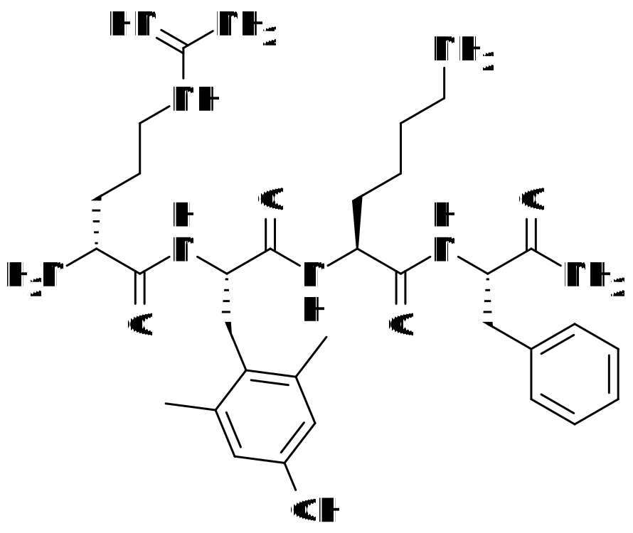
The whole molecule is four residues long: D-arginine, then 2',6'-dimethyl-L-tyrosine, then L-lysine, then L-phenylalanine ending in a carboxamide rather than a free acid. Two departures from ordinary peptide chemistry matter. The first residue is the D enantiomer, which is what the lower-case d in d-Arg denotes and which removes the molecule from the substrate space of most peptidases. The second is a tyrosine methylated at both ring positions flanking its phenol, which is not one of the twenty proteinogenic amino acids; analogs of this series lacking it failed to inhibit mitochondrial reactive oxygen species generation or swelling in the 2004 report.[1,14,15,2]
The charge arithmetic carries the targeting argument. Three groups are protonated at physiological pH, the arginine guanidinium, the lysine side-chain amine and the free N-terminal alpha-amine, and the C-terminal amide leaves nothing to offset them, so the same residue set was characterised as a 3+ net charge tetrapeptide. Against that sit two aromatic rings, one of them the dimethylated phenol. That is what the literature means by the alternating cationic and aromatic motif, and it is why the proposed binding partner is a lipid rather than a protein: cardiolipin carries two phosphates and is confined to the inner mitochondrial membrane.[19,1,4,2]
There is no 3D view here because there is nothing authoritative to show: nothing has been deposited, and PubChem holds no computed conformer for this CID. Models do exist, from a 2022 nuclear magnetic resonance and molecular dynamics study, and what they report is itself a reason one rendering would mislead. The free peptide is a disordered extended monomer, and on a cardiolipin-containing bicelle three of the four analogs tested formed hydrogen-bonded reverse turns while SS-31 alone stayed extended. That paper attributes the extended form to steric restraint from the two methyl groups on the position 2 dimethyltyrosine, which it reports preclude the turn from forming, so the residue the class was designed around is also what keeps this member the least ordered of the four. Its nuclear Overhauser effects to lipid protons came mostly from the aromatic residues to protons near the head groups, placing it at the interface rather than in the bilayer core.[25,1,6]
Modifications
- 1 D-arginine: the N-terminal residue is the D enantiomer, which is what the lower-case d in the literature's d-Arg denotes. The guanidinium side chain is retained.[15,1,14]
- 2 2',6'-dimethyl-L-tyrosine (Dmt), a non-proteinogenic residue. The two ring methyl groups flank the phenolic hydroxyl. The series' originators report that analogs lacking this residue did not inhibit mitochondrial reactive oxygen species generation or swelling, so it is the residue the class was designed around.[15,2]
- 4 C-terminal phenylalaninamide: the chain terminates in a primary carboxamide, removing the negative charge a free C-terminal carboxylate would carry.[1,14]
Four stereocentres, one per residue, with position 1 in the D configuration and positions 2, 3 and 4 in the L configuration; the PubChem and FDA records agree on all four. There is no cysteine, so no disulfide; the peptide is linear and uncyclised, with no lipidation, no PEG, no glycosylation and no metal coordination. Three groups are protonated at physiological pH, the arginine guanidinium, the lysine side-chain amine and the free N-terminal alpha-amine, and a study of the same residue set in the opioid-active order describes it as a 3+ net charge tetrapeptide; the C-terminal amide leaves no compensating negative charge. That charge, set against two aromatic rings, is the alternating cationic and aromatic motif the whole peptide class is defined by.[1,14,19,2]
What studies on SS-31 show
Each card names the experimental system and what was measured. A result in one system is not evidence about another.
Solution and bicelle NMR with isothermal titration calorimetry and all-atom molecular dynamics · Free peptide in water; bicelles of phosphatidylcholine with tetraoleoyl cardiolipin; large unilamellar vesicles for calorimetry; HK-2 cells under serum withdrawal
The first structural models of this peptide class. Free peptides are extended, disordered monomers. On cardiolipin-containing bicelles, every analog except SS-31 formed a hydrogen-bonded reverse turn; SS-31 showed only sequential backbone effects and no main-chain hydrogen bonds. All four bound cardiolipin membranes with roughly equal dissociation constants, with binding dominated by favourable entropy, and the calculated membrane footprint per peptide ran 270 square angstroms for SPN10, 434 for SS-31, 466 for SPN4 and 605 for SS-20. The analogs differed markedly in their effect on membrane surface charge.[6]
- Material
- SS-31 benchmarked against the analogs SS-20, SPN4 and SPN10
- Measured
- Nuclear Overhauser effects and derived structures for the free and membrane-bound peptide; translational diffusion; equilibrium binding constants, enthalpy and lipid-to-peptide stoichiometry; membrane surface charge; mitochondrial membrane potential and ATP content in cells
- Compared with
- The three analogs, and membranes without cardiolipin
- Limitation
- Model membranes and bicelles, not mitochondria, and the cell work is a serum-withdrawal stress model. The structures are NMR ensembles refined by simulation rather than deposited coordinates, and the molecular dynamics ensembles matched them only infrequently until distance restraints were imposed. The inventor of the peptide class is an author with financial interests in the licensee company.
Mitchell W et al., 2022, eLife
Fluorescence binding with isolated mitochondria and a rodent ischemia model · Polarity-sensitive fluorescent analog of the peptide; isolated mitochondria; rat kidney after ischemia and reperfusion
The study that established the cardiolipin mechanism. It reported high-affinity binding to cardiolipin, and that the complex inhibited cytochrome c peroxidase activity by protecting the heme iron, with cristae membranes preserved and mitochondrial swelling prevented in rat kidney after ischemia.[17]
- Material
- SS-31 and a fluorescent analog of it
- Measured
- Binding to cardiolipin; cytochrome c peroxidase activity of the peptide-cardiolipin complex; cristae membrane morphology and mitochondrial swelling; recovery of ATP after reperfusion
- Compared with
- Untreated ischemic animals
- Limitation
- The binding measurement uses a fluorescent analog, not the unlabelled peptide, and the same group had already shown that a fluorophore can change where a tetrapeptide of this class goes and what it does to a mitochondrion. The rodent readouts are measured endpoints in rats. Read as an abstract: the open-access full text was not served.
Birk AV et al., 2013, Journal of the American Society of Nephrology
Model membrane biophysics with electron transfer and respirometry · Liposomes and bicelles of phosphatidylcholine alone or with cardiolipin; cytochrome c reduction in vitro; oxygen consumption in mitoplasts and in frozen and fresh mitochondria
Interaction occurred only with cardiolipin-containing liposomes and bicelles, in about a one-to-one ratio, and NMR placed the aromatic residues deep within those bilayers. In the presence of added cardiolipin the peptide restored cytochrome c reduction and mitochondrial oxygen consumption, and in fresh mitochondria it increased state 3 respiration and the efficiency of ATP synthesis.[4]
- Material
- SS-31
- Measured
- Interaction by fluorescence, turbidity and NMR; cytochrome c reduction kinetics; oxygen consumption and state 3 respiration
- Compared with
- Membranes lacking cardiolipin; cytochrome c with and without added cardiolipin
- Limitation
- Model bilayers and isolated organelles throughout, with the stoichiometry specific to the lipid compositions used. The author list includes the inventor of the compound class. Read as an abstract.
Birk AV et al., 2014, British Journal of Pharmacology
Quantitative membrane biophysics with computational analysis · Model bilayers of varying surface charge; isolated mitochondrial membranes
The peptide partitions into the interfacial region with affinity and binding density directly related to membrane surface charge, and does not destabilise lamellar bilayers even at the highest binding concentrations, though it changes lipid packing saturably. It modulated the surface electrostatics of model and of mitochondrial membranes, shifted calcium distribution within the interfacial region, and reduced the energetic burden of calcium stress in mitochondria.[5]
- Material
- SS-31 (elamipretide)
- Measured
- Partition coefficient and lipid binding density against surface charge; bilayer stability and lipid packing; membrane surface potential; distribution of divalent calcium in the interfacial region
- Compared with
- Membranes of differing anionic lipid content; calcium-loaded mitochondria
- Limitation
- The electrostatic account is offered as a set of non-exclusive mechanisms rather than demonstrated in a cell, and the systems are model membranes plus isolated mitochondria. The inventor of the peptide class is an author with financial interests in the licensee.
Mitchell W et al., 2020, Journal of Biological Chemistry
Chemical cross-linking with mass spectrometry · Mitochondria
Every protein identified was an already known cardiolipin binder, and they fell into two groups, proteins of the oxidative phosphorylation pathway and enzymes of 2-oxoglutarate metabolism. The cross-linked residues lay, in many cases, close to the regions those proteins use to contact cardiolipin.[26]
- Material
- SS-31
- Measured
- Protein partners cross-linked to the peptide, and the residues at which the cross-links formed
- Compared with
- Not established from the abstract
- Limitation
- Cross-linking reports spatial proximity, not affinity and not functional engagement, so this does not establish that any of these proteins is a receptor for the peptide. Read as an abstract: the open-access full text was not served by the endpoint used.
Chavez JD et al., 2020, Proceedings of the National Academy of Sciences of the United States of America
Mitochondrial respirometry with proteomics in aged tissue · Isolated skeletal muscle mitochondria from young and old mice; mouse heart and muscle function; proteomic measurement of abundance, phosphorylation and S-glutathionylation
ADP-stimulated respiration in old muscle mitochondria increased, which the authors attribute to greater ADP uptake through the adenine nucleotide translocator. Protein abundance across the ADP and ATP pathway was unchanged while S-glutathionylation decreased, including on the translocator itself, and the paper states that the peptide binds that transporter and ATP synthase directly.[27]
- Material
- Elamipretide
- Measured
- Respiratory response to ADP; abundance and modification state of ADP and ATP transport and synthesis proteins; muscle force and cardiac systolic function
- Compared with
- Young against old mitochondria, treated against untreated
- Limitation
- Mouse tissue, with organism-level readouts as measured endpoints in aged mice. The direct-binding statement rests on the earlier cross-linking work rather than on a structure or an affinity measurement made here.
Pharaoh G et al., 2023, GeroScience
Rodent ischemia-reperfusion study with 3D electron microscopy and lipidomics · Rat heart after ischemia and reperfusion; permeabilised ventricular fibres; serial block face scanning electron microscopy; lipid mass spectrometry; biomimetic membranes
The decrements in complex I, II and IV activity after ischemia and reperfusion were smaller with the peptide, and the reconstructions showed less fragmentation of cristae networks. Mass spectrometry showed the peptide did not prevent the fall in cardiolipin concentration, and in biomimetic membranes it aggregated cardiolipin.[28]
- Material
- Elamipretide
- Measured
- Activity of respiratory complexes I, II and IV; three-dimensional reconstruction of cristae networks; cardiolipin concentration; biophysical properties of model membranes
- Compared with
- Vehicle-treated ischemic hearts
- Limitation
- A rat model, so the functional readings are measured endpoints in that species. The negative lipidomic result is the informative part and bounds the mechanism: whatever the peptide does, it is not restoring cardiolipin content. Two authors consulted for the developer and one became its employee during review. Read as an abstract.
Allen ME et al., 2020, Communications Biology
Rodent infarct study comparing two analogs · Rat left anterior descending coronary artery ligation and reperfusion, eight animals per group
The study designed to separate the antioxidant contribution of this chemistry from the opioid activity of the parent scaffold, and the one source located that states plainly that SS-31 lacks opioid activity. Infarct area as a fraction of area at risk was 53.9 percent with SS-31 and 47.1 percent with SS-20 against 59.9 percent in controls, with lipid peroxidation reduced by both. The authors note that SS-20 does not scavenge reactive oxygen species.[21]
- Material
- SS-31 and SS-20, both described by the authors as analogs lacking opioid activity
- Measured
- Infarct area as a fraction of the area at risk; myocardial lipid peroxidation; arrhythmia scoring
- Compared with
- SS-20 and saline placebo
- Limitation
- A rat model with small groups, and the readouts are measured endpoints in that species. The absence of opioid activity is asserted in the authors' framing rather than shown by a binding assay in this paper, which leaves the cleanest statement about this molecule's receptor pharmacology resting on an assertion. Read as an abstract.
Cho J et al., 2007, Coronary Artery Disease
Randomised, double-blind, placebo-controlled phase 3 trial · 218 participants with genetically confirmed primary mitochondrial myopathy, randomised 1:1, mean age 45.6 years, 64 percent women and 94 percent White, 74 percent carrying a mitochondrial DNA alteration and the remainder nuclear DNA defects; 24 weeks
The trial did not meet either primary endpoint. The difference in least squares mean change from baseline to week 24 was -3.2 metres on the six-minute walk test (95 percent confidence interval -18.7 to 12.3, p = 0.69) and -0.07 on the total fatigue score (95 percent confidence interval -0.10 to 0.26, p = 0.37). At baseline the mean distance walked was 336.7 metres and the mean total fatigue score 10.6. The authors record the treatment as well tolerated with most adverse events mild to moderate in severity, and the paper carries a Class I evidence classification for the conclusion that the compound does not improve either of those two measures at 24 weeks.[16]
- Material
- Elamipretide, which is this molecule under its approved name, across the single dose arm tested
- Measured
- Change from baseline to week 24 in distance walked on the six-minute walk test and in the total fatigue score of the Primary Mitochondrial Myopathy Symptom Assessment, as co-primary endpoints; most bothersome symptom score, a fatigue short-form score and patient and clinician global impressions as secondary endpoints
- Compared with
- Placebo, 109 participants against 109
- Limitation
- This is the largest human trial of the molecule located here and it is a negative result on its primary endpoints, in a different condition from the one the 2025 accelerated approval covers, so it is evidence neither for nor against the approved indication: the population, the endpoints and the duration all differ. One trial, one duration, a 94 percent White population, and an author list including an affiliate of the sponsor.
Karaa A et al., 2023, Neurology
What binding to cardiolipin has been measured, and what that binding has not explained
The mechanistic account is unusual among peptides in that the proposed binding partner is a lipid, not a receptor. Cardiolipin is a doubly phosphorylated phospholipid found, in healthy cells, essentially only in the inner mitochondrial membrane, where it supports cristae curvature and the organisation of respiratory complexes. A tetrapeptide with three positive charges and two aromatic rings has the chemistry to find an anionic lipid interface, and the 2013 and 2014 experiments tested that: interaction occurred with cardiolipin-containing liposomes and bicelles and not with equivalent membranes lacking it, at about one peptide per lipid, with NMR placing the aromatic residues inside those bilayers.[17,4]
Two consequences were measured rather than assumed. The complex of cytochrome c with cardiolipin can act as a peroxidase instead of an electron carrier, and the 2013 work reported that the peptide-cardiolipin complex inhibited that peroxidase activity, while the 2014 work reported restored cytochrome c reduction and increased state 3 respiration in fresh mitochondria. The quantitative biophysics of 2020 then showed that what changes is the interface itself: partitioning scales with surface charge, lipid packing changes saturably, the bilayer is not destabilised, and divalent calcium is redistributed within the interfacial region. Simulations of four analogs reproduced the surface and dipole potential changes.[17,4,5,29,30]
The account is less settled at the protein level. Cross-linking in mitochondria placed the peptide beside proteins of the oxidative phosphorylation pathway and of 2-oxoglutarate metabolism, all known cardiolipin binders and often at residues near their lipid-contacting regions, which is consistent with a lipid-mediated mechanism and does not demonstrate a protein receptor. A later study read that proximity as direct binding to the adenine nucleotide translocator and ATP synthase, reporting increased ADP uptake through the translocator in old mouse muscle mitochondria with no change in protein abundance and less S-glutathionylation. No affinity for any of those proteins was located here, which leaves two readings of the same data.[26,27,5]
What is still unknown about SS-31
Why this member of the series is the one that advanced is not addressed anywhere read. The 2022 nuclear magnetic resonance and molecular dynamics study, whose ensembles are the only structural information that exists on the molecule and whose limits are set out under Molecular structure, reports SS-31 as the one analog of four that did not form a reverse turn in the membrane-bound state, while the analog with the strongest effect on membrane properties and the best showing in that study's cell assays was SPN10. The structural cause is accounted for, as steric restraint from the position 2 methyl groups, but the question of why the least ordered member of the series went into clinical development is left open. Those nearest chemical relatives, SS-01, SS-20, SPN4 and SPN10, have no entries of their own in this library and appear here only in the list of molecules this one is distinct from.[25,1,6]
The binding evidence has a weakness its own authors identified earlier. The original cardiolipin affinity measurement used a polarity-sensitive fluorescent analog rather than the unlabelled peptide, and a 2005 paper from the same group showed that two fluorophores on one tetrapeptide of this class gave opposite results, one depolarising mitochondria and opening the permeability transition pore, the other staying in the inner membrane and protecting against swelling; its recommendation was to confirm such results with the original or radiolabelled molecule. The later calorimetry on unlabelled peptide reports dissociation constants roughly equal for SS-31 and three analogs that behave differently in cells, so affinity for cardiolipin does not by itself distinguish members of the series.[17,31,6]
Receptor-level pharmacology is thin in a specific way. The one clear statement that this peptide lacks opioid activity comes from the framing of a 2007 rodent infarct study rather than from a binding assay on SS-31, which matters because the molecule shares its four residues with a mu opioid receptor ligand of 0.199 nanomolar affinity and differs only in their order. The independent curated record for this ligand requires an API key and was not read, so no outside annotation of its targets was consulted, and no radioligand binding constant, enzyme inhibition constant or receptor assay for this compound was located.[21,20,1]
Two further gaps. The mechanism is not cardiolipin replacement: a 2020 rat study with lipid mass spectrometry found the fall in cardiolipin concentration after ischemia and reperfusion was not prevented while cristae fragmentation was reduced, and in model membranes the peptide aggregated cardiolipin instead. And nothing located here lets anyone check what a preparation contains: no published method for identifying this peptide in a matrix was found, and two substance records exist, the parent and the trihydrochloride, differing by 109 in formula weight, with no public statement of which form a non-pharmaceutical preparation holds.[28,14,32]
Key papers on SS-31
- Structure-activity relationships of mitochondria-targeted tetrapeptide pharmacological compounds.
Mitchell W, Tamucci JD, Ng EL, Liu S, Birk AV, Szeto HH, May ER, Alexandrescu AT, Alder NN (2022), eLife · NMR and molecular dynamics structure-activity study
The only structural work on this peptide class and the most informative single paper on it: NMR structures of the free and membrane-bound peptide, calorimetric binding constants, and three analogs measured beside it. Open access.
- The mitochondrial-targeted compound SS-31 re-energizes ischemic mitochondria by interacting with cardiolipin.
Birk AV, Liu S, Soong Y, Mills W, Singh P, Warren JD, Seshan SV, Pardee JD, Szeto HH (2013), Journal of the American Society of Nephrology · Fluorescence binding with isolated mitochondria and a rodent model
The paper that established cardiolipin as the binding partner and peroxidase inhibition as the consequence. Everything written since traces back here, including the fluorescent-analog caveat.
- The mitochondria-targeted peptide SS-31 binds lipid bilayers and modulates surface electrostatics as a key component of its mechanism of action.
Mitchell W, Ng EA, Tamucci JD, Boyd KJ, Sathappa M, Coscia A, Pan M, Han X, Eddy NA, May ER, Szeto HH, Alder NN (2020), Journal of Biological Chemistry · Membrane biophysics with computational analysis
The most careful quantitative account of what the peptide does at a membrane, and the source of the surface-electrostatics reading of the mechanism. Open access.
- Mitochondrial protein interaction landscape of SS-31.
Chavez JD, Tang X, Campbell MD, Reyes G, Kramer PA, Stuppard R, Keller A, Zhang H, Rabinovitch PS, Marcinek DJ, Bruce JE (2020), Proceedings of the National Academy of Sciences of the United States of America · Chemical cross-linking with mass spectrometry
The only survey of which mitochondrial proteins the peptide ends up next to, and a good illustration of how proximity differs from affinity.
- The cardiolipin-binding peptide elamipretide mitigates fragmentation of cristae networks following cardiac ischemia reperfusion in rats.
Allen ME, Pennington ER, Perry JB, Dadoo S, Makrecka-Kuka M, Dambrova M, Moukdar F, Patel HD, Han X, Kidd GK, Benson EK, Raisch TB, Poelzing S, Brown DA, Shaikh SR (2020), Communications Biology · Rodent ischemia-reperfusion with 3D electron microscopy and lipidomics
Read for its negative result: cardiolipin concentration was not restored while cristae structure was preserved, which bounds what the mechanism can be.
- Potent mitochondria-targeted peptides reduce myocardial infarction in rats.
Cho J, Won K, Wu D, Soong Y, Liu S, Szeto HH, Hong MK (2007), Coronary Artery Disease · Rodent infarct comparison of two analogs
The study that separates this chemistry from the opioid activity of the scaffold it came from, and the only source located stating that SS-31 lacks that activity.
- First-in-class cardiolipin-protective compound as a therapeutic agent to restore mitochondrial bioenergetics.
Szeto HH (2014), British Journal of Pharmacology · Narrative review
The class-level account by the inventor, useful for the design history and for seeing how the mechanism was argued at the time. Read with its authorship in mind.
References
- 1.National Center for Biotechnology Information. PubChem Compound Summary for CID 11764719, Elamipretide. PubChem. source · database record
- 2.Zhao K, Zhao GM, Wu D, Soong Y, Birk AV, Schiller PW, Szeto HH. (2004). Cell-permeable peptide antioxidants targeted to inner mitochondrial membrane inhibit mitochondrial swelling, oxidative cell death, and reperfusion injury. Journal of Biological Chemistry. 279:34682-90. doi:10.1074/jbc.M402999200 · abstract only
- 3.Shirley M. (2026). Elamipretide: First Approval. Drugs. 86:377-383. doi:10.1007/s40265-025-02269-8 · abstract only
- 4.Birk AV, Chao WM, Bracken C, Warren JD, Szeto HH. (2014). Targeting mitochondrial cardiolipin and the cytochrome c/cardiolipin complex to promote electron transport and optimize mitochondrial ATP synthesis. British Journal of Pharmacology. 171:2017-28. doi:10.1111/bph.12468 · abstract only
- 5.Mitchell W, Ng EA, Tamucci JD, Boyd KJ, Sathappa M, Coscia A, Pan M, Han X, Eddy NA, May ER, Szeto HH, Alder NN. (2020). The mitochondria-targeted peptide SS-31 binds lipid bilayers and modulates surface electrostatics as a key component of its mechanism of action. Journal of Biological Chemistry. 295:7452-7469. doi:10.1074/jbc.RA119.012094 · full text read
- 6.Mitchell W, Tamucci JD, Ng EL, Liu S, Birk AV, Szeto HH, May ER, Alexandrescu AT, Alder NN. (2022). Structure-activity relationships of mitochondria-targeted tetrapeptide pharmacological compounds. eLife. 11:e75531. doi:10.7554/eLife.75531 · full text read
- 7.U.S. Food and Drug Administration. (2026). Drugs@FDA: elamipretide hydrochloride, NDA 215244. Drugs@FDA database. source · database record
- 8.U.S. National Library of Medicine. (2026). DailyMed: elamipretide hydrochloride (Stealth BioTherapeutics Inc.) prescribing information, Indications and Usage. DailyMed. source · full text read
- 9.European Medicines Agency. (2026). Medicines: search for human medicines (centrally authorised medicines). European Medicines Agency medicines database. source · database record
- 10.Health Canada. (2026). Drug Product Database online query. Health Canada Drug Product Database. source · database record
- 11.World Anti-Doping Agency. (2025). World Anti-Doping Code International Standard: Prohibited List 2026. World Anti-Doping Agency. source · full text read
- 12.Kanehisa Laboratories. KEGG DRUG entry D11542, Elamipretide hydrochloride (USAN). KEGG DRUG. source · database record
- 13.US Food and Drug Administration. (2026). Certain Bulk Drug Substances for Use in Compounding that May Present Significant Safety Risks. FDA human drug compounding. source · full text read
- 14.US Food and Drug Administration, Global Substance Registration System. Substance records ELAMIPRETIDE (UNII 87GWG91S09) and ELAMIPRETIDE HYDROCHLORIDE (UNII E40WZ3BK2D). FDA GSRS. source · database record
- 15.Zhao K, Luo G, Giannelli S, Szeto HH. (2005). Mitochondria-targeted peptide prevents mitochondrial depolarization and apoptosis induced by tert-butyl hydroperoxide in neuronal cell lines. Biochemical Pharmacology. 70:1796-806. doi:10.1016/j.bcp.2005.08.022 · abstract only
- 16.Karaa A, Bertini E, Carelli V, Cohen BH, Enns GM, Falk MJ, Goldstein A, Gorman GS, Haas R, Hirano M, Klopstock T, Koenig MK, Kornblum C, Lamperti C, Lehman A, Longo N, Molnar MJ, Parikh S, Phan H, Pitceathly RDS, Saneto R, Scaglia F, Servidei S, Tarnopolsky M, Toscano A, Van Hove JLK, Vissing J, Vockley J, Finman JS, Brown DA, Shiffer JA, Mancuso M; MMPOWER-3 Trial Investigators. (2023). Efficacy and Safety of Elamipretide in Individuals With Primary Mitochondrial Myopathy: The MMPOWER-3 Randomized Clinical Trial. Neurology. 101:e238-e252. doi:10.1212/WNL.0000000000207402 · abstract only
- 17.Birk AV, Liu S, Soong Y, Mills W, Singh P, Warren JD, Seshan SV, Pardee JD, Szeto HH. (2013). The mitochondrial-targeted compound SS-31 re-energizes ischemic mitochondria by interacting with cardiolipin. Journal of the American Society of Nephrology. 24:1250-61. doi:10.1681/ASN.2012121216 · abstract only
- 18.Szeto HH, Schiller PW. (2011). Novel therapies targeting inner mitochondrial membrane--from discovery to clinical development. Pharmaceutical Research. 28:2669-79. doi:10.1007/s11095-011-0476-8 · abstract only
- 19.Zhao K, Luo G, Zhao GM, Schiller PW, Szeto HH. (2003). Transcellular transport of a highly polar 3+ net charge opioid tetrapeptide. Journal of Pharmacology and Experimental Therapeutics. 304:425-32. doi:10.1124/jpet.102.040147 · abstract only
- 20.Zhao GM, Qian X, Schiller PW, Szeto HH. (2003). Comparison of [Dmt1]DALDA and DAMGO in binding and G protein activation at mu, delta, and kappa opioid receptors. Journal of Pharmacology and Experimental Therapeutics. 307:947-54. doi:10.1124/jpet.103.054775 · abstract only
- 21.Cho J, Won K, Wu D, Soong Y, Liu S, Szeto HH, Hong MK. (2007). Potent mitochondria-targeted peptides reduce myocardial infarction in rats. Coronary Artery Disease. 18:215-20. doi:10.1097/01.mca.0000236285.71683.b6 · abstract only
- 22.Russo S, De Rasmo D, Rossi R, Signorile A, Lobasso S. (2024). SS-31 treatment ameliorates cardiac mitochondrial morphology and defective mitophagy in a murine model of Barth syndrome. Scientific Reports. 14:13655. doi:10.1038/s41598-024-64368-y · abstract only
- 23.Anzmann AF, Sniezek OL, Pado A, Busa V, Vaz FM, Kreimer SD, DeVine LR, Cole RN, Le A, Kirsch BJ, Claypool SM, Vernon HJ. (2021). Diverse mitochondrial abnormalities in a new cellular model of TAFFAZZIN deficiency are remediated by cardiolipin-interacting small molecules. Journal of Biological Chemistry. 297:101005. doi:10.1016/j.jbc.2021.101005 · abstract only
- 24.Tung C, Varzideh F, Farroni E, Mone P, Kansakar U, Jankauskas SS, Santulli G. (2025). Elamipretide: A Review of Its Structure, Mechanism of Action, and Therapeutic Potential. International Journal of Molecular Sciences. 26:944. doi:10.3390/ijms26030944 · abstract only
- 25.RCSB Protein Data Bank. Full-text and chemical descriptor searches for elamipretide, SS-31 and Szeto-Schiller, returning no entry containing this molecule. RCSB PDB search service. source · database record
- 26.Chavez JD, Tang X, Campbell MD, Reyes G, Kramer PA, Stuppard R, Keller A, Zhang H, Rabinovitch PS, Marcinek DJ, Bruce JE. (2020). Mitochondrial protein interaction landscape of SS-31. Proceedings of the National Academy of Sciences of the United States of America. 117:15363-15373. doi:10.1073/pnas.2002250117 · abstract only
- 27.Pharaoh G, Kamat V, Kannan S, Stuppard RS, Whitson J, Martin-Perez M, Qian WJ, MacCoss MJ, Villen J, Rabinovitch P, Campbell MD, Sweet IR, Marcinek DJ. (2023). The mitochondrially targeted peptide elamipretide (SS-31) improves ADP sensitivity in aged mitochondria by increasing uptake through the adenine nucleotide translocator (ANT). GeroScience. 45:3529-3548. doi:10.1007/s11357-023-00861-y · full text read
- 28.Allen ME, Pennington ER, Perry JB, Dadoo S, Makrecka-Kuka M, Dambrova M, Moukdar F, Patel HD, Han X, Kidd GK, Benson EK, Raisch TB, Poelzing S, Brown DA, Shaikh SR. (2020). The cardiolipin-binding peptide elamipretide mitigates fragmentation of cristae networks following cardiac ischemia reperfusion in rats. Communications Biology. 3:389. doi:10.1038/s42003-020-1101-3 · abstract only
- 29.Tamucci JD, Alder NN, May ER. (2023). Peptide Power: Mechanistic Insights into the Effect of Mitochondria-Targeted Tetrapeptides on Membrane Electrostatics from Molecular Simulations. Molecular Pharmaceutics. 20:6114-6129. doi:10.1021/acs.molpharmaceut.3c00480 · abstract only
- 30.Szeto HH. (2014). First-in-class cardiolipin-protective compound as a therapeutic agent to restore mitochondrial bioenergetics. British Journal of Pharmacology. 171:2029-50. doi:10.1111/bph.12461 · abstract only
- 31.Szeto HH, Schiller PW, Zhao K, Luo G. (2005). Fluorescent dyes alter intracellular targeting and function of cell-penetrating tetrapeptides. FASEB Journal. 19:118-20. doi:10.1096/fj.04-1982fje · abstract only
- 32.Li M, Kong D, Meng L, Wang Z, Bai Z, Wu G. (2024). Discovery of novel SS-31 (d-Arg-dimethylTyr-Lys-Phe-NH2) derivatives as potent agents to ameliorate inflammation and increase mitochondrial ATP synthesis. RSC Advances. 14:29789-29799. doi:10.1039/d4ra05517a · abstract only
- 33.ChEBI, European Molecular Biology Laboratory European Bioinformatics Institute. CHEBI:233331, elamipretide. ChEBI, read through the EBI Ontology Lookup Service. source · database record
- 34.Kanehisa Laboratories. KEGG DRUG entry D10925, Elamipretide (USAN/INN). KEGG DRUG. source · database record
Related entries
- MOTS-cMOTS-c is a 16-residue peptide, MRWQEMGYIFYPRKLR, encoded by a 51 base pair reading frame inside the human mitochondrial 12S ribosomal RNA gene. It has no identified receptor, no published affinity constant and no deposited three-dimensional structure.
- Nicotinamide adenine dinucleotide (NAD+)NAD+ is the oxidised form of nicotinamide adenine dinucleotide, in which nicotinamide riboside and adenosine are joined through a pyrophosphate bridge. Cells use it both as a carrier of hydride in redox reactions and as a substrate that sirtuins, PARPs, CD38 and SARM1 cleave.
- Glutathione (reduced, GSH)Glutathione is the cysteine-containing tripeptide that cells build for themselves, and the most abundant low-molecular-weight thiol in animal cells. It exists as a reduced form carrying a free thiol, written GSH, and as its disulfide dimer GSSG, and almost all of its chemistry runs through that one sulfur.
