Glutathione in brief
Glutathione is the tripeptide gamma-L-glutamyl-L-cysteinylglycine, 307.3 g/mol as reduced GSH, unusual because its glutamate is linked through the side-chain carboxyl. It is the most abundant low-molecular-weight thiol in cells, at about 1 to 10 mM in the cytosol against micromolar concentrations in plasma, and it is built in two cytosolic steps whose rate-limiting enzyme is glutamate cysteine ligase. Two GSH molecules joined by a disulfide form GSSG, a separate compound, which glutathione reductase returns to two GSH using NADPH. Its chemistry is that of one cysteine thiol, whose pKa near 8.7 to 9 was measured at 6.6 in a glutathione transferase site. Experimental structures show it bound to human enzymes near 1 angstrom resolution, and free glutathione also has its own small-molecule crystal structure. Measured GSSG values are easily inflated by oxidation during sample handling, and while an unqualified label means the reduced form, the GSSG content and salt state of a particular lot are not on public record.[1,2,3,4,5,6,7,8,9]
Glutathione regulatory status
No FDA, EMA or Health Canada marketing authorisation for glutathione as a medicine was found in Drugs@FDA (queried through openFDA), the EMA medicines dataset or the Health Canada Drug Product Database (searched November 2026); the only authorised products in the FDA and Health Canada records that list a glutathione ingredient are multi-ingredient solutions containing the disulfide GSSG, not GSH. In the FDA list of bulk drug substances nominated for compounding under section 503A (updated 14 May 2026), glutathione is in Category 1, substances under evaluation. Glutathione is named nowhere on the 2026 WADA Prohibited List, and it appears in neither the category 2 table nor the table of withdrawn nominations on the FDA page of bulk substances that may present significant safety risks under the 503A and 503B interim policies (content current as of 22 April 2026). No retraction or expression of concern was found for any study cited here; the erratum on R09 and the erratum on R14 are noted where those works are used.[10,11,12,13,14,15]
Glutathione at a glance
- A tripeptide of glutamate, cysteine and glycine, 307.3 g/mol as reduced GSH (C10H17N3O6S), unusual in that the glutamate is attached through its side-chain carboxyl. Two molecules joined by a disulfide give GSSG at 612.6 g/mol, a separate compound with its own identifiers.[1,16,2]
- Its chemistry is the chemistry of one sulfur. The free thiol has a pKa near 8.7 to 9; a glutathione transferase site was measured to lower it to 6.6, so the bound molecule is largely the more reactive thiolate.[17,4]
- The GSSG/2GSH couple has a standard reduction potential of about -240 mV, a value confirmed independently by protein-protein redox equilibria. Because two GSH make one GSSG, the actual potential depends on total concentration as well as on the ratio.[18,19,20]
- It is the most abundant low-molecular-weight thiol inside cells: cytosolic GSH is in the range of 1 to 10 mM, nearer 1 to 2 mM in most cells and about 10 mM in hepatocytes, against micromolar concentrations in plasma, and the cytosolic ratio of GSH to GSSG stays very high. Cells make it in two cytosolic steps whose rate-limiting enzyme is glutamate cysteine ligase, with the availability of cysteine the other major determinant.[7,8]
- Measured GSSG values are easily inflated by oxidation during sample handling; reported blood GSSG figures span more than two orders of magnitude for this reason.[6,21]
- Reduced glutathione has its own small-molecule crystal structure, first determined in 1958, redetermined at 120 K in 1987 and measured again at ambient pressure in 2010: orthorhombic, space group P212121, a 5.6163 Å, b 8.7795 Å, c 27.999 Å, cell volume 1380.6 Å3 at 293 K, Z = 4, calculated density 1.478 g/cm3. The experimental-properties record gives a melting point of 195 degrees and a water solubility of 292.5 mg/mL.[9,22,23,24]
What is Glutathione?
Glutathione is a small thiol made inside cells from three amino acids. Chemically it is a tripeptide, but the first bond is an isopeptide: the glutamate is joined to cysteine through the carboxyl at the end of its side chain (the gamma carboxyl) instead of the backbone carboxyl. That leaves the glutamate with a free amino group and a free carboxyl. The enzyme that cleaves this gamma-glutamyl bond, gamma-glutamyl transpeptidase 1, sits on the outer surface of animal cells.[16,25]
It exists mainly in two forms. Reduced glutathione (GSH) carries a free thiol, written -SH. Oxidised glutathione (GSSG) is two GSH molecules linked through a sulfur-sulfur bond. The two interconvert by thiol-disulfide exchange, and the flavoenzyme glutathione reductase uses NADPH to turn one GSSG back into two GSH. A third family, mixed disulfides between glutathione and protein cysteines, is formed by the same exchange chemistry. Inside a cell almost all of it is the reduced form: cytosolic GSH runs from about 1 to 10 mM, close to 1 to 2 mM in most cells and about 10 mM in hepatocytes, while plasma concentrations are micromolar, and the cytosolic GSH to GSSG ratio stays very high even under oxidative stress. The endoplasmic reticulum is the one compartment reported with a relatively high GSSG to GSH ratio.[3,2,21,7]
Its synthesis takes two enzymatic steps in the cytosol. Glutamate cysteine ligase, also called gamma-glutamylcysteine synthetase, joins glutamate to cysteine; a review of the regulation of glutathione synthesis calls this the rate-limiting enzyme and names the availability of cysteine, the sulfur amino acid precursor, as the other major determinant, which is what connects this entry to N-acetylcysteine next door. The enzyme is inhibited by glutathione itself in a purified-enzyme assay, so the pathway is also feedback-controlled. Glutathione synthetase then adds glycine in an ATP-dependent reaction, and the human enzyme has been crystallised with glutathione bound. Mitochondria cannot make it and import it, a step that depends on the carrier SLC25A39 in mammalian cell lines.[8,7,26,27,28]
Not to be confused with
- Glutathione disulfide (GSSG, oxidized glutathione)
- Two glutathione molecules joined through a disulfide bond between their cysteine sulfurs: C20H32N6O12S2 at 612.6 g/mol, with its own PubChem record (CID 65359) and CAS number (27025-41-8). It is the oxidised partner of GSH in the GSSG/2GSH redox couple, not an alternative name, and a label reading only 'glutathione' does not say which of the two is meant.[2,1,19]
- alpha-glutamyl tripeptide Glu-Cys-Gly
- Glutathione's glutamate is joined to cysteine through its side-chain (gamma) carboxyl, not through the backbone alpha carboxyl used by ordinary peptide bonds. This is why the molecule is described as tripeptide-like, and why its N-terminal bond is the substrate of a dedicated enzyme, gamma-glutamyl transpeptidase 1.[16,25]
- S-substituted glutathione conjugates (for example S-hexylglutathione)
- Products and analogues in which the cysteine sulfur carries another group. They keep the peptide part, and are used in crystallography to mark the glutathione-binding site, but they no longer have the free thiol on which glutathione's own chemistry depends.[31]
- Ophthalmic acid (gamma-glutamyl-alpha-aminobutyryl-glycine)
- The same tripeptide skeleton with the cysteine sulfur replaced by a methyl group. In an enzyme assay it was only a weak inhibitor where glutathione was a strong one, which locates part of glutathione's binding behaviour on the thiol.[26]
Glutathione structure and sequence
The sequence is written gamma-Glu-Cys-Gly. Its two stereocentres are the alpha carbons of L-glutamate and L-cysteine (named 2S and 2R by the priority rules, both L). There is no ring, no metal and no disulfide in GSH itself, so the molecule is flexible: the conformation it adopts depends on what holds it. For GSSG, NMR found more than one antiparallel arrangement of the two halves in solution and a parallel one in the solid state, consistent with a deposited crystal structure. Free glutathione has been crystallised in its own right: the structure was determined in 1958, redetermined at 120 K in 1987 and measured again across a pressure series in 2010, and the ambient-pressure coordinates are orthorhombic, space group P212121, with a 5.6163 Å, b 8.7795 Å, c 27.999 Å, a cell volume of 1380.6 Å3 at 293 K, four molecules per cell and a calculated density of 1.478 g/cm3. That is a lattice conformation, so it is no more the solution shape than a protein-bound pose is.[1,16,29,9,22,23]
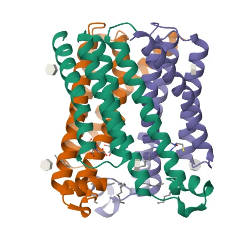
The 3D view shows GSH in human microsomal prostaglandin E synthase-1 at 1.16 Å. All 20 heavy atoms are modelled at full occupancy. The two carboxylates and the free amine are each paired with charged or polar enzyme side chains, one of them (Arg38, holding the glutamate carboxylate) from the neighbouring subunit of the trimer, which the file generates by crystal symmetry. The sulfur points toward Ser127. Hydrogens are not modelled, so the picture cannot show whether the sulfur is a thiol or a thiolate, and it is one bound pose, not the shape of the free molecule in water.[30,5]
Glutathione transferases illustrate the same split between the peptide and the sulfur. In the human pi-class enzyme, crystallised with S-hexylglutathione, the peptide part occupies a G-site and the sulfur substituent points into an adjacent hydrophobic H-site lined by Tyr7 and other residues.[31]
How differently one molecule can be held is clear from a second enzyme of the same MAPEG family. In human leukotriene C4 synthase, crystallised with glutathione bound at 2.15 Å, the active site enforces a horseshoe-shaped conformation on the tripeptide and positions the thiol next to an arginine at the membrane-enzyme interface, which is where the sulfur is activated. The mPGES-1 site shown on this page holds the same molecule in a different shape.[32,5]
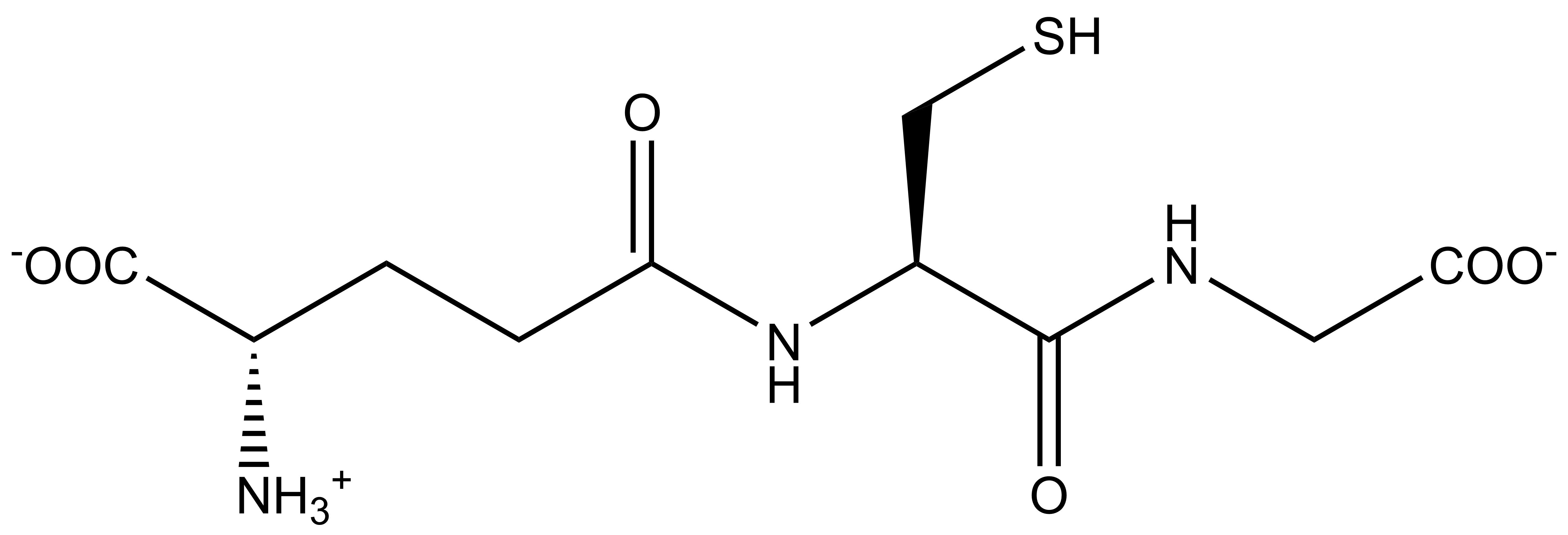
- gamma1
- Glu2
- Cys3
- Gly4
Modifications
- Glu1 side chain to Cys2 amino group An isopeptide (gamma-glutamyl) bond in place of a backbone peptide bond. The glutamate therefore keeps a free alpha-amino group and a free alpha-carboxyl group, which in the deposited mPGES-1 structure are both held by enzyme side chains. The gamma-glutamyl bond is the one hydrolysed or transferred by gamma-glutamyl transpeptidase 1.[16,30,25]
- Cys2 side chain (thiol, -SH) The reactive group. In free solution its pKa is roughly 8.7 to 9, so at neutral pH most of it is protonated. Oxidation joins two thiols into the disulfide GSSG; reaction with protein cysteines gives mixed disulfides (S-glutathionylation); and in some glutaredoxins the sulfur coordinates iron in a [2Fe-2S] cluster.[17,4,2,37]
- Gly3 C-terminus A free carboxyl group. With the glutamate alpha-carboxyl it gives the molecule two carboxylates and one amine at neutral pH, so the net charge there is close to minus one; the computed record is the neutral, fully protonated form.[1,16]
Two defined stereocentres, the alpha carbons of the glutamate and the cysteine, both from L-amino acids. PubChem names them (2S) for the glutamate and (2R) for the cysteine; the R label is a consequence of the sulfur outranking the carboxyl in the priority rules and does not mean D-cysteine. Glycine has no stereocentre. GSSG doubles this to four. The same two centres are defined in the small-molecule crystal structure of reduced glutathione, which is orthorhombic in space group P212121 with four molecules in the cell.[1,16,2,9]
What studies on Glutathione show
Each card names the experimental system and what was measured. A result in one system is not evidence about another.
X-ray crystallography · Purified recombinant human microsomal prostaglandin E synthase-1, crystallised as the trimer
Three active-site cavities sit at the subunit interfaces within the membrane-spanning region, and the authors proposed a catalytic mechanism in which Ser127 plays a key role.[5]
- Material
- Enzyme with bound glutathione (PDB 4AL0) and, separately, with a glutathione-based analogue
- Measured
- Atomic model at about 1.2 Å, active-site architecture; the deposited model places one glutathione in each protomer
- Compared with
- Other MAPEG-family membrane proteins of known structure
- Limitation
- Read as abstract only, so the authors' description of the glutathione contacts was not transcribed; the contacts named on this page were measured from the deposited coordinates. The proposed role of Ser127 is a model from the structure, not a measured rate.
Sjögren T et al., 2013, Proceedings of the National Academy of Sciences of the USA
X-ray crystallography · Purified human leukotriene C4 synthase, a MAPEG-family membrane enzyme
The enzyme is a homotrimer of four-transmembrane-segment subunits, and the substrate complex shows the active site forcing glutathione into a horseshoe shape that places its thiol beside an arginine at the membrane-enzyme interface, which the authors identify as the activating group. A hydrophobic cleft pins the other end of the lipid co-substrate so that its reactive epoxide is aligned with the glutathione sulfur.[32]
- Material
- Apo enzyme and the glutathione complex
- Measured
- Atomic models at 2.00 Å (apo) and 2.15 Å (glutathione complex); active-site architecture
- Compared with
- Apo against glutathione-bound; other homologous MAPEG proteins
- Limitation
- Read as abstract only. A static picture of substrate binding rather than a measured rate, and the conclusion that this binding mode is shared across the family is the authors' inference from homology.
Martinez Molina D et al., 2007, Nature
Cryo X-ray crystallography at 0.95 to 1.1 Å · Crystals of human erythrocyte glutathione reductase
Soaking with GSH reduced the enzyme's active-site disulfide, producing the Cys63 thiolate to flavin charge-transfer state. The authors stated that the new resolution gave no new insight into how GSSG or GSH bind, and showed that disulfides cleaved by the X-ray beam do not reproduce chemically reduced ones.[3]
- Material
- Enzyme alone and soaked with GSSG plus NADP+, with GSH, or with NADPH
- Measured
- Atomic models of four catalytic states
- Compared with
- Earlier room-temperature structures at 2 Å; synchrotron-reduced against chemically reduced enzyme
- Limitation
- The glutathione models were not the paper's focus. In the deposited GSH complex the ligand is partly a mixed disulfide with the enzyme, so it does not show free GSH.
Berkholz DS et al., 2008, Journal of Molecular Biology
UV difference spectroscopy and binding titration · Purified rat liver glutathione transferase isozyme 4-4
The band was assigned to the thiolate of bound GSH, and its titration gave a pKa of 6.6 for the enzyme-bound thiol, about 2.4 units below that of free GSH, corresponding to roughly 3.3 kcal/mol of binding energy spent on ionisation.[4]
- Material
- GSH, with the oxygen analogue gamma-glutamyl-serylglycine (GOH) as control
- Measured
- A 239 nm absorption band titrated over pH 5 to 8
- Compared with
- Enzyme with GOH; GSH free in solution
- Limitation
- Abstract only, truncated in PubMed; an erratum to the paper exists and was not read. One isozyme of one species.
Graminski GF et al., 1989, Biochemistry
Site-directed mutagenesis with activity and binding assays · Recombinant human glutathione transferase pi expressed from a cDNA vector
Every substitution reduced activity below 2% of wild type. The Tyr7 to Phe mutant kept 27% of glutathione binding, which the authors read as Tyr7 acting mainly in chemical activation of the bound glutathione rather than in holding it.[33]
- Material
- Tyr7, Arg13, Gln62 and Asp96 each replaced non-conservatively
- Measured
- Specific activity and glutathione binding
- Compared with
- Wild-type enzyme
- Limitation
- Abstract only. Binding and activation are inferred from two coarse measurements, not from a structure of each mutant.
Manoharan TH et al., 1992, Journal of Molecular Biology
Purified enzyme kinetics · Gamma-glutamylcysteine synthetase, the first enzyme of glutathione synthesis
Glutathione inhibited its own first biosynthetic step non-allosterically, apparently by binding the glutamate site and a second site that needed the thiol; ophthalmic acid was only a weak inhibitor. The apparent Km for L-cysteine was 0.35 mM.[26]
- Material
- Glutathione as inhibitor
- Measured
- Inhibition of enzyme activity and its pattern
- Compared with
- Ophthalmic acid, the sulfur-free analogue
- Limitation
- Abstract only. A 1975 in vitro enzyme study; the enzyme source and assay conditions were not transcribed.
Richman PG et al., 1975, Journal of Biological Chemistry
Genetically encoded sensor with thiol alkylation and gel shift · HeLa cells expressing an ER-targeted yeast glutaredoxin mutant (sCGrx1pER)
92% of the sensor was glutathionylated, giving an ER ratio below 7:1. Combined with a previously measured [GSH]^2:[GSSG] of 83 mM, this implies total ER glutathione above 15 mM against about 7 mM for the whole cell.[20]
- Material
- Sensor whose single cysteine equilibrates with GSH/GSSG with a known equilibrium constant
- Measured
- Fraction of sensor glutathionylated, converted to the ER [GSH]:[GSSG] ratio
- Compared with
- DTT-treated cells; an earlier glycopeptide probe measurement
- Limitation
- Assumes the sensor reaches equilibrium in the ER and depends on an earlier, separately measured quantity; densitometry above 90% oxidation is imprecise, as the authors note.
Montero D et al., 2013, Redox Biology
Analytical method comparison · Human blood samples
Acid deproteinisation alone produced 30 to 150 micromol/L of GSSG, linked to oxygenated haemoglobin; with artefacts controlled, GSSG in blood from healthy donors was 2 to 6 micromol/L.[6]
- Material
- GSH and GSSG measured by an enzymatic recycling assay and by HPLC of monobromobimane derivatives
- Measured
- GSSG generated during sample processing
- Compared with
- Different deproteinisation and handling procedures, oxygen deprivation, carbon monoxide atmosphere
- Limitation
- Abstract only. It establishes how fragile the measurement is, not the value in any particular tissue.
Rossi R et al., 2002, Clinical Chemistry
How glutathione's thiol is activated and exchanged
Almost every reaction of glutathione runs through the sulfur. In water at neutral pH the thiol (pKa about 8.7) is mostly protonated, and the thiolate (GS-) is the form that attacks. A glutathione transferase site was measured to lower that pKa to 6.6, so the bound molecule is largely thiolate. Human thioltransferase (a glutaredoxin) behaved differently: the pH dependence of its glutathione-dependent rate followed the titration of glutathione's own thiol, which the authors read as rate-limiting attack by the glutathione thiolate, and GSH reacted faster than its pKa alone predicted.[17,4]
In thiol-disulfide exchange a thiolate attacks a disulfide, taking one sulfur partner and releasing the other. This is how GSSG forms, how glutathione reductase returns it to two GSH using NADPH, and how proteins become S-glutathionylated. Selenium-dependent glutathione peroxidases consume GSH to reduce peroxides: rat erythrocytes lacking selenium had almost no glutathione peroxidase activity, and in cancer cell lines depleting glutathione inactivated these enzymes, one route to the cell death form called ferroptosis. Several enzyme families consume or carry glutathione through this same chemistry: glutathione transferases conjugate it to electrophilic xenobiotics and to endogenous electrophiles such as 4-hydroxynonenal, which is how many such compounds are eliminated; glyoxalase I isomerises the hemithioacetal that glutathione forms spontaneously with methylglyoxal and other alpha-oxoaldehydes into an S-2-hydroxyacylglutathione thioester; and leukotriene C4 synthase conjugates glutathione to the epoxide leukotriene A4.[3,21,34,35,7,36,32]
The sulfur can also act as a metal ligand. In poplar glutaredoxin C1, X-ray crystallography and spectroscopy showed a [2Fe-2S] cluster held between two protein subunits by two protein cysteines and the cysteines of two glutathione molecules.[37]

What is still unknown about Glutathione
What a particular lot contains is a lot property, not a naming question. An unqualified glutathione is the reduced tripeptide, which is what CAS 70-18-8, the UNII and CHEBI:16856 all denote and what the catalogue rows therefore mean, but GSH oxidises readily to GSSG during handling and in solution, so the GSSG content and the salt or free-acid state belong to a sample, not to the name.[1,2,16,6]
Measured ratios of GSH to GSSG are method-dependent. Blood GSSG values in the literature span from about 1 to more than 500 micromol/L, and in cultured cells GSSG and protein-glutathione mixed disulfides were 3 to 5 and 10 to 20 times higher when samples were processed without immediately blocking thiols with N-ethylmaleimide. Older ratios should be read with that in mind.[6,21]
Compartment values disagree depending on the probe. A glycopeptide probe put the secretory-pathway GSH:GSSG ratio at 1:1 to 3:1 against 30:1 to 100:1 for whole cells, while sensors coupled to glutaredoxin report a potential, not concentrations, and need an independent total-glutathione figure to give one. Fused glutaredoxin sensors detect nanomolar changes in GSSG against millimolar GSH, which is why a single 'redox state' figure for a cell is an approximation.[38,39,20,19]
How intact glutathione crosses membranes is only partly mapped. The mitochondrial carrier was identified in 2021, and its own abundance falls when glutathione is plentiful. Outside animal cells, gamma-glutamyl transpeptidase 1, expressed on the cell surface, cleaves the gamma-glutamyl bond; the first eukaryotic structure of that enzyme was solved with glutamate, the released product, bound.[28,25]
Key papers on Glutathione
- Redox potentials of glutaredoxins and other thiol-disulfide oxidoreductases of the thioredoxin superfamily determined by direct protein-protein redox equilibria.
Aslund F, Berndt KD, Holmgren A (1997), Journal of Biological Chemistry · Protein-protein redox equilibria
Fixes the standard reduction potential of the GSSG/2GSH couple at about -240 mV by a route that avoids glutathione buffers, which is the number every later redox calculation rests on.
- Spectroscopic and kinetic evidence for the thiolate anion of glutathione at the active site of glutathione S-transferase.
Graminski GF, Kubo Y, Armstrong RN (1989), Biochemistry · UV difference spectroscopy
The clearest single measurement of how an enzyme turns glutathione's thiol into a thiolate: a direct spectroscopic pKa for the bound sulfur.
- Catalytic cycle of human glutathione reductase near 1 A resolution.
Berkholz DS, Faber HR, Savvides SN, Karplus PA (2008), Journal of Molecular Biology · Cryo X-ray crystallography
Near-atomic structures of the enzyme that regenerates GSH from GSSG, with an open discussion of what X-ray damage does to disulfides.
- Blood glutathione disulfide: in vivo factor or in vitro artifact?
Rossi R, Milzani A, Dalle-Donne I, Giustarini D, Lusini L, Colombo R, Di Simplicio P (2002), Clinical Chemistry · Analytical method comparison
Explains why so many published GSSG numbers are too high, and what sample handling causes it.
- Intracellular glutathione pools are heterogeneously concentrated.
Montero D, Tachibana C, Rahr Winther J, Appenzeller-Herzog C (2013), Redox Biology · Genetically encoded sensor
Shows how a sensor ratio becomes a concentration estimate, and why total glutathione differs between the ER and the rest of the cell.
- SLC25A39 is necessary for mitochondrial glutathione import in mammalian cells.
Wang Y, Yen FS, Zhu XG, Timson RC, Weber R, Xing C, et al. (2021), Nature · Organellar proteomics and metabolomics
Identified the mitochondrial glutathione carrier, separating where glutathione is made from where it is used.
- Regulation of glutathione synthesis
Lu SC (2009), Molecular aspects of medicine · Review
Sets out which step of glutathione synthesis is rate-limiting and why cysteine supply is the other determinant, which is the regulation question most readers arrive with.
References
- 1.National Center for Biotechnology Information. (2026). PubChem Compound Summary for CID 124886, Glutathione. PubChem. source · database record
- 2.National Center for Biotechnology Information. (2026). PubChem Compound Summary for CID 65359, Glutathione disulfide. PubChem. source · database record
- 3.Berkholz DS, Faber HR, Savvides SN, Karplus PA. (2008). Catalytic cycle of human glutathione reductase near 1 A resolution. Journal of Molecular Biology. 382:371-384. doi:10.1016/j.jmb.2008.06.083 · full text read
- 4.Graminski GF, Kubo Y, Armstrong RN. (1989). Spectroscopic and kinetic evidence for the thiolate anion of glutathione at the active site of glutathione S-transferase. Biochemistry. 28:3562-3568. doi:10.1021/bi00434a062 · abstract only · corrected: PubMed links an erratum: Biochemistry 1989;28(17):7138. The erratum text was not read.
- 5.Sjögren T, Nord J, Ek M, Johansson P, Liu G, Geschwindner S. (2013). Crystal structure of microsomal prostaglandin E2 synthase provides insight into diversity in the MAPEG superfamily. Proceedings of the National Academy of Sciences of the USA. 110:3806-3811. doi:10.1073/pnas.1218504110 · abstract only
- 6.Rossi R, Milzani A, Dalle-Donne I, Giustarini D, Lusini L, Colombo R, Di Simplicio P. (2002). Blood glutathione disulfide: in vivo factor or in vitro artifact?. Clinical Chemistry. 48:742-753. PMID 11978601 · abstract only
- 7.Forman HJ, Zhang H, Rinna A. (2009). Glutathione: overview of its protective roles, measurement, and biosynthesis. Molecular aspects of medicine. 30:1-12. doi:10.1016/j.mam.2008.08.006 · full text read
- 8.Lu SC. (2009). Regulation of glutathione synthesis. Molecular aspects of medicine. 30:42-59. doi:10.1016/j.mam.2008.05.005 · abstract only
- 9.Moggach SA, Lennie AR, Morrison CA, Richardson P, Stefanowicz FA, Warren JE. (2010). Pressure induced phase transitions in the tripeptide glutathione to 5.24 GPa: the crystal structure of glutathione-II at 2.94 GPa and glutathione-III at 3.70 GPa. CrystEngComm. 12:2587. doi:10.1039/c001254h · abstract only
- 10.U.S. Food and Drug Administration. (2026). openFDA drugsfda endpoint, query for active ingredients matching GLUTATHIONE. openFDA. source · database record
- 11.European Medicines Agency. (2026). Medicines output: medicines report (JSON), all EPARs for human and veterinary medicines. European Medicines Agency. source · database record
- 12.Health Canada. (2026). Drug Product Database API, active ingredient search: GLUTATHIONE. Health Canada Drug Product Database. source · database record
- 13.U.S. Food and Drug Administration. (2026). Bulk Drug Substances Nominated for Use in Compounding Under Section 503A of the Federal Food, Drug, and Cosmetic Act (updated May 14, 2026). U.S. Food and Drug Administration. source · full text read
- 14.World Anti-Doping Agency. (2026). The 2026 Prohibited List: International Standard. World Anti-Doping Agency. source · full text read
- 15.U.S. Food and Drug Administration. (2026). Certain Bulk Drug Substances for Use in Compounding that May Present Significant Safety Risks (content current as of 22 April 2026). U.S. Food and Drug Administration, Human Drug Compounding. source · full text read
- 16.European Bioinformatics Institute. (2026). ChEBI entry CHEBI:16856, glutathione. Chemical Entities of Biological Interest (ChEBI). source · database record
- 17.Srinivasan U, Mieyal PA, Mieyal JJ. (1997). pH profiles indicative of rate-limiting nucleophilic displacement in thioltransferase catalysis. Biochemistry. 36:3199-3206. doi:10.1021/bi962017t · abstract only
- 18.Aslund F, Berndt KD, Holmgren A. (1997). Redox potentials of glutaredoxins and other thiol-disulfide oxidoreductases of the thioredoxin superfamily determined by direct protein-protein redox equilibria. Journal of Biological Chemistry. 272:30780-30786. doi:10.1074/jbc.272.49.30780 · abstract only
- 19.Schafer FQ, Buettner GR. (2001). Redox environment of the cell as viewed through the redox state of the glutathione disulfide/glutathione couple. Free Radical Biology and Medicine. 30:1191-1212. doi:10.1016/s0891-5849(01)00480-4 · abstract only
- 20.Montero D, Tachibana C, Rahr Winther J, Appenzeller-Herzog C. (2013). Intracellular glutathione pools are heterogeneously concentrated. Redox Biology. 1:508-513. doi:10.1016/j.redox.2013.10.005 · full text read
- 21.Giustarini D, Galvagni F, Tesei A, Farolfi A, Zanoni M, Pignatta S, et al. (2015). Glutathione, glutathione disulfide, and S-glutathionylated proteins in cell cultures. Free Radical Biology and Medicine. 89:972-981. doi:10.1016/j.freeradbiomed.2015.10.410 · abstract only
- 22.Crystallography Open Database. (2010). COD entry 7212513, L-glutamyl-L-cysteinylglycine, ambient pressure. Crystallography Open Database. source · database record
- 23.Goerbitz CH. (1987). A Redetermination of the Crystal and Molecular Structure of Glutathione (gamma-L-Glutamyl-L-cysteinylglycine) at 120 K. Acta Chemica Scandinavica. 41b:362-366. doi:10.3891/acta.chem.scand.41b-0362 ·
- 24.National Center for Biotechnology Information. (2026). PubChem Compound Summary for CID 124886, Glutathione: Experimental Properties. PubChem. source · database record
- 25.West MB, Chen Y, Wickham S, Heroux A, Cahill K, Hanigan MH, Mooers BH. (2013). Novel insights into eukaryotic γ-glutamyltranspeptidase 1 from the crystal structure of the glutamate-bound human enzyme. Journal of Biological Chemistry. 288:31902-31913. doi:10.1074/jbc.M113.498139 · abstract only · corrected: PubMed links an erratum: J Biol Chem 2014;289(16):11569. The erratum text was not read.
- 26.Richman PG, Meister A. (1975). Regulation of gamma-glutamyl-cysteine synthetase by nonallosteric feedback inhibition by glutathione. Journal of Biological Chemistry. 250:1422-1426. PMID 1112810 · abstract only
- 27.Polekhina G, Board PG, Gali RR, Rossjohn J, Parker MW. (1999). Molecular basis of glutathione synthetase deficiency and a rare gene permutation event. EMBO Journal. 18:3204-3213. doi:10.1093/emboj/18.12.3204 · abstract only
- 28.Wang Y, Yen FS, Zhu XG, Timson RC, Weber R, Xing C, et al. (2021). SLC25A39 is necessary for mitochondrial glutathione import in mammalian cells. Nature. 599:136-140. doi:10.1038/s41586-021-04025-w · abstract only
- 29.Senapati DK, Yarava JR, Ramanathan KV, Raghothama S. (2025). Deciphering the Conformations of Glutathione Oxidized Peptide: A Comparative NMR Study in Solution and Solid-State Environments. Magnetic Resonance in Chemistry. 63:24-35. doi:10.1002/mrc.5486 · abstract only
- 30.Sjogren T, Nord J, Ek M, Johansson P, Liu G, Geschwindner S. (2013). Crystal structure of Human PS-1. RCSB Protein Data Bank, entry 4AL0. doi:10.2210/pdb4al0/pdb · database record
- 31.Reinemer P, Dirr HW, Ladenstein R, Huber R, Lo Bello M, Federici G, Parker MW. (1992). Three-dimensional structure of class pi glutathione S-transferase from human placenta in complex with S-hexylglutathione at 2.8 A resolution. Journal of Molecular Biology. 227:214-226. doi:10.1016/0022-2836(92)90692-d · abstract only
- 32.Martinez Molina D, Wetterholm A, Kohl A, McCarthy AA, Niegowski D, Ohlson E, et al. (2007). Structural basis for synthesis of inflammatory mediators by human leukotriene C4 synthase. Nature. 448:613-6. doi:10.1038/nature06009 · abstract only
- 33.Manoharan TH, Gulick AM, Reinemer P, Dirr HW, Huber R, Fahl WE. (1992). Mutational substitution of residues implicated by crystal structure in binding the substrate glutathione to human glutathione S-transferase pi. Journal of Molecular Biology. 226:319-322. doi:10.1016/0022-2836(92)90949-k · abstract only
- 34.Rotruck JT, Pope AL, Ganther HE, Swanson AB, Hafeman DG, Hoekstra WG. (1973). Selenium: biochemical role as a component of glutathione peroxidase. Science. 179:588-590. doi:10.1126/science.179.4073.588 · abstract only
- 35.Yang WS, SriRamaratnam R, Welsch ME, Shimada K, Skouta R, Viswanathan VS, et al. (2014). Regulation of ferroptotic cancer cell death by GPX4. Cell. 156:317-331. doi:10.1016/j.cell.2013.12.010 · abstract only
- 36.Thornalley PJ. (2003). Glyoxalase I: structure, function and a critical role in the enzymatic defence against glycation. Biochemical Society transactions. 31:1343-8. doi:10.1042/bst0311343 · abstract only
- 37.Rouhier N, Unno H, Bandyopadhyay S, Masip L, Kim SK, Hirasawa M, et al. (2007). Functional, structural, and spectroscopic characterization of a glutathione-ligated [2Fe-2S] cluster in poplar glutaredoxin C1. Proceedings of the National Academy of Sciences of the USA. 104:7379-7384. doi:10.1073/pnas.0702268104 · abstract only
- 38.Hwang C, Sinskey AJ, Lodish HF. (1992). Oxidized redox state of glutathione in the endoplasmic reticulum. Science. 257:1496-1502. doi:10.1126/science.1523409 · abstract only
- 39.Gutscher M, Pauleau AL, Marty L, Brach T, Wabnitz GH, Samstag Y, Meyer AJ, Dick TP. (2008). Real-time imaging of the intracellular glutathione redox potential. Nature Methods. 5:553-559. doi:10.1038/nmeth.1212 · abstract only
- 40.Berkholz DS, Faber HR, Savvides SN, Karplus PA. (2008). Catalytic cycle of human glutathione reductase near 1 A resolution. RCSB Protein Data Bank, entries 3DK8 and 3DK4. doi:10.2210/pdb3dk8/pdb · database record
