NAD+ in brief
NAD+ is the oxidised form of nicotinamide adenine dinucleotide (C21H27N7O14P2, 663.4 g/mol as the neutral inner salt), two nucleotides joined through a pyrophosphate, registered under the INN nadide. Cells use it in two ways: as a hydride carrier that cycles with NADH without being consumed, and as a substrate that sirtuins, PARPs, CD38 and SARM1 cleave, which consumes it. The evidence base is mostly purified enzymes, crystal and cryo-EM structures and cultured cells, with one small human pilot. Whether intact NAD+ crosses the plasma membrane is unsettled, since several cell systems cleave it outside the cell to smaller precursors before anything enters.[1,2,3,4,5,6]
NAD+ regulatory status
NAD+ carries the nonproprietary name nadide, and a name is not an authorisation: no marketing authorisation as a medicine was found for it in Drugs@FDA, the EMA medicines data or the Health Canada Drug Product Database (searched November 2026), querying both nadide and nicotinamide adenine dinucleotide. It is an endogenous cofactor and a laboratory reagent in those three jurisdictions. It is not named on the World Anti-Doping Agency 2026 Prohibited List, and it appears in neither table of the FDA's list of bulk drug substances for use in compounding that may present significant safety risks, which is also where that agency records withdrawn nominations and its stated concern about each (queried November 2026). No work cited on this page carries a retraction or expression-of-concern flag.[7,8,9,10,11,12]
NAD+ at a glance
- Cells rebuild it constantly, because consuming enzymes break it apart. The salvage route runs from nicotinamide through nicotinamide mononucleotide, with nicotinamide phosphoribosyltransferase the rate-limiting step in mouse fibroblasts, and nicotinamide riboside enters through its own kinase.[13,14,15]
- Two separate chemistries live in one molecule: hydride transfer at C4 of the nicotinamide ring, which interconverts NAD+ and NADH without consuming either, and cleavage of the nicotinamide-ribose bond by sirtuins, PARPs, CD38 and SARM1, which does consume it.[3,16,4]
- Free NAD+ was measured with a genetically encoded sensor at about 106 micromolar in the cytoplasm, 109 in the nucleus and 230 in mitochondria of a human cell line, values the authors report as approximating the Michaelis constants of sirtuins and PARPs.[16]
- In several cell systems extracellular NAD+ is degraded to smaller precursors before anything enters the cell, while intact NAD+ is carried into mitochondria by the transporter SLC25A51.[17,5,18]
- Measuring it is harder than it looks. Total NAD+ in an extract and free NAD+ reported by a sensor are different quantities, and because a degradation product can carry either ring, an absorbance reading can report a stability the sample does not have.[16,19,20]
What is NAD+?
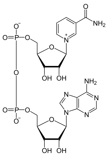
NAD+ is a coenzyme present in all living cells. Chemically it is two nucleotides joined head to head: nicotinamide riboside 5'-phosphate (NMN) and adenosine 5'-phosphate (AMP), linked through their phosphates into a pyrophosphate. The nicotinamide end does the chemistry; the adenosine end is largely a handle that enzymes grip.[1,21,3]
It was long studied as a redox carrier, accepting a hydride from a substrate to become NADH and handing it on. In 2000 two groups showed that the Sir2 family of proteins deacetylates histones in a reaction that absolutely requires NAD+ and breaks it apart, establishing NAD+ as a consumed substrate as well as a recycled cofactor. It is also cleaved by PARPs, by the CD38 ectoenzyme and by SARM1.[22,4,23,24,3]
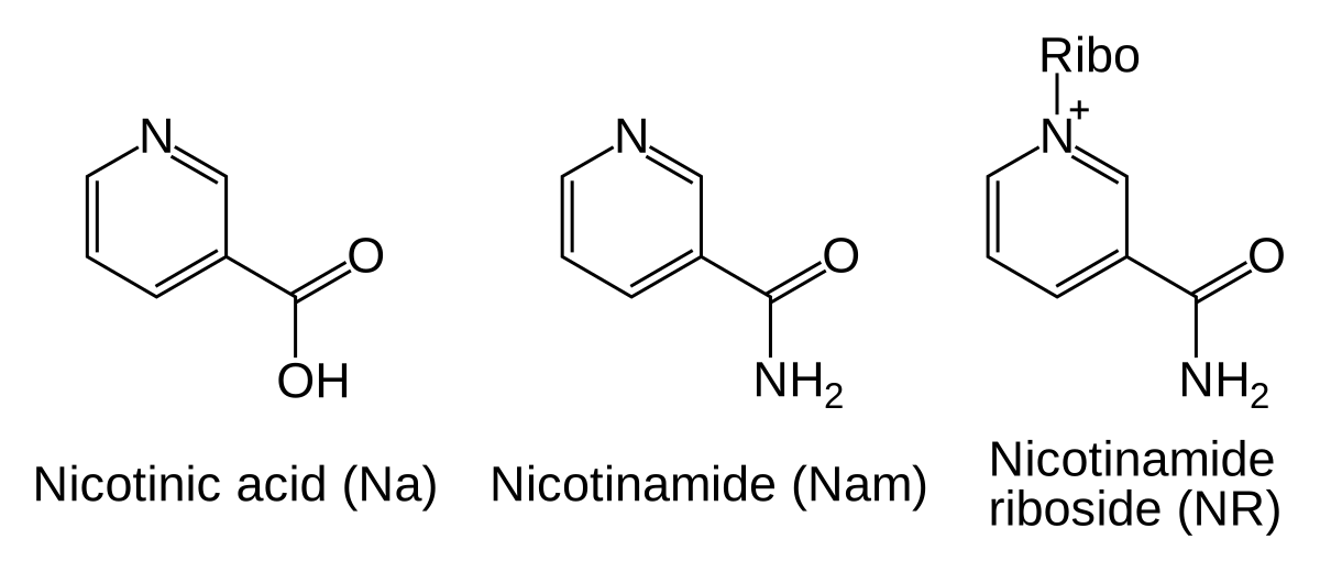
Because consumption is continuous, cells rebuild NAD+ from smaller pieces. The salvage route runs from nicotinamide through NMN, with nicotinamide phosphoribosyltransferase as the rate-limiting step in mouse fibroblasts, and nicotinamide riboside is phosphorylated by its own kinase. Nicotinamide has its own entry (as niacinamide); NMN and nicotinamide riboside are discussed here only as precursors, and this entry covers the dinucleotide itself.[13,14,15]
The name is used loosely. NAD sometimes means the oxidised form and sometimes the NAD+/NADH pair, and the plus sign follows the pyridinium nitrogen rather than the overall charge, which is why ChEBI keeps three entries for three charge states of one molecule.[2,25]
Status, in one sentence: the nonproprietary name nadide exists but no marketing authorisation as a medicine was found in the three jurisdictions searched, the DrugBank record and FDA unique ingredient identifier are catalogue entries rather than approvals, and the compound is named neither on the World Anti-Doping Agency 2026 Prohibited List nor in either table of the FDA bulk-substance list.[7,8,9,10,11,12,1]
Not to be confused with
- NADH
- The reduced form of the same dinucleotide, C21H29N7O14P2 at 665.4 g/mol: two hydrogens heavier, with the nicotinamide ring no longer aromatic or positively charged. The two interconvert in every redox reaction the molecule takes part in, and the library treats NADH as a separate entry.[1,26]
- alpha-NAD+
- The anomer with the opposite configuration at the carbon joining the nicotinamide ring to its ribose. It has the same formula and mass and a separate PubChem record; the biologically used form, and the one this page describes, is beta.[1]
- NADP+
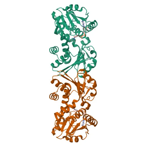
- Carries an extra phosphate on the 2' hydroxyl of the adenosine ribose. Enzymes discriminate sharply between the two: the dehydrogenase whose structure is shown on this page is strictly NAD(H) dependent, and its structures were used to explain why it rejects the phosphorylated cofactor.[32,25]
- Nicotinamide mononucleotide (NMN) and nicotinamide riboside (NR)
- Half-molecules and biosynthetic precursors, not alternative names. Several cell studies find that extracellular NAD+ is cut down to these before anything enters the cell, which is a reason not to treat a statement about a precursor as a statement about NAD+ itself, or the reverse.[17,5,15,14]
NAD+ molecular structure
Read from one end to the other, the molecule is an adenine on a ribose, a 5' phosphate, a second phosphate, a second ribose, and a nicotinamide ring attached to that ribose through its nitrogen. Attaching the ring nitrogen to a sugar quaternises it, so the ring is an aromatic pyridinium carrying a positive charge. Reduction adds a hydride at C4, removes the aromaticity and the charge, and gives NADH, two hydrogens heavier. The two glycosidic bonds are beta, and inverting just the nicotinamide one gives alpha-NAD+, a distinct compound with the same formula.[1,2,26]
Protein-bound NAD+ has been surveyed across the Protein Data Bank, and the shapes sort by what the enzyme does to it. Across 23 oxidoreductases most bound conformations were broadly alike and almost all of those enzymes bind the cofactor at the C-terminal end of a parallel beta sheet; in diphtheria toxin, which cleaves the molecule rather than reducing it, the nicotinamide half was sharply folded and the ring twisted outside its usual orientations. Sirtuins add a third arrangement, exposing one face of the nicotinamide ribose to the acetyl-lysine carbonyl. The cards below give each case.[27,28,29]
The ring chemistry is visible in a spectrophotometer, and that is how the two redox states are told apart: the aromatic pyridinium of NAD+ absorbs at 260 nm and not at 340 nm, while the dihydropyridine of NADH absorbs at both. Handling follows from the same ring. The solid is highly hygroscopic, and in solution the buffer matters more than one would expect: over 43 days at 19 °C in Tris the 260 nm peak fell by only 4%, in sodium phosphate the compound degraded at a rate comparable to NADH in the same study, and a mild rise to 25 °C was significant. That study is careful about its own method, and the caution carries: NAD+ cannot be quantified cleanly at 260 nm because NADH degradation products absorb there, and a product keeping either ring can make an absorbance reading report a stability the sample does not have.[20,30]
The viewer shows NAD+ from PDB 3JYO, a 1.00 angstrom structure of a bacterial quinate/shikimate dehydrogenase in which every heavy atom of the ligand is modelled at full occupancy. It shows one enzyme-bound shape, with no hydrogens and no information about which phosphates carry a charge. The nicotinamide ring is the least ordered part of the ligand in this crystal.[31,32]
Modifications
- Nicotinamide N1, glycosidic bond to the ribose The pyridine nitrogen is bonded to C1' of a ribose, which makes it a quaternary pyridinium and gives NAD+ its formal positive charge. This N-glycosidic bond is the one that sirtuins, PARPs, CD38 and SARM1 break, releasing nicotinamide and an ADP-ribose unit, so it is the bond on which the molecule's non-redox chemistry turns.[16,28,27]
- Pyrophosphate bridge between the two 5' carbons Two phosphates joined by an anhydride oxygen link the nicotinamide riboside to adenosine. Each phosphate has one ionisable oxygen: in the inner-salt formula one is ionised and the other is not, and ChEBI keeps separate entries for the forms with neither, one and both ionised.[2,1]
- Nicotinamide C4 The carbon that accepts or donates hydride. Reduction here converts NAD+ to NADH and turns the aromatic pyridinium into a non-aromatic dihydropyridine; at 1 angstrom an alcohol dehydrogenase structure shows that reduced ring puckered into a twisted boat.[26,19]
Eight defined stereocentres, four in each of the two D-ribose rings, with the (2R,3S,4R,5R) configuration in both rings as named by PubChem. Both glycosidic bonds are beta, the nicotinamide and the adenine each sitting on the same face of its ribose as the 5' carbon. Inverting only the nicotinamide-ribose anomeric carbon gives alpha-NAD+, a separate record with a different InChIKey stereo block. The phosphorus atoms are not assigned a configuration in the record.[1]
What studies on NAD+ show
Each card names the experimental system and what was measured. A result in one system is not evidence about another.
Enzyme kinetics with atomic-resolution X-ray crystallography · Purified recombinant quinate/shikimate dehydrogenase from Corynebacterium glutamicum
The enzyme was strictly NAD(H) dependent and preferred quinate. The binary structure gives a complete model of bound NAD+, and the comparison with related enzymes was used to explain the rejection of NADP(H).[32]
- Material
- The enzyme crystallised with NAD+ alone (binary complex, PDB 3JYO) and with NADH plus quinate or shikimate (ternary complexes)
- Measured
- Substrate and cosubstrate preference across pH, and three structures at atomic resolution
- Compared with
- Shikimate against quinate as substrate; NAD(H) against NADP(H) as cosubstrate; closely related shikimate/quinate dehydrogenases by sequence and structure
- Limitation
- Read as abstract only, so the residue contacts that discriminate NAD+ from NADP+ are not transcribed here. A bacterial enzyme in a crystal; it describes this binding site, not NAD+ in solution.
Höppner A et al., 2013, Biological chemistry
Comparative analysis of deposited crystal structures · NAD(P) conformations from 23 oxidoreductases in the Protein Data Bank, against NAD bound to diphtheria toxin
Oxidoreductase-bound conformations were mostly similar and grouped by structural class. Toxin-bound NAD was distinct: a highly folded NMN half, a short phosphorus-to-ring-nitrogen distance and a near-zero torsion about the scissile bond, which the authors proposed could orient or strain that bond for cleavage.[27]
- Material
- NAD and NADP as observed in each deposited complex
- Measured
- Superposition, atomic deviation, principal components and torsion angles
- Compared with
- Oxidoreductases, which reduce the ring, against an ADP-ribosylating toxin, which cleaves the glycosidic bond
- Limitation
- Abstract only. A survey of what crystals happened to capture in 1997, and the catalytic role of the strained conformation is a proposal, not a measurement.
Bell CE et al., 1997, Protein science : a publication of the Protein Society
X-ray crystallography of ternary complexes · Sir2Tm, a bacterial sirtuin, and its catalytic mutant H116Y
With the peptide present, NAD+ bound in a different conformation from earlier structures, exposing the alpha face of the nicotinamide ribose to the acetyl-lysine carbonyl oxygen, identically in wild type and mutant. The authors proposed that an invariant phenylalanine promotes the O-alkylamidate intermediate and blocks nicotinamide from re-entering.[28]
- Material
- Each enzyme with NAD+ and an acetylated peptide; a third structure with deacetylated peptide and 3'-O-acetyl ADP-ribose
- Measured
- The conformation of bound NAD+ relative to the acetyl-lysine
- Compared with
- Earlier sirtuin structures with NAD+ bound without peptide, and wild type against mutant
- Limitation
- Abstract only. A bacterial enzyme and a catalytic mutant; the mechanism is inferred from static structures.
Hoff KG et al., 2006, Structure (London, England : 1993)
Purified enzyme assays · Yeast SIR2 and homologs from other organisms
The enzymes catalysed an NAD-nicotinamide exchange reaction that required acetylated lysine, and deacetylated histones only in the presence of NAD+. No histone ADP-ribosylation was detected. A paper published the same year reached the NAD-dependence independently with yeast and mouse Sir2.[4]
- Material
- Recombinant SIR2-family proteins with acetylated histone substrates
- Measured
- Histone deacetylation, NAD-nicotinamide exchange and histone ADP-ribosylation
- Compared with
- Previously characterised histone deacetylases; reactions without NAD+; later structural work on nicotinamide inhibition and on substrate-induced conformational change
- Limitation
- Abstract only. Established the cosubstrate requirement; the chemical mechanism was worked out later from structures, where free nicotinamide was shown to bind a conserved pocket in the same site and drive the exchange reaction that inhibits the enzyme, and calorimetry on human SIRT3 indicated that the acetylated peptide binds before NAD+ does.
Landry J et al., 2000, Proceedings of the National Academy of Sciences of the United States of America
Genetically encoded fluorescent biosensor, flow cytometry and imaging · HEK293T and HeLa human cell lines, sensor targeted to nucleus, cytoplasm or mitochondria
Only NAD+ among the related nucleotides tested moved the sensor. Free NAD+ was about 106 micromolar in the cytoplasm, 109 in the nucleus and 230 in mitochondria of HEK293T cells. Knocking down NMNAT3 lowered the mitochondrial value, and in HeLa cells cytoplasmic NMNAT2 knockdown lowered both cytoplasmic and mitochondrial NAD+.[16]
- Material
- Circularly permuted Venus fused to an NAD+-binding domain from bacterial DNA ligase, mutated so it does not consume NAD+
- Measured
- Free NAD+ concentration in each compartment, calibrated in permeabilised cells
- Compared with
- cpVenus alone; related nucleotides and precursors for specificity; knockdown of each NMNAT isoform
- Limitation
- Measures free, not total, NAD+. The mitochondrial figure was read off the cytoplasmic calibration curve, and two cultured lines do not describe tissue.
Cambronne XA et al., 2016, Science (New York, N.Y.)
1H-NMR metabolomics of culture medium · HEK293 cells in medium with and without fetal bovine serum, and medium without cells
With cells present, under 40 percent of the added NAD+ remained at 12 hours and none was detectable at 48. Serum alone carried degrading activity; without serum NAD+ was relatively stable in medium, but cells still cleaved it to NMN. Transport inhibitors indicated that cleavage to nicotinamide riboside was a prerequisite for the nucleotides to sustain intracellular NAD.[5]
- Material
- NAD+ and related intermediates added to the medium (100 micromolar, assay condition)
- Measured
- The remaining compound and its breakdown products in the medium over time, and intracellular NAD maintenance
- Compared with
- Serum-containing against serum-free medium; with against without cells; plasma-membrane transport inhibitors
- Limitation
- One cell line in culture. The authors stress that the result depends on culture conditions, which is itself the point: a cell experiment adding NAD+ may be delivering its breakdown products.
Kulikova V et al., 2019, Metabolites
Isolated-organelle uptake assays with genetic knockout and complementation · Mitochondria isolated from human cells lacking or overexpressing SLC25A51, and from yeast lacking both native NAD+ carriers
Mammalian mitochondria took up intact NAD+, the uptake disappeared without SLC25A51 and returned on re-expression, and neither nicotinamide nor NMN reproduced or competed with it. Human SLC25A51 restored uptake in transporter-deficient yeast mitochondria. Two other groups independently reported the same transporter the same year.[18]
- Material
- Intact NAD+, including tritium-labelled NAD+ (100 micromolar, assay condition)
- Measured
- Matrix NAD+ content after incubation and initial rates of labelled uptake
- Compared with
- Nicotinamide and NMN; unlabelled NAD+ as competitor; yeast NDT1 as a known transporter
- Limitation
- No transport constants were determined, and the authors do not exclude other routes into mitochondria.
Luongo TS et al., 2020, Nature
Cryo-electron microscopy with mutagenesis · SARM1 homo-octamer, maps at 2.9 and 2.7 angstroms
An extra density in the NAD+-supplemented map sits at the tips of the first two armadillo repeats, a region the authors call the ARM allosteric site, away from the catalytic NADase domain, and holds one NAD+ molecule: its nicotinamide stacks on the Trp103 side chain, the adjacent ribose contacts Leu152 and His190, Arg110 forms a salt bridge to the phosphate nearer the nicotinamide while the far phosphate bridges to Arg110 and Arg157, and the weaker density at the distal ribose and adenosine involves Arg322, Gly323 and Asp326 of the second repeat. Occupying that site stabilises a packed, auto-inhibited octamer. In cells, L152A, R157E and R322E each made the enzyme constitutively active, matching a construct missing the whole armadillo domain, while D314A and Q320A, whose side chains point away from the ligand, and the control substitutions E94R and K363A did not. W103A had only a small effect but W103D activated strongly, so contact with that tryptophan appears dispensable while an acidic side chain in its place is not tolerated. Because nicotinamide mononucleotide lacks the distal phosphate, ribose and adenosine, it can bind the first repeat without making the bridging contacts to the second, which is how the authors explain its activating effect.[33]
- Material
- Octameric hSARM1 carrying the NADase-attenuated E642Q substitution, imaged with and without a 5 mM NAD+ supplement, plus point mutants of the site
- Measured
- The location of bound NAD+ and the activity of site mutants
- Compared with
- Mutants of the NAD+ site
- Limitation
- Read in full text. The ligand is fitted into a cryo-EM density at 2.7 Å rather than resolved atom by atom, the map is weaker at the adenosine end than at the nicotinamide end, and the activity readout is cell death on transfection rather than a rate. It shows NAD+ acting as a regulatory ligand in this protein; it does not establish the free NAD+ concentration at which the switch operates in a neuron.
Sporny M et al., 2020, eLife
How NAD+ is studied as a hydride carrier and as a cleaved substrate
In its redox role NAD+ accepts a hydride at C4 of the nicotinamide ring and leaves as NADH; the reverse step regenerates it, so the pair cycles without net loss. The structure on this page shows the cofactor bound to one such dehydrogenase, and a survey of oxidoreductase structures found most bound conformations broadly alike within each structural class.[19,3,27,32]
In its second role the same molecule is broken. Sirtuins, PARPs, CD38 and SARM1 all cut the bond between nicotinamide and its ribose, releasing nicotinamide and transferring or freeing the ADP-ribose unit; in sirtuins that cut is coupled to removing an acyl group from a lysine, and free nicotinamide inhibits the enzyme by binding a conserved pocket in the same site and driving the reaction backwards. Because the free NAD+ measured in cell compartments approximates the Michaelis constants reported for sirtuins and PARPs, changes in its level have been proposed to change their rates; that proposal comes from a comparison of figures, not from a rate measurement in the same cells. The enzyme-level detail sits in the study cards.[16,4,34,28,35,23,24,36]
A third, newer role is regulatory: in SARM1, NAD+ occupies an allosteric site away from the catalytic one and holds the enzyme inactive, and nicotinamide mononucleotide competes for it.[33,24,37]
What is still unknown about NAD+
Whether intact NAD+ crosses the plasma membrane is not settled. Several groups find that outside the cell it is cleaved by serum enzymes and ectoenzymes such as CD73 and CD38 to NMN and nicotinamide riboside, which then enter. Others report uptake of low concentrations in two cell types, or entry into neurons through P2X7-gated channels. The carriers that would do it at the plasma membrane have not been identified, in contrast to mitochondria, where SLC25A51 has been.[17,5,38,15,39,40,18,41,42]
Measurement is a persistent problem. Total NAD+ measured in extracts and free NAD+ measured by a sensor are different quantities, and ultraviolet detection after chromatography lacks the specificity a mixture of related nucleotides demands. Reported falls in tissue NAD+ with age in human skin and in mouse tissues are correlational measurements in those specific tissues.[16,19,43,44]
The fate of the intact molecule in people was examined in a published pilot with eight participants and three controls, which reported no plasma rise in NAD+ or its measured metabolites for the first two hours and a breakdown profile consistent with glycohydrolase and pyrophosphatase activity. One author was affiliated with a company in this area. No larger controlled pharmacokinetic study of intact NAD+ was found in the searches run for this entry, and most of the literature on raising NAD+ concerns precursors rather than the dinucleotide itself, so the human evidence about the molecule on this page rests on that one small study.[6,45]
How much of the structural account was read in full is worth stating. The SARM1 cryo-EM work was opened for this pass and its contact residues are in the card above; the diphtheria toxin survey is not open access and the atomic-resolution dehydrogenase paper behind the 3D view is paywalled, so both were read at abstract level, neither paper is transcribed at residue level here, and the geometry of the view was taken from the deposited coordinates instead.[27,32,31]
Key papers on NAD+
- Biosensor reveals multiple sources for mitochondrial NAD⁺.
Cambronne XA, Stewart ML, Kim D, Jones-Brunette AM, Morgan RK, Farrens DL, et al. (2016), Science (New York, N.Y.) · Genetically encoded biosensor
Measured free NAD+ separately in nucleus, cytoplasm and mitochondria, and set those figures beside the Michaelis constants of the consuming enzymes. The clearest single paper on how much NAD+ a cell compartment actually holds.
- The silencing protein SIR2 and its homologs are NAD-dependent protein deacetylases.
Landry J, Sutton A, Tafrov ST, Heller RC, Stebbins J, Pillus L, et al. (2000), Proceedings of the National Academy of Sciences of the United States of America · Purified enzyme assays
One of the two 2000 papers that made NAD+ a consumed substrate as well as a redox carrier, with the exchange reaction that later explained nicotinamide inhibition.
- Unusual conformation of nicotinamide adenine dinucleotide (NAD) bound to diphtheria toxin: a comparison with NAD bound to the oxidoreductase enzymes.
Bell CE, Yeates TO, Eisenberg D (1997), Protein science : a publication of the Protein Society · Comparative structure analysis
A structural survey showing what shapes NAD+ takes in enzymes that reduce it against one that cleaves it.
- SLC25A51 is a mammalian mitochondrial NAD(+) transporter.
Luongo TS, Eller JM, Lu MJ, Niere M, Raith F, Perry C, et al. (2020), Nature · Organelle uptake with knockout and complementation
Settled how intact NAD+ reaches the mitochondrial matrix in mammalian cells, with the controls that rule out a precursor route.
- Degradation of Extracellular NAD(+) Intermediates in Cultures of Human HEK293 Cells.
Kulikova V, Shabalin K, Nerinovski K, Yakimov A, Svetlova M, Solovjeva L, et al. (2019), Metabolites · NMR metabolomics of culture medium
Shows how quickly NAD+ added to a culture is broken down and what it becomes, which bears on how any cell experiment with added NAD+ should be read.
- Insights into the sirtuin mechanism from ternary complexes containing NAD+ and acetylated peptide.
Hoff KG, Avalos JL, Sens K, Wolberger C (2006), Structure (London, England : 1993) · Ternary complex crystallography
The sirtuin structures with both NAD+ and an acetylated peptide bound, showing the conformation in which the nicotinamide bond is cleaved.
- Structural basis for SARM1 inhibition and activation under energetic stress.
Sporny M, Guez-Haddad J, Khazma T, Yaron A, Dessau M, Shkolnisky Y, et al. (2020), eLife · Cryo-EM with mutagenesis
NAD+ as an allosteric ligand rather than a substrate, in cryo-EM maps of SARM1, with the contact residues and the mutations that break the site.
- NAD(+) metabolism and its roles in cellular processes during ageing.
Covarrubias AJ, Perrone R, Grozio A, Verdin E (2021), Nature reviews. Molecular cell biology · Review
A broad, referenced review of NAD+ synthesis, consumption and compartments, useful for orientation once the primary papers above have been read.
References
- 1.National Center for Biotechnology Information. (2026). PubChem Compound Summary for CID 5892, NAD+ (nadide). PubChem. source · database record
- 2.European Bioinformatics Institute. (2026). ChEBI entry CHEBI:15846, NAD(+). Chemical Entities of Biological Interest (ChEBI). source · database record
- 3.Covarrubias AJ, Perrone R, Grozio A, Verdin E. (2021). NAD(+) metabolism and its roles in cellular processes during ageing. Nature reviews. Molecular cell biology. 22:119-141. doi:10.1038/s41580-020-00313-x · abstract only
- 4.Landry J, Sutton A, Tafrov ST, Heller RC, Stebbins J, Pillus L, et al. (2000). The silencing protein SIR2 and its homologs are NAD-dependent protein deacetylases. Proceedings of the National Academy of Sciences of the United States of America. 97:5807-11. doi:10.1073/pnas.110148297 · abstract only
- 5.Kulikova V, Shabalin K, Nerinovski K, Yakimov A, Svetlova M, Solovjeva L, et al. (2019). Degradation of Extracellular NAD(+) Intermediates in Cultures of Human HEK293 Cells. Metabolites. 9. doi:10.3390/metabo9120293 · full text read
- 6.Grant R, Berg J, Mestayer R, Braidy N, Bennett J, Broom S, et al. (2019). A Pilot Study Investigating Changes in the Human Plasma and Urine NAD+ Metabolome During a 6 Hour Intravenous Infusion of NAD. Frontiers in aging neuroscience. 11:257. doi:10.3389/fnagi.2019.00257 · full text read
- 7.U.S. Food and Drug Administration. (2026). Drugs@FDA data via the openFDA drugsfda endpoint: active-ingredient and generic-name query for "nadide", "nicotinamide adenine dinucleotide", no matching application returned (queried November 2026). openFDA. source · database record
- 8.European Medicines Agency. (2026). Medicines output: medicines, the agency's JSON data report of centrally evaluated human and veterinary medicines (queried November 2026). European Medicines Agency. source · database record
- 9.Health Canada. (2026). Drug Product Database active-ingredient query for "nadide", no matching product returned (queried November 2026). Drug Product Database. source · database record
- 10.Health Canada. (2026). Drug Product Database active-ingredient query for "nicotinamide adenine", no matching product returned (queried November 2026). Drug Product Database. source · database record
- 11.World Anti-Doping Agency. (2025). World Anti-Doping Code International Standard: Prohibited List 2026. World Anti-Doping Agency. source · full text read
- 12.U.S. Food and Drug Administration. (2026). Certain Bulk Drug Substances for Use in Compounding that May Present Significant Safety Risks (queried November 2026). U.S. Food and Drug Administration. source · full text read
- 13.Revollo JR, Grimm AA, Imai S. (2004). The NAD biosynthesis pathway mediated by nicotinamide phosphoribosyltransferase regulates Sir2 activity in mammalian cells. The Journal of biological chemistry. 279:50754-63. doi:10.1074/jbc.M408388200 · abstract only
- 14.Bieganowski P, Brenner C. (2004). Discoveries of nicotinamide riboside as a nutrient and conserved NRK genes establish a Preiss-Handler independent route to NAD+ in fungi and humans. Cell. 117:495-502. doi:10.1016/s0092-8674(04)00416-7 · abstract only
- 15.Ratajczak J, Joffraud M, Trammell SA, Ras R, Canela N, Boutant M, et al. (2016). NRK1 controls nicotinamide mononucleotide and nicotinamide riboside metabolism in mammalian cells. Nature communications. 7:13103. doi:10.1038/ncomms13103 · abstract only
- 16.Cambronne XA, Stewart ML, Kim D, Jones-Brunette AM, Morgan RK, Farrens DL, et al. (2016). Biosensor reveals multiple sources for mitochondrial NAD⁺. Science (New York, N.Y.). 352:1474-7. doi:10.1126/science.aad5168 · full text read
- 17.Nikiforov A, Dölle C, Niere M, Ziegler M. (2011). Pathways and subcellular compartmentation of NAD biosynthesis in human cells: from entry of extracellular precursors to mitochondrial NAD generation. The Journal of biological chemistry. 286:21767-78. doi:10.1074/jbc.M110.213298 · abstract only
- 18.Luongo TS, Eller JM, Lu MJ, Niere M, Raith F, Perry C, et al. (2020). SLC25A51 is a mammalian mitochondrial NAD(+) transporter. Nature. 588:174-179. doi:10.1038/s41586-020-2741-7 · full text read
- 19.Trammell SA, Brenner C. (2013). Targeted, LCMS-based Metabolomics for Quantitative Measurement of NAD(+) Metabolites. Computational and structural biotechnology journal. 4:e201301012. doi:10.5936/csbj.201301012 · abstract only
- 20.Wolfe KD, Alahuhta M, Himmel ME, Bomble YJ, Jennings GK, Cliffel DE. (2024). Long-Term Stability of Nicotinamide Cofactors in Common Aqueous Buffers: Implications for Cell-Free Biocatalysis. Molecules. 29:5453. doi:10.3390/molecules29225453 · full text read
- 21.Verdin E. (2015). NAD⁺ in aging, metabolism, and neurodegeneration. Science (New York, N.Y.). 350:1208-13. doi:10.1126/science.aac4854 · abstract only
- 22.Imai S, Armstrong CM, Kaeberlein M, Guarente L. (2000). Transcriptional silencing and longevity protein Sir2 is an NAD-dependent histone deacetylase. Nature. 403:795-800. doi:10.1038/35001622 · abstract only
- 23.Liu Q, Kriksunov IA, Graeff R, Munshi C, Lee HC, Hao Q. (2005). Crystal structure of human CD38 extracellular domain. Structure (London, England : 1993). 13:1331-9. doi:10.1016/j.str.2005.05.012 · abstract only
- 24.Jiang Y, Liu T, Lee CH, Chang Q, Yang J, Zhang Z. (2020). The NAD(+)-mediated self-inhibition mechanism of pro-neurodegenerative SARM1. Nature. 588:658-663. doi:10.1038/s41586-020-2862-z · abstract only
- 25.Ying W. (2008). NAD+/NADH and NADP+/NADPH in cellular functions and cell death: regulation and biological consequences. Antioxidants & redox signaling. 10:179-206. doi:10.1089/ars.2007.1672 · abstract only
- 26.Meijers R, Adolph HW, Dauter Z, Wilson KS, Lamzin VS, Cedergren-Zeppezauer ES. (2007). Structural evidence for a ligand coordination switch in liver alcohol dehydrogenase. Biochemistry. 46:5446-54. doi:10.1021/bi6023594 · abstract only
- 27.Bell CE, Yeates TO, Eisenberg D. (1997). Unusual conformation of nicotinamide adenine dinucleotide (NAD) bound to diphtheria toxin: a comparison with NAD bound to the oxidoreductase enzymes. Protein science : a publication of the Protein Society. 6:2084-96. doi:10.1002/pro.5560061004 · abstract only
- 28.Hoff KG, Avalos JL, Sens K, Wolberger C. (2006). Insights into the sirtuin mechanism from ternary complexes containing NAD+ and acetylated peptide. Structure (London, England : 1993). 14:1231-40. doi:10.1016/j.str.2006.06.006 · abstract only
- 29.Min J, Landry J, Sternglanz R, Xu RM. (2001). Crystal structure of a SIR2 homolog-NAD complex. Cell. 105:269-79. doi:10.1016/s0092-8674(01)00317-8 · abstract only
- 30.National Center for Biotechnology Information. PubChem experimental-properties record for CID 5892, NAD+ (the hygroscopicity attributed there to the Merck Index; the melting point and solubility entries carry no named source). PubChem. source · database record
- 31.Hoppner A, Schomburg D, Niefind K. (2010). Quinate dehydrogenase from Corynebacterium glutamicum in complex with NAD. RCSB Protein Data Bank, entry 3JYO. doi:10.2210/pdb3jyo/pdb · database record
- 32.Höppner A, Schomburg D, Niefind K. (2013). Enzyme-substrate complexes of the quinate/shikimate dehydrogenase from Corynebacterium glutamicum enable new insights in substrate and cofactor binding, specificity, and discrimination. Biological chemistry. 394:1505-16. doi:10.1515/hsz-2013-0170 · abstract only
- 33.Sporny M, Guez-Haddad J, Khazma T, Yaron A, Dessau M, Shkolnisky Y, et al. (2020). Structural basis for SARM1 inhibition and activation under energetic stress. eLife. 9. doi:10.7554/eLife.62021 · full text read
- 34.Avalos JL, Bever KM, Wolberger C. (2005). Mechanism of sirtuin inhibition by nicotinamide: altering the NAD(+) cosubstrate specificity of a Sir2 enzyme. Molecular cell. 17:855-68. doi:10.1016/j.molcel.2005.02.022 · abstract only
- 35.Jin L, Wei W, Jiang Y, Peng H, Cai J, Mao C, et al. (2009). Crystal structures of human SIRT3 displaying substrate-induced conformational changes. The Journal of biological chemistry. 284:24394-405. doi:10.1074/jbc.M109.014928 · abstract only
- 36.Feldman JL, Dittenhafer-Reed KE, Denu JM. (2012). Sirtuin catalysis and regulation. The Journal of biological chemistry. 287:42419-27. doi:10.1074/jbc.R112.378877 · abstract only
- 37.Figley MD, Gu W, Nanson JD, Shi Y, Sasaki Y, Cunnea K, et al. (2021). SARM1 is a metabolic sensor activated by an increased NMN/NAD(+) ratio to trigger axon degeneration. Neuron. 109:1118-1136.e11. doi:10.1016/j.neuron.2021.02.009 · abstract only
- 38.Grozio A, Sociali G, Sturla L, Caffa I, Soncini D, Salis A, et al. (2013). CD73 protein as a source of extracellular precursors for sustained NAD+ biosynthesis in FK866-treated tumor cells. The Journal of biological chemistry. 288:25938-25949. doi:10.1074/jbc.M113.470435 · abstract only
- 39.Billington RA, Travelli C, Ercolano E, Galli U, Roman CB, Grolla AA, et al. (2008). Characterization of NAD uptake in mammalian cells. The Journal of biological chemistry. 283:6367-74. doi:10.1074/jbc.M706204200 · abstract only
- 40.Alano CC, Garnier P, Ying W, Higashi Y, Kauppinen TM, Swanson RA. (2010). NAD+ depletion is necessary and sufficient for poly(ADP-ribose) polymerase-1-mediated neuronal death. The Journal of neuroscience : the official journal of the Society for Neuroscience. 30:2967-78. doi:10.1523/JNEUROSCI.5552-09.2010 · abstract only
- 41.Kory N, Uit de Bos J, van der Rijt S, Jankovic N, Güra M, Arp N, et al. (2020). MCART1/SLC25A51 is required for mitochondrial NAD transport. Science advances. 6. doi:10.1126/sciadv.abe5310 · abstract only
- 42.Girardi E, Agrimi G, Goldmann U, Fiume G, Lindinger S, Sedlyarov V, et al. (2020). Epistasis-driven identification of SLC25A51 as a regulator of human mitochondrial NAD import. Nature communications. 11:6145. doi:10.1038/s41467-020-19871-x · abstract only
- 43.Massudi H, Grant R, Braidy N, Guest J, Farnsworth B, Guillemin GJ. (2012). Age-associated changes in oxidative stress and NAD+ metabolism in human tissue. PloS one. 7:e42357. doi:10.1371/journal.pone.0042357 · abstract only
- 44.Camacho-Pereira J, Tarragó MG, Chini CCS, Nin V, Escande C, Warner GM, et al. (2016). CD38 Dictates Age-Related NAD Decline and Mitochondrial Dysfunction through an SIRT3-Dependent Mechanism. Cell metabolism. 23:1127-1139. doi:10.1016/j.cmet.2016.05.006 · abstract only
- 45.Rajman L, Chwalek K, Sinclair DA. (2018). Therapeutic Potential of NAD-Boosting Molecules: The In Vivo Evidence. Cell metabolism. 27:529-547. doi:10.1016/j.cmet.2018.02.011 · abstract only
