MOTS-c in brief
MOTS-c is an unmodified 16-residue peptide, MRWQEMGYIFYPRKLR, whose 51 base pair reading frame sits inside the mitochondrial 12S ribosomal RNA gene, MT-RNR1; it was named in 2015 as a mitochondrial-derived peptide. No receptor, affinity constant or three-dimensional structure has been reported. The evidence is mostly cell-line overexpression, co-immunoprecipitation and pull-down work, mouse models and a few human studies, and the two proposed mechanisms, a folate and AICAR route and a nuclear stress response, remain unconnected. Human measurements rest almost entirely on commercial immunoassays with no independently validated method, and how the peptide is translated outside the mitochondrion is still unexplained.[1,2,3,4,5,6]
MOTS-c regulatory status
No marketing authorisation as a medicine was found for MOTS-c in Drugs@FDA, the EMA medicines data or the Health Canada Drug Product Database (searched November 2026). It is investigational: ClinicalTrials.gov records a phase 2a study in adults with prediabetes and overweight or obesity, entered in 2026 and recruiting (NCT07505745), and no completed controlled study of the peptide has posted results. The WADA 2026 Prohibited List names it under S4.4.1, metabolic modulators that activate AMP-activated protein kinase, prohibited at all times. The FDA also lists it among bulk drug substances nominated for compounding whose nominations the nominators later withdrew, and that entry carries the agency's stated concerns: that such compounded products may pose significant risk for immunogenicity and may have complexities with regard to peptide-related impurities and substance characterisation, that FDA has identified no human exposure data for them, and that the agency lacks important information about any safety issues the peptide raises, including whether it would cause harm in people. One cited primary study on bone has been retracted and is flagged as such in the reference list; no other work cited here carries a retraction or expression-of-concern flag.[7,8,9,10,11,12,13]
MOTS-c at a glance
- A 16-residue peptide, MRWQEMGYIFYPRKLR, encoded by a 51 base pair open reading frame inside the mitochondrial 12S ribosomal RNA gene MT-RNR1. The discovery paper argues the reading frame can only be translated in the cytoplasm, because the mitochondrial genetic code reads it as tandem start and stop codons.[1,2,5]
- No receptor has been identified. Every molecular interaction reported for it is an association found by co-immunoprecipitation or by a biotinylated-peptide pull-down, or a computational docking prediction. There is no published affinity constant and no structure of a bound form.[3,14,15,16]
- Two mechanisms have been proposed and they remain unconnected: a metabolic one, in which extra peptide depletes 5-methyl-tetrahydrofolate, blocks de novo purine synthesis, raises the intermediate AICAR and increases AMPK-alpha Thr172 phosphorylation; and a nuclear one, in which peptide reaches the nucleus within minutes of metabolic stress and associates with antioxidant-response promoter fragments and with the transcription factors NRF2 and ATF1. The mechanism section sets out both with their figures and their gaps.[1,3]
- A 2026 study reads the sequence as a host defence peptide, cationic and amphipathic, and reports it aggregating and permeabilising Escherichia coli and methicillin-resistant Staphylococcus aureus and reprogramming monocyte differentiation. The active concentrations are tens of micromolar.[17]
- Human measurement is the weakest link in the whole literature. The published numbers come almost entirely from commercial immunoassays and they disagree on direction: one cohort has circulating levels falling with age while muscle levels rise, another has plasma levels higher in metabolic syndrome and rising with liver fat.[4,18]
- No experimental 3D structure exists, and no computed conformer either. Published conformational models are sequence-based predictions described by their authors as flexible.[19,20,21]
What is MOTS-c?
The human mitochondrial genome is small and was long thought to be fully annotated at 37 genes: 22 transfer RNAs, 2 ribosomal RNAs and 13 messenger RNAs. MOTS-c belongs to a set of claimed additional products, the mitochondrial-derived peptides or mitochondrial microproteins, read from short open reading frames inside the two ribosomal RNA genes. Humanin and the six small humanin-like peptides are read from the 16S gene; this peptide is the one read from the 12S gene, MT-RNR1, and a 2023 review counts ten such microproteins in all.[22,23,24]
It was named and characterised in 2015 from an in silico search of the human 12S ribosomal RNA for short reading frames with a strong Kozak sequence. The frame found is 51 base pairs, which is 16 codons plus a stop, and was deposited as GenBank KP715230. One structural fact about the sequence is load-bearing for the whole account: read with the mitochondrion's own genetic code the frame contains tandem start and stop codons, so if a peptide is made from it at all, translation has to happen in the cytoplasm, which in turn requires the transcript to leave the mitochondrion. The curated UniProt entry records that argument as a CAUTION, stating that the peptide has been shown to be biologically active but that the mechanisms allowing its production and secretion remain unclear.[1,2,5]
What distinguishes it in the literature is not a receptor but a proposed direction of travel. Nuclear-encoded proteins regulating mitochondrial gene expression were already familiar; the 2018 study reported the reverse, a mitochondrially encoded peptide entering the nucleus and associating with promoter DNA, which its authors frame as retrograde mitonuclear signalling. That framing is why most of the evidence is correlative: a peptide proposed to coordinate a stress response is easy to measure and hard to pin to a molecular event.[3,25,4]
Status belongs beside the identity, because it is the fact most often searched after the sequence. No medicines regulator has authorised the peptide for any indication, and the one registered interventional study of the peptide itself was still recruiting in November 2026, so no completed controlled human trial has reported safety outcomes for it. The World Anti-Doping Agency's 2026 Prohibited List names it under S4.4.1, among metabolic modulators that activate AMP-activated protein kinase, prohibited at all times. The FDA lists it among bulk drug substances that were nominated for compounding and then withdrawn by the nominators, and records its own concerns in that entry: compounded products containing the peptide may pose significant risk for immunogenicity and may have complexities with regard to peptide-related impurities and substance characterisation; the agency has identified no human exposure data for such products; and it states that it lacks important information about any safety issues the peptide raises, including whether it would cause harm in people.[7,8,9,10,11,12]
Not to be confused with
- Humanin and the small humanin-like peptides SHLP1 to SHLP6
- The other named members of the mitochondrial-derived peptide family. They are encoded in the 16S ribosomal RNA gene (MT-RNR2), whereas this peptide is encoded in the 12S gene (MT-RNR1), and their sequences are unrelated to MRWQEMGYIFYPRKLR. Several studies measure this peptide and humanin side by side in the same samples, which is a comparison and not an identity.[23,33,34]
- SHMOOSE and Gau
- Two later mitochondrial microproteins, encoded over a serine tRNA and antisense in the mitochondrial genome respectively. A review listing ten such microproteins names them as separate entries beside this one.[24]
- K14Q MOTS-c
- A different peptide by one residue. The mitochondrial variant m.1382A>C (rs111033358) replaces lysine 14 with glutamine, lowering the net charge at pH 7.0 and raising the hydrophobic moment. In matched assays the wild-type peptide raised insulin-stimulated AKT Ser473 phosphorylation in C2C12 myotubes and the variant peptide did not, so the two are not interchangeable in the literature.[21,35]
- MotS (Bacillus subtilis flagellar stator protein)
- An unrelated bacterial membrane protein with a confusingly similar name. It is the only thing a full-text search of the Protein Data Bank for “MOTS-c” returns: the entries 9LJK, 9LJL and 9LJM are X-ray structures of its periplasmic domain. Anyone searching a structure database for this peptide will meet those three first and they characterise nothing about it.[19]
- The 12S ribosomal RNA itself
- MT-RNR1 is a structural RNA of the mitochondrial ribosome. The peptide's 51 base pair open reading frame lies inside that gene, so the gene has two reported products and the peptide is the minor, contested one rather than a renaming of the rRNA.[2,4]
- CB4211
- A different molecule: a modified peptide that its sponsor described as "a novel and improved analog of MOTS-c" when it entered clinical testing. It was studied in a randomised, double-blind, placebo-controlled phase 1a/1b programme in healthy participants and in people with non-alcoholic fatty liver disease, which enrolled 88 participants and completed in April 2021 with no results posted to the registry. Nothing measured for it is a measurement of this peptide, and the sequence difference is not stated in either public record.[32,36]
- Elamipretide (SS-31, MTP-131)
- A different molecule that market material repeatedly confuses with this peptide, to the point of attributing its clinical trials to this one. It is a synthetic, nuclear-independent tetrapeptide of 639.8 g/mol (PubChem CID 11764719, C32H49N9O5) carrying a D-arginine and a dimethyltyrosine, and it works by binding cardiolipin in the inner mitochondrial membrane, which is a lipid target rather than a sequence encoded in mitochondrial DNA. Nothing measured for it is a measurement of this peptide, and the only thing the two share is the word mitochondrial.[37,38]
MOTS-c structure and sequence
The molecule is as simple as a peptide gets: sixteen L-amino acids in one chain, a free amine at Met1, a free carboxylate at Arg16, no cysteine and therefore no disulfide, no lipid arm, no linker, no sugar and no metal. PubChem computes C101H152N28O22S2 for the free acid, an average mass of 2174.6 and a monoisotopic mass of 2173.1077. Nothing in the sequence needs curated notation, so the identity question here is whether the molecule is made in the body at all rather than what it is.[20,5]
Two features of the sequence carry most of the published chemistry. Positions 13 to 16 are Arg-Lys-Leu-Arg, a cluster of basic residues resembling a classical nuclear localisation signal, and positions 8 to 11 are Tyr-Ile-Phe-Tyr, a hydrophobic and aromatic patch. The 2018 study replaced each of those four-residue blocks with alanines and ran the resulting peptides in a gel-shift assay: binding to antioxidant-response-element promoter fragments was lost in both cases. The two blocks separate when nuclear entry is measured instead. Replacing the basic cluster did not block nuclear localisation of an EGFP fusion, while replacing the hydrophobic patch did, so the basic cluster is required for the DNA binding and not for the transport, and the hydrophobic patch is required for both.[3]
A 2026 study profiled the same sequence from the chemistry rather than from the cell biology, on the Kyte-Doolittle and Sweet-Eisenberg hydrophobicity scales, and arrived at the same two-domain reading: a hydrophobic core at Tyr8-Ile-Phe-Tyr11 and a cationic tail at Arg13-Lys-Leu-Arg16, with the peptide holding a net charge of +3 at physiological pH, mostly from the tail. The authors also note that the tail resembles a heparin-binding motif. Quadruple-alanine substitution of either block removed the bacterial aggregation and growth inhibition they measured, which are the same two substitutions that removed promoter-fragment binding in the 2018 nuclear work. So the two blocks are doing double duty in the literature, and that is a point in favour of reading them as a plain charge-and-hydrophobicity feature of a short peptide rather than as a dedicated recognition surface.[17,3]
The one natural sequence variant is a charge change in that same cluster. Mitochondrial m.1382A>C replaces lysine 14 with glutamine, lowering the net charge at pH 7.0 and raising hydrophobicity, and modelling of the 12S ribosomal RNA secondary structure around the site showed very little difference between the two alleles, so the authors read the consequence as a property of the peptide rather than of the RNA. As for shape, there is nothing measured to report: no crystal, cryo-EM or NMR structure has been deposited, PubChem serves no computed conformer, and the only published models are PEP-FOLD3 and I-TASSER predictions that are mostly loop with a short N-terminal helix. A three-dimensional view here would be a drawing of an assumption, which is why there is none.[21,19,20]
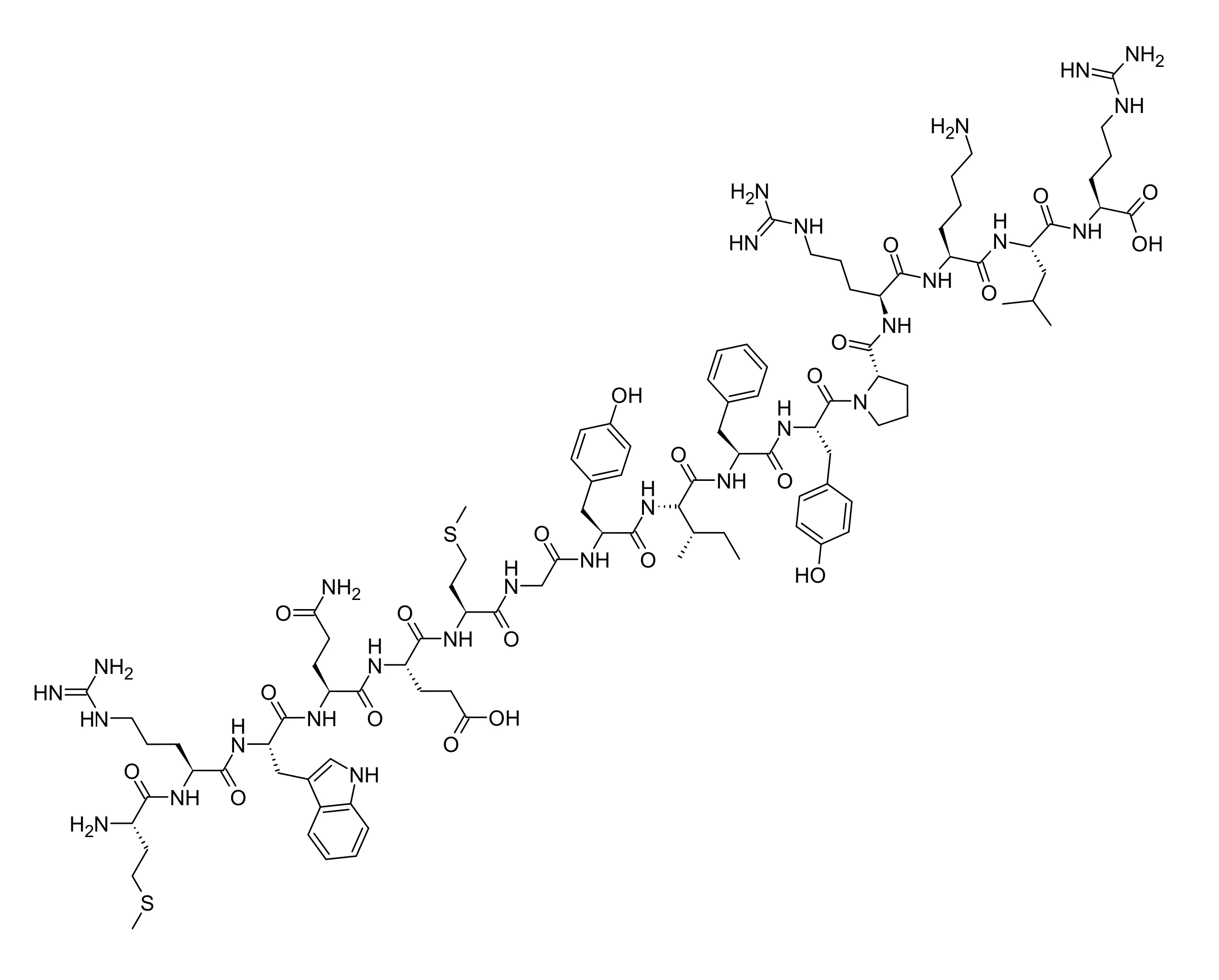
All stereocentres are L; glycine at position 7 is achiral and proline at position 12 is the only cyclic residue. There is no disulfide, no lipid arm, no linker, no glycosylation and no metal coordination, so the molecule is fully specified by its sequence and its termini. Three features of that sequence matter chemically. Three arginines and one lysine against a single glutamate give the peptide a net positive charge near neutral pH, and the basic cluster at positions 13 to 16 (Arg-Lys-Leu-Arg) resembles a classical nuclear localisation signal without being one. Two methionines, at positions 1 and 6, and the tryptophan at position 3 are oxidation-labile side chains, which is a handling property of the synthetic material rather than a finding about the molecule. The variant arising from mtDNA m.1382A>C replaces lysine 14, so it changes the charge of exactly that basic cluster.[5,20,3,21]
What studies on MOTS-c show
Each card names the experimental system and what was measured. A result in one system is not evidence about another.
Untargeted metabolomics with microarray and extracellular flux assays · HEK293 cells stably overexpressing the peptide, and HEK293 cells treated with synthetic peptide
In overexpressing cells 5-methyl-tetrahydrofolate and methionine fell while homocysteine rose, de novo purine intermediates fell and AICAR rose more than 20-fold, with increased phosphorylation of AMPK-alpha Thr172. Adding 100 nM folic acid to the medium reversed the increase in extracellular acidification rate, and neither E5A, G7A nor the scrambled peptide produced that increase.[1]
- Material
- Synthetic human 16-mer, plus the alanine point mutants E5A and G7A and a scrambled-sequence control peptide
- Measured
- Folate and methionine cycle metabolites, de novo purine intermediates, AICAR, AMPK-alpha Thr172 phosphorylation, and extracellular acidification rate
- Compared with
- Vector-only and untreated cells; folic acid add-back; the AMPK inhibitor compound C and AMPK-alpha knockdowns
- Limitation
- The experiment did not establish a direct molecular target. No folate-cycle enzyme was shown to be inhibited by the peptide; the pathway assignment rests on the pattern of metabolites and transcripts. Removing AMPK accounted for only part of the glycolytic readout: knockdown of AMPK-alpha2 gave a 16 percent decrease and of AMPK-alpha1/2 a 30 percent decrease, with the inhibitor compound C giving 40 percent, so AMPK is a partial route at best.
Lee C et al., 2015, Cell Metabolism
Comparative sequence analysis · Deduced peptide sequences from 14 species
The first 11 residues are the conserved part of the sequence. Positions 4, 5, 7 and 9 returned dN/dS above 1, and positions 3 and 10 below 1.[1]
- Material
- The 51 base pair reading frame within the 12S ribosomal RNA gene
- Measured
- Residue-level dN/dS ratios and a random-effects likelihood test
- Compared with
- Synonymous against non-synonymous substitution rates across the 14 alignments
- Limitation
- The experiment did not establish that the peptide is translated. Selection signatures inside a gene that also encodes a structural RNA can reflect constraint on the RNA, and the test was run on deduced rather than observed peptide sequences.
Lee C et al., 2015, Cell Metabolism
Subcellular fractionation, co-immunoprecipitation, electrophoretic mobility shift assay, ChIP-qPCR and RNA sequencing · HEK293 and HepG2 cells under glucose restriction, serum deprivation or tert-butyl hydroperoxide
Labelled peptide reached both mitochondria and nucleus within 30 minutes, with nuclear levels peaking around 3 hours after stress and returning to baseline by 24 hours. NRF2 co-precipitated from nuclear extracts only in stressed cells at 3 hours, and ATF1 co-precipitation strengthened after glucose restriction. In the gel-shift assay the peptide bound promoter fragments of HO-1, NQO1, UGT1A1, UGT1A6, TXN, FTL and GPX2 in a concentration-dependent way, and binding was abolished by either alanine substitution. Under glucose restriction 802 transcripts changed, 390 up and 412 down.[3]
- Material
- FITC-labelled synthetic peptide, an EGFP fusion, and a synthetic wild-type peptide beside quadruple-alanine mutants of Tyr8-Ile-Phe-Tyr11 and Arg13-Lys-Leu-Arg16
- Measured
- Nuclear versus mitochondrial localisation over time, association with NRF2, ATF1 and NRF1, direct binding to antioxidant-response-element promoter fragments, and transcript changes
- Compared with
- Unstressed cells at 0 and 24 hours; the two alanine mutants; vector-only transfection
- Limitation
- The experiment did not establish sequence-specific recognition. A gel-shift assay with a charged, arginine-rich peptide shows binding under cell-free conditions and no consensus motif for the peptide was derived; the ChIP and reporter readouts came from overexpressing cells, and the paper notes that primary human cells were not used.
Kim KH et al., 2018, Cell Metabolism
Human exercise trial with muscle biopsy, plus mouse metabolomics and RNA sequencing · Healthy young men on a cycle ergometer; C57BL/6J and CD-1 mice
Endogenous peptide in human skeletal muscle rose about 11.9-fold after exercise and was still elevated after four hours of rest, with circulating levels also rising.[6]
- Material
- Endogenous peptide measured by western blot in muscle and by immunoassay in plasma; synthetic peptide given to the mice
- Measured
- Endogenous peptide in skeletal muscle and plasma before, during and after exercise; muscle metabolite sets and gene-set enrichment in mice
- Compared with
- Each participant's own pre-exercise sample; vehicle-treated mice
- Limitation
- The experiment did not establish what the peptide does when it rises. It is an association between a stressor and a measured level, in a small single-sex cohort, and the plasma arm depends on an immunoassay whose specificity is not independently characterised.
Reynolds JC et al., 2021, Nature Communications
Matched-peptide cell assays with cohort genotyping and computational variant assessment · C2C12 myotubes, L6 myotubes and HEK293 cells; three cohorts totalling 27,527 participants
The single lysine-to-glutamine change at position 14 lowered the net charge and raised hydrophobicity while barely altering the predicted 12S ribosomal RNA secondary structure. The wild-type peptide raised insulin-stimulated AKT Ser473 phosphorylation in C2C12 myotubes and increased glucose uptake in L6 myotubes and transfected HEK293; the variant peptide did neither.[21]
- Material
- Synthetic wild-type 16-mer against the K14Q variant peptide encoded by mtDNA m.1382A>C
- Measured
- Insulin-stimulated AKT Ser473 phosphorylation, glucose uptake, net peptide charge at pH 7.0, PROVEAN score and predicted conformers
- Compared with
- The two peptides head to head, with untreated and vector-only controls
- Limitation
- The experiment did not establish a structural reason for the difference. The conformer models are sequence-based predictions, the cell readouts use peptide at assay concentrations, and a one-residue loss of activity is equally consistent with a lost interaction surface and with changed uptake or stability.
Zempo H et al., 2021, Aging
Co-immunoprecipitation with domain mapping, in a membrane-damage assay · Cultured cells under hypotonic membrane damage; TRIM72 knockout mice; human participants after eccentric exercise
The peptide co-precipitated with the TRIM72 C-terminus and not with the N-terminus, and the effect on membrane repair was abolished by TRIM72 knockdown but not by the AMPK inhibitor. The authors report that the peptide alone had negligible effect on membrane repair.[14]
- Material
- Synthetic peptide; separate TRIM72 N-terminal and C-terminal fragments
- Measured
- Which TRIM72 fragment the peptide co-precipitates with, trafficking of TRIM72 to the damaged membrane, and membrane integrity
- Compared with
- TRIM72 small interfering RNA, an AMPK inhibitor, and TRIM72 knockout animals
- Limitation
- The experiment did not establish binding in the quantitative sense. A co-immunoprecipitation shows association in a lysate, with no affinity, no stoichiometry and no structural contact, and read only from the abstract here.
Jia H et al., 2024, Theranostics
Biotinylated-peptide pull-down with mass spectrometry and functional substitution · Human umbilical vein endothelial cells exposed to taurocholic acid; a cholic-acid mouse model
The pull-down nominated the transcriptional repressor BACH1 as a binding partner, with competition reported at BACH1 residue D560 that displaces USP7 and shifts BACH1 toward K33-linked polyubiquitination.[15]
- Material
- Biotinylated synthetic peptide
- Measured
- Which proteins the biotinylated peptide retrieves, and competition at a named BACH1 residue
- Compared with
- Unconjugated controls; BACH1 residue substitution; the deubiquitinase USP7
- Limitation
- The experiment did not establish a contact. A pull-down nominates a partner, the residue assignment rests on functional substitution rather than on a solved structure, and this was read from the abstract only.
Li S et al., 2026, Cell Communication and Signaling
Antibacterial assays, a mouse infection model, and monocyte transcriptomics · Escherichia coli and methicillin-resistant Staphylococcus aureus in culture; C57BL/6J mice in an acute peritonitis model; THP-1 human monocytes and primary mouse monocytes
Across a 0 to 100 µM series the peptide aggregated both organisms in water, and a single 100 µM addition retarded Escherichia coli growth, with two additions of 50 µM comparable to it. Bacteria carried into the mouse peritonitis model after exposure to 100 µM peptide gave complete survival against 16.7% for the water control and a 3.4-fold lower colony count in the inoculum, with a male replicate at 100% against 20% and a 19.8-fold reduction. Interferon gamma, lipopolysaccharide and differentiation signals each raised expression of the endogenous peptide in THP-1 cells, and peptide present during primary mouse monocyte differentiation produced macrophages with antigen-presentation and interferon transcriptomic signatures, faster bacterial clearance and a shifted metabolism.[17]
- Material
- Synthetic 16-mer, with quadruple-alanine substitutions of Tyr8-Ile-Phe-Tyr11 and of Arg13-Lys-Leu-Arg16
- Measured
- Bacterial aggregation, growth by optical density, membrane integrity by SYTOX Green, bacterial ATP, survival and colony counts in the mouse model, expression of the endogenous peptide after immune stimuli, and macrophage transcriptomes
- Compared with
- Water-exposed and heat-killed bacteria; the two alanine mutants; monocytes differentiated without peptide
- Limitation
- Read in full text. No minimum inhibitory concentration was determined, and the active concentrations are tens of micromolar, roughly six orders of magnitude above the plasma concentrations immunoassays report for the endogenous peptide, so this is a property of the synthetic peptide at assay concentrations and not a demonstrated physiological role. Most of the induction work is one cell line, THP-1, which the authors say they chose for tractability, and they flag the single-cell arm's limited sample number themselves. The senior author declares that he is a consultant and shareholder in the company that developed the peptide analogue.
Rice MC et al., 2026, eLife
How MOTS-c is studied when no receptor has been found
The original account is indirect and remains unresolved at the molecular level. Cells carrying extra peptide showed depletion of 5-methyl-tetrahydrofolate, a block in de novo purine synthesis, a large rise in the intermediate AICAR, and AMPK-alpha Thr172 phosphorylation; adding folic acid back to the medium reversed the glycolytic readout. That is a coherent chain, and no step of it includes the peptide touching a protein. Eleven years on, no folate-cycle enzyme has been shown to be inhibited directly by it. The arrangement is also partly circular: a later study reported that AMPK inhibition and PGC-1-alpha knockdown both lower measured peptide levels while PGC-1-alpha overexpression raises them, which puts the peptide downstream of the pathway it is said to activate.[1,26]
The second account is nuclear. Within 30 minutes of glucose restriction or oxidative stress, labelled peptide is recovered from the nuclear fraction, peaking at about three hours; it co-precipitates with NRF2 and with ATF1 only in stressed cells, and in a cell-free gel-shift assay it binds antioxidant-response-element promoter fragments in a way that depends on both the Tyr8-Ile-Phe-Tyr11 and the Arg13-Lys-Leu-Arg16 blocks. How it gets there is contested within its own literature. The 2018 study found that destroying the basic cluster did not prevent nuclear entry, so the resemblance to a nuclear localisation signal does not explain the transport; a 2025 study instead proposes a carrier route in which CK2-alpha phosphorylation of MYH9 at Ser1943 lets the peptide ride MYH9 and gamma-actin complexes into the nucleus.[3,27]
Beyond those two, the reported interactions are a list of named proteins rather than a mechanism. Co-immunoprecipitation places an association on the TRIM72 C-terminus; a biotinylated pull-down with mass spectrometry nominates BACH1, with competition assigned to one residue; a kinase-cascade paper reports a CK2 to PTEN to mTORC2 to AKT route lowering FOXO1 activity and myostatin levels; a macrophage study reports lowered MAPK phosphorylation with raised aryl hydrocarbon receptor and STAT3 expression. None is anchored to a measured affinity or a solved contact, and they do not obviously reduce to one another.[14,15,28,29]
A third account arrived in 2026 and is the only one that does not require the peptide to act on a human protein at all. Read purely as chemistry the sequence is a short cationic amphipathic peptide, which is the composition of a host defence peptide, and the study reports it aggregating and permeabilising Escherichia coli and methicillin-resistant Staphylococcus aureus, neutralising the latter's infectivity in a mouse peritonitis model, being induced in monocytes by interferon gamma and lipopolysaccharide, and reprogramming monocyte differentiation toward an interferon and antigen-presentation profile. It carries its own limit, stated in the card above: the concentrations are tens of micromolar. It also unifies rather than adds, because the same two sequence blocks the 2018 work mapped are the ones whose substitution removes the antibacterial activity.[17,3]
What is still unknown about MOTS-c
The most basic open question is measurement. Human work depends almost entirely on commercial immunoassays for a 16-residue peptide, and no independently validated analytical method for the circulating form turned up in this search. The consequences show in the published numbers. One cohort reports circulating levels falling with age while muscle levels rise about 1.5-fold, correlating with each other only in young men; another reports plasma levels around 315 pg/ml that are higher in metabolic syndrome and rise with liver fat, the opposite direction. In an Alzheimer disease cohort, transcript measures separated the groups and plasma protein measures did not, in the same participants. Levels also move with stressors other than exercise, repeated heat exposure among them. Until an assay is pinned down, the literature cannot settle which way the number moves.[4,18,30,6,31]
The second is that there is no receptor, no affinity constant and no structure. Every interaction on record is an association from a lysate or a prediction from software, and one recent report docks the 16-mer against MAPK, mTOR, AMPK, NRF2, PI3K and caspase 3 together, which illustrates how freely a small flexible peptide can be computationally assigned partners. The two accounts of nuclear entry also disagree, one finding the basic cluster dispensable and the other proposing a phosphorylation-gated carrier. A deposited structure would discipline this and none exists; a sequence search of the Protein Data Bank returns nothing and PubChem has no computed conformer.[16,3,27,19,20]
The third is the production question that UniProt's own curators flag. Because the mitochondrial genetic code reads the frame as tandem start and stop codons, the peptide can only be made in the cytoplasm, which requires the transcript to be exported from the organelle by a route that is still described as poorly understood. One human study adds evidence that the transcript may be regulated independently of the full-length 12S ribosomal RNA gene it lies inside, and the same study found that muscle expression was not associated with antioxidant-response-element genes in the way the cell-culture work predicted. So the step from gene to peptide, and the step from cell line to human tissue, both remain open.[5,1,4]
Finally, the breadth of the claimed effects is worth noting on its own. Reports span skeletal muscle, bone, adipose tissue, lung, placenta, heart, macrophages, senescent fibroblasts and now bacterial membranes, which is wide for one unmodified 16-mer with no identified target, so individual papers carry more weight than usual. Three caveats belong with that. One 2019 report on bone has been retracted. A leading review in the field, the 2026 host-defence paper and a company-developed analogue of the peptide share authorship and a commercial interest: the review carries a declared conflict with one author a consultant and stockholder in a company working on these peptides, the host-defence paper's senior author declares the same relationship with the company that developed the analogue CB4211, and that analogue's phase 1a/1b programme completed in 2021 without posting results. And no independent assay for the circulating peptide has been established, so the field's own measurements cannot arbitrate between its accounts. None of this settles anything about the molecule, and all of it belongs in a reader's judgement of how firm the picture is.[13,24,17,32,29,33]
Key papers on MOTS-c
- The mitochondrial-derived peptide MOTS-c promotes metabolic homeostasis and reduces obesity and insulin resistance.
Lee C, Zeng J, Drew BG, Sallam T, Martin-Montalvo A, Wan J, Kim SJ, Mehta H, Hevener AL, de Cabo R, Cohen P (2015), Cell Metabolism · Primary study, metabolomics and cell assays
The discovery paper. It establishes the sequence, the 51 base pair reading frame, the cytoplasmic-translation argument and the folate to AICAR to AMPK account that everything later either builds on or works around.
- The Mitochondrial-Encoded Peptide MOTS-c Translocates to the Nucleus to Regulate Nuclear Gene Expression in Response to Metabolic Stress.
Kim KH, Son JM, Benayoun BA, Lee C (2018), Cell Metabolism · Primary study, localisation, co-immunoprecipitation, gel-shift and ChIP
The nuclear study, and the best molecular work on the peptide. It is where the two functional residue blocks come from, and where the gap between reaching the nucleus and binding DNA is separated.
- MOTS-c is a mitochondrial-encoded interferon-linked host defense peptide.
Rice MC, Imun M, Jung SW, Park CY, Kim JS, Lai RW, Barr CR, Son JM, Tor K, Kim E, Lu RJ, Cohen I, Benayoun BA, Lee C (2026), eLife · Primary study, antibacterial assays, mouse infection model and monocyte transcriptomics
The newest reading of the molecule and the only one derived from its chemistry rather than from a cell assay: a cationic amphipathic 16-mer behaving as a host defence peptide, with the two functional sequence blocks arrived at independently.
- A pro-diabetogenic mtDNA polymorphism in the mitochondrial-derived peptide, MOTS-c.
Zempo H, Kim SJ, Fuku N, Nishida Y, Higaki Y, Wan J, Yen K, Miller B, Vicinanza R, Miyamoto-Mikami E, Kumagai H, Naito H, Xiao J, Mehta HH, Lee C, Hara M, Patel SA, Longo VD, Lee HM, Stoyneva M, Yeh JR, Nakamichi I, Taira K, Takeuchi K, Hirose N, Tanaka H, Shimokata H, Tanaka K, Arai Y, Cohen P (2021), Aging · Primary study, matched peptides with cohort genotyping
The one natural sequence variant, treated properly: a single lysine-to-glutamine change assayed against the wild-type peptide side by side, with the charge consequence spelled out and the conformational models honestly labelled as predictions.
- Increased expression of the mitochondrial derived peptide, MOTS-c, in skeletal muscle of healthy aging men is associated with myofiber composition.
D'Souza RF, Woodhead JST, Hedges CP, Zeng N, Wan J, Kumagai H, Lee C, Cohen P, Cameron-Smith D, Mitchell CJ, Merry TL (2020), Aging · Primary study, human tissue and plasma
The corrective. Human muscle and plasma do not behave like the cell lines, muscle expression was not associated with the antioxidant-response genes the culture work predicted, and the transcript may be regulated apart from the rRNA gene around it.
- UniProtKB A0A0C5B5G6: Mitochondrial-derived peptide MOTS-c, Homo sapiens.
UniProt Consortium, UniProtKB · Database record
The curated identity record, and the one place the production problem is stated plainly: biologically active, mitochondrially encoded, and with no clear mechanism for how it is made or secreted.
- Mitochondrial-derived peptides in energy metabolism.
Merry TL, Chan A, Woodhead JST, Reynolds JC, Kumagai H, Kim SJ, Lee C (2020), American Journal of Physiology. Endocrinology and Metabolism · Review
Read for placement rather than for findings. It sets out which peptide comes from which ribosomal RNA gene, which is the quickest way to stop confusing this one with humanin and the small humanin-like peptides.
References
- 1.Lee C, Zeng J, Drew BG, Sallam T, Martin-Montalvo A, Wan J, Kim SJ, Mehta H, Hevener AL, de Cabo R, Cohen P. (2015). The mitochondrial-derived peptide MOTS-c promotes metabolic homeostasis and reduces obesity and insulin resistance. Cell Metabolism. 21:443-454. doi:10.1016/j.cmet.2015.02.009 · full text read
- 2.Lee C, Cohen P. (2015). GenBank KP715230.1: Homo sapiens MOTS-c gene, complete cds. GenBank. source · database record
- 3.Kim KH, Son JM, Benayoun BA, Lee C. (2018). The Mitochondrial-Encoded Peptide MOTS-c Translocates to the Nucleus to Regulate Nuclear Gene Expression in Response to Metabolic Stress. Cell Metabolism. 28:516-524.e7. doi:10.1016/j.cmet.2018.06.008 · full text read
- 4.D'Souza RF, Woodhead JST, Hedges CP, Zeng N, Wan J, Kumagai H, Lee C, Cohen P, Cameron-Smith D, Mitchell CJ, Merry TL. (2020). Increased expression of the mitochondrial derived peptide, MOTS-c, in skeletal muscle of healthy aging men is associated with myofiber composition. Aging. 12:5244-5258. doi:10.18632/aging.102944 · abstract only
- 5.UniProt Consortium. UniProtKB A0A0C5B5G6: Mitochondrial-derived peptide MOTS-c, Homo sapiens. UniProtKB. source · database record
- 6.Reynolds JC, Lai RW, Woodhead JST, Joly JH, Mitchell CJ, Cameron-Smith D, Lu R, Cohen P, Graham NA, Benayoun BA, Merry TL, Lee C. (2021). MOTS-c is an exercise-induced mitochondrial-encoded regulator of age-dependent physical decline and muscle homeostasis. Nature Communications. 12:470. doi:10.1038/s41467-020-20790-0 · full text read
- 7.U.S. Food and Drug Administration. (2026). Drugs@FDA data via the openFDA drugsfda endpoint: active-ingredient and generic-name query for "MOTS-c", no matching application returned (queried November 2026). openFDA. source · database record
- 8.European Medicines Agency. (2026). Medicines output: medicines, the agency's JSON data report of centrally evaluated human and veterinary medicines (queried November 2026). European Medicines Agency. source · database record
- 9.Health Canada. (2026). Drug Product Database active-ingredient query for "mots", no matching product returned (queried November 2026). Drug Product Database. source · database record
- 10.ClinicalTrials.gov (U.S. National Library of Medicine). (2026). A Phase 2a, Randomized, Double-blind, Placebo-controlled Study to Evaluate the Efficacy, Safety, and Pharmacodynamics of MOTS-c (a Mitochondrial-Derived Peptide) in Adults With Prediabetes and Overweight/Obesity (NCT07505745). ClinicalTrials.gov. source · database record
- 11.World Anti-Doping Agency. (2025). World Anti-Doping Code International Standard: Prohibited List 2026. World Anti-Doping Agency. source · full text read
- 12.U.S. Food and Drug Administration. (2026). Certain Bulk Drug Substances for Use in Compounding that May Present Significant Safety Risks (page content current as of 22 April 2026). U.S. Food and Drug Administration. source · full text read
- 13.Weng FB, Zhu LF, Zhou JX, Shan Y, Tian ZG, Yang LW. (2019). MOTS-c accelerates bone fracture healing by stimulating osteogenesis of bone marrow mesenchymal stem cells via positively regulating FOXF1 to activate the TGF-β pathway. European Review for Medical and Pharmacological Sciences. 23:10623-10630. doi:10.26355/eurrev_201912_19759 · abstract only · retracted: Retracted. Retraction notice: European Review for Medical and Pharmacological Sciences 2021;25(6):2459 (PMID 33829422). Cited here only as the one retraction found in this literature; no finding from it is used.
- 14.Jia H, Zhou LC, Chen YF, Zhang W, Qi W, Wang P, Huang X, Guo JW, Hou WF, Zhang RR, Zhou JJ, Zhang DW. (2024). Mitochondria-encoded peptide MOTS-c participates in plasma membrane repair by facilitating the translocation of TRIM72 to membrane. Theranostics. 14:5001-5021. doi:10.7150/thno.100321 · abstract only
- 15.Li S, Ai L, Ge L, Chen D, Chen S, Zhang W, Zhang Y, Sun J, Chen Y, Wu Y, Zhu W, Han S, Pang Q. (2026). MOTS-c attenuates placental vascular endothelial cell apoptosis in intrahepatic cholestasis of pregnancy by inhibiting K33-linked deubiquitination of BACH1. Cell Communication and Signaling. 24. doi:10.1186/s12964-026-03109-2 · abstract only
- 16.Santhanam SS, Jayaraman S, Kurian GA. (2026). Exogenous MOTS-c mitigates myocardial ischemia-reperfusion injury: experimental and in silico evidence from rat heart models. Naunyn-Schmiedeberg's Archives of Pharmacology. 399:9969-9984. doi:10.1007/s00210-026-05018-0 · abstract only
- 17.Rice MC, Imun M, Jung SW, Park CY, Kim JS, Lai RW, Barr CR, Son JM, Tor K, Kim E, Lu RJ, Cohen I, Benayoun BA, Lee C. (2026). MOTS-c is a mitochondrial-encoded interferon-linked host defense peptide. eLife. 12:RP87615. doi:10.7554/eLife.87615 · full text read
- 18.Sequeira IR, Woodhead JST, Chan A, D'Souza RF, Wan J, Hollingsworth KG, Plank LD, Cohen P, Poppitt SD, Merry TL. (2021). Plasma mitochondrial derived peptides MOTS-c and SHLP2 positively associate with android and liver fat in people without diabetes. Biochimica et Biophysica Acta. General Subjects. 1865:129991. doi:10.1016/j.bbagen.2021.129991 · abstract only
- 19.RCSB Protein Data Bank. RCSB PDB search service: full-text query “MOTS-c” and sequence query MRWQEMGYIFYPRKLR, with entry records 9LJK, 9LJL and 9LJM. RCSB PDB. source · database record
- 20.PubChem. PubChem Compound Summary CID 146675088, Mots-c. PubChem. source · database record
- 21.Zempo H, Kim SJ, Fuku N, Nishida Y, Higaki Y, Wan J, Yen K, Miller B, Vicinanza R, Miyamoto-Mikami E, Kumagai H, Naito H, Xiao J, Mehta HH, Lee C, Hara M, Patel SA, Longo VD, Lee HM, Stoyneva M, Yeh JR, Nakamichi I, Taira K, Takeuchi K, Hirose N, Tanaka H, Shimokata H, Tanaka K, Arai Y, Cohen P. (2021). A pro-diabetogenic mtDNA polymorphism in the mitochondrial-derived peptide, MOTS-c. Aging. 13:1692-1717. doi:10.18632/aging.202529 · full text read
- 22.Lee C, Kim KH, Cohen P. (2016). MOTS-c: A novel mitochondrial-derived peptide regulating muscle and fat metabolism. Free Radical Biology and Medicine. 100:182-187. doi:10.1016/j.freeradbiomed.2016.05.015 · abstract only
- 23.Merry TL, Chan A, Woodhead JST, Reynolds JC, Kumagai H, Kim SJ, Lee C. (2020). Mitochondrial-derived peptides in energy metabolism. American Journal of Physiology. Endocrinology and Metabolism. 319:E659-E666. doi:10.1152/ajpendo.00249.2020 · abstract only
- 24.Kumagai H, Miller B, Kim SJ, Leelaprachakul N, Kikuchi N, Yen K, Cohen P. (2023). Novel Insights into Mitochondrial DNA: Mitochondrial Microproteins and mtDNA Variants Modulate Athletic Performance and Age-Related Diseases. Genes. 14:286. doi:10.3390/genes14020286 · abstract only
- 25.Benayoun BA, Lee C. (2019). MOTS-c: A Mitochondrial-Encoded Regulator of the Nucleus. BioEssays. 41:e1900046. doi:10.1002/bies.201900046 · abstract only
- 26.Yang B, Yu Q, Chang B, Guo Q, Xu S, Yi X, Cao S. (2021). MOTS-c interacts synergistically with exercise intervention to regulate PGC-1α expression, attenuate insulin resistance and enhance glucose metabolism in mice via AMPK signaling pathway. Biochimica et Biophysica Acta. Molecular Basis of Disease. 1867:166126. doi:10.1016/j.bbadis.2021.166126 · abstract only
- 27.Li X, Zhan F, Qiu G, Lu P, Shen Z, Qi Y, Wu M, Chu M, Feng J, Wen Z, Yao X, Wang A. (2025). MOTS-c attenuates lung ischemia-reperfusion injury via MYH9-Dependent nuclear translocation and transcriptional activation of antioxidant genes. Redox Biology. 84:103681. doi:10.1016/j.redox.2025.103681 · abstract only
- 28.Kumagai H, Coelho AR, Wan J, Mehta HH, Yen K, Huang A, Zempo H, Fuku N, Maeda S, Oliveira PJ, Cohen P, Kim SJ. (2021). MOTS-c reduces myostatin and muscle atrophy signaling. American Journal of Physiology. Endocrinology and Metabolism. 320:E680-E690. doi:10.1152/ajpendo.00275.2020 · abstract only
- 29.Zhai D, Ye Z, Jiang Y, Xu C, Ruan B, Yang Y, Lei X, Xiang A, Lu H, Zhu Z, Yan Z, Wei D, Li Q, Wang L. (2017). MOTS-c peptide increases survival and decreases bacterial load in mice infected with MRSA. Molecular Immunology. 92:151-160. doi:10.1016/j.molimm.2017.10.017 · abstract only
- 30.Rodríguez-Esparragón F, Cazorla-Rivero SE, Torrealba E, Cánovas-Molina Á, González-Hernández AN, Martín-Alfaro R, Afonso-Medina MP, Martínez de Saavedra-Álvarez MT, Pérez-Santana CG, Bartolomé C, Estupiñán L, González-Martín JM, Clavo B. (2025). Insights into the Biomarker Potential of Humanin and Mots-c Expression and Telomere Length in Alzheimer's Disease. International Journal of Molecular Sciences. 26:10866. doi:10.3390/ijms262210866 · abstract only
- 31.Elhusseiny R, Ihsan M, Labidi M, Alhammoud M, Mtibaa K, Nader N, Nasir N, Farooq A, Papakostas E, Olory B, Cruz F, D'Hooghe P, Racinais S, Deldicque L. (2025). Repeated Heat Stress Modulates the Levels of the Mitokines MOTS-C and FGF21 in Active Men during Calf Muscle Immobilization. Medicine and Science in Sports and Exercise. 57:2764-2774. doi:10.1249/MSS.0000000000003825 · abstract only
- 32.ClinicalTrials.gov (U.S. National Library of Medicine). (2021). NCT03998514: A Phase 1a/1b Study of CB4211 in Healthy Non-obese Subjects and Subjects With Nonalcoholic Fatty Liver Disease (completed 19 April 2021; 88 participants; no results posted as of November 2026). ClinicalTrials.gov. source · database record
- 33.Kim SJ, Mehta HH, Wan J, Kuehnemann C, Chen J, Hu JF, Hoffman AR, Cohen P. (2018). Mitochondrial peptides modulate mitochondrial function during cellular senescence. Aging. 10:1239-1256. doi:10.18632/aging.101463 · abstract only
- 34.Goetzl EJ, Wolkowitz OM, Srihari VH, Reus VI, Goetzl L, Kapogiannis D, Heninger GR, Mellon SH. (2021). Abnormal levels of mitochondrial proteins in plasma neuronal extracellular vesicles in major depressive disorder. Molecular Psychiatry. 26:7355-7362. doi:10.1038/s41380-021-01268-x · abstract only
- 35.Fuku N, Pareja-Galeano H, Zempo H, Alis R, Arai Y, Lucia A, Hirose N. (2015). The mitochondrial-derived peptide MOTS-c: a player in exceptional longevity?. Aging Cell. 14:921-923. doi:10.1111/acel.12389 · abstract only
- 36.CohBar, Inc. (2020). CohBar Announces First Subjects Dosed in its Phase 1b Clinical Trial for CB4211 Under Development for NASH and Obesity (company announcement, 24 August 2020). GlobeNewswire, republished by BioSpace. source · full text read
- 37.National Center for Biotechnology Information. PubChem Compound Summary for CID 11764719, Elamipretide. PubChem. source · database record
- 38.Birk AV, Liu S, Soong Y, Mills W, Singh P, Warren JD, Seshan SV, Pardee JD, Szeto HH. (2013). The mitochondrial-targeted compound SS-31 re-energizes ischemic mitochondria by interacting with cardiolipin. Journal of the American Society of Nephrology. 24:1250-1261. doi:10.1681/ASN.2012121216 · abstract only
- 39.U.S. Food and Drug Administration, National Center for Advancing Translational Sciences. (2026). Global Substance Registration System record for UNII A5CV6JFB78, mitochondria-derived peptide MOTS-c (queried November 2026). Global Substance Registration System. source · database record
