Somatropin in brief
Somatropin is recombinant human growth hormone: a single 191-residue chain matching the mature pituitary hormone, with two disulfide bonds and no carbohydrate. It folds as a four-helix bundle and binds two copies of its receptor's extracellular domain in sequence, through two surfaces with sharply different binding energetics. Recombinant material made in Escherichia coli replaced pituitary-derived preparations from 1984. The binding step is unusually well characterised, almost entirely with purified extracellular domains rather than whole cells. No deposited structure of the free protein with this exact sequence exists, and materials with the same sequence have differed measurably in oxidation, deamidation and backbone cleavage.[1,2,3,4,5,6]
Somatropin regulatory status
Somatropin is an approved prescription biologic: in the United States licensed somatropin products are indicated for growth failure in children due to growth hormone deficiency and several other named conditions and for growth hormone deficiency in adults, two somatropin medicines are centrally authorised in the EU for comparable indications (a third authorisation was withdrawn and a fourth lapsed), and Health Canada lists marketed somatropin products as prescription drugs on Schedule D (all searched November 2026). United States federal law, 21 U.S.C. 333(e), makes it a criminal offence to distribute or possess with intent to distribute human growth hormone for any use in humans other than the treatment of a disease or other recognised medical condition, where that use has been authorised by the Secretary of Health and Human Services and is pursuant to the order of a physician. The WADA 2026 Prohibited List names growth hormone, its analogues and fragments under S2.2.3, in the class prohibited at all times. It is named neither in the FDA list of bulk drug substances that may present significant safety risks, which therefore records no agency concern against it, nor among the nominations that page lists as withdrawn (checked November 2026), and no retraction or expression of concern was found on any work cited here.[7,8,9,10,11,12,13]
Somatropin at a glance
- One polypeptide chain of 191 L-amino acids, no carbohydrate, two disulfide bonds (Cys53 to Cys165 and Cys182 to Cys189), average mass about 22.1 kDa. The sequence is that of the mature pituitary hormone, which is why the protein is routinely called 22 kDa growth hormone.[1,14,15]
- The fold is a four-helix bundle with an unusual topology, first described in the 1992 crystal structure of the hormone bound to its receptor. One hormone molecule binds two copies of the receptor extracellular domain, and it does so sequentially, a first receptor at site 1 and then a second at site 2. The two surfaces of the hormone have no structural similarity to each other, yet both receptors contribute essentially the same residues.[2,3]
- The two interfaces are built on different energetic principles. At site 1 a small hydrophobic patch on the receptor, dominated by Trp104 and Trp169, carries more than three quarters of the binding free energy; that concentration is what a hot spot means, a few side chains accounting for most of the energy of an interface many residues wide. At site 2 no comparable hot spot exists and the important residues are spread across the whole surface. Both readings come from alanine scanning, which replaces one side chain at a time with the small methyl group of alanine and measures the change in binding, so it reports what a side chain contributes energetically rather than what it touches.[4,16,17,18]
- Zinc is part of the molecular chemistry: two zinc ions bind per protein dimer through His18, His21 and Glu174, and adding zinc raised affinity for the prolactin receptor binding domain roughly 8000-fold in a binding assay.[19,20]
- Because the sequence is short and unglycosylated, the identity of a given preparation is settled analytically rather than by inference, and materials that are nominally the same protein have been shown to differ measurably in methionine oxidation, deamidation at Asn149 and backbone cleavage.[6,15]
What is Somatropin?
Somatropin is the name given to recombinant human growth hormone when its chain matches the 191-residue mature pituitary protein exactly. The hormone itself was sequenced from pituitary material in the 1960s, and the published primary structure was revised in 1971 by two groups working at once, which is a useful reminder that the sequence now treated as settled was corrected before it was fixed. The same 1971 comparison showed that human placental lactogen matched growth hormone at around 80 percent of the residues examined, and that all three members of the family carry internal repeats, read then as evidence of descent from a smaller ancestral peptide by gene duplication.[21,22]
Where the hormone sits in the body is the context the rest of this page assumes. Growth hormone is made and released by the anterior pituitary under opposing hypothalamic control: growth hormone-releasing hormone drives its secretion and somatostatin restrains it, with ghrelin acting through its own receptor on the releasing-hormone neurones as a third input, and the output is pulsatile rather than steady. Downstream, the curated protein record states that the hormone's major role in stimulating body growth is to make the liver and other tissues secrete insulin-like growth factor 1, and that it also stimulates the differentiation and proliferation of myoblasts and amino acid uptake and protein synthesis in muscle and other tissues. The step between the receptor and those changes runs through the receptor-associated kinase JAK2 and the STAT transcription factors, which is why a mechanistic account of this receptor is an account of how ligand binding repositions a kinase rather than of how it opens a channel. That axis is also what joins this entry to its neighbours: growth hormone-releasing hormone analogues act one step upstream of this molecule and insulin-like growth factor 1 analogues one step downstream.[23,1,24]
The protein belongs to the ligands of the class 1 cytokine receptor family, and structurally it is a four-helix bundle: four long helices packed against each other with an unusual up-up-down-down connectivity, with two long crossover loops, one of which is pinned back by the larger disulfide bond. Its binding partner, the growth hormone receptor, is a single-pass membrane protein whose extracellular region of roughly 250 residues contains a fibronectin type-III domain and three disulfide bonds; the soluble fragment used in almost all of the structural work is that extracellular region, called the growth hormone binding protein in the older literature.[2,25,1]
Its production history is unusually relevant to its identity. The gene was expressed directly in Escherichia coli in 1979, from a hybrid construct combining chemically synthesised DNA with enzymatically prepared complementary DNA, and recombinant material replaced preparations purified from cadaveric pituitary tissue from 1984. Because growth hormone carries no sugars and only two disulfide bonds, a bacterial host can reproduce the natural covalent structure, and the molecule was well characterised physicochemically early: by the 1990s a series of collaborative studies had established that physicochemical methods detected structural degradation more selectively than the available bioassays, and a 1994 consensus replaced routine bioassay with physicochemical analysis for batch characterisation. Somatropin is therefore one of the few proteins whose identity in practice rests on chromatography and mass spectrometry rather than on a potency measurement.[5,15]
One further consequence of the gene is why identity is a real question here rather than a formality. The GH1 gene yields more than one protein: the 191-residue chain this entry is about, routinely called 22 kDa growth hormone, and a 20 kDa form made by an alternative splice acceptor site inside exon 3 that removes 45 base pairs and therefore 15 residues. A separate gene in the same cluster on chromosome 17 encodes the placental variant, also 22 kDa but differing at 13 residues scattered through the chain. Serum measurement by mass spectrometry treats the 22 kDa and 20 kDa forms as separate measurands for exactly that reason. Two further substances share the description without sharing the molecule: the 192-residue methionyl form, somatrem, and a polymer conjugate, each with its own registry record.[26,27,15,14,28]
The supplier catalogue rows behind this entry list the material only as HGH, in international units per vial. An international unit is a potency convention referred to a reference preparation, not a statement of sequence, mass or purity, so a listing of that kind identifies nothing on its own: the 20 kDa isoform, the 192-residue methionyl form and a chain carrying a thioether in place of its small disulfide would all be described the same way. Nothing on this page should be read as a statement about any particular material.[15,26,29]
Not to be confused with
- Somatrem
- A 192-residue methionyl form of the same protein, carrying an extra N-terminal methionine left over from bacterial expression. The review that defines somatropin as the 191-residue molecule explicitly treats somatrem as a separate substance outside its scope. One residue is the whole difference, and it is at the end of the chain that site 1 binding uses.[15,18]
- The 20 kDa growth hormone isoform
- A shorter natural form produced from the same gene by an alternative splice acceptor site within exon 3, which removes 45 base pairs and therefore 15 residues. It is a different protein, not a degradation product of somatropin, and mass spectrometry of serum treats the 22 kDa and 20 kDa forms as separate measurands precisely because their presence distinguishes one pool from another.[26,27]
- Placental growth hormone variant (hGH-V)
- The product of a separate gene in the same cluster. It is also 22 kDa but differs from the pituitary protein at 13 residues scattered through the chain, and in a side-by-side in vitro comparison its 20 kDa form showed reduced prolactin-receptor-mediated activity relative to the pituitary forms.[27]
- Human prolactin and human placental lactogen
- Relatives rather than the same molecule. Placental lactogen matched growth hormone at roughly 80 percent of the residues examined in the 1971 sequence comparison, and the three hormones carry internal homologies read as evidence of a shared ancestor. Growth hormone binds the prolactin receptor; prolactin does not bind the growth hormone receptor.[22,37,38]
- hGH fragment 176-191
- A short synthetic peptide corresponding to a stretch near the C-terminus of this protein. A 16-residue fragment cannot form the four-helix bundle, cannot present either receptor-binding surface and carries neither disulfide intact, so nothing established for the whole protein transfers to it.[2,1]
- Engineered growth hormone variants used in the structural literature
- Much of the highest-resolution work uses deliberate mutants: G120R, which binds one receptor instead of two, and phage-display variants carrying 5 to 15 substitutions. They are the molecules in several of the deposited structures and they are not somatropin, which is why the structure shown here is the wild-type complex.[16,33,36,35]
- Long-acting and conjugated growth hormone analogues
- The substance registry holds separate records for modified forms such as somatropin pegol, which carries a polymer conjugate. A conjugate has a different mass, a different hydrodynamic size and a modified surface, so it is a distinct substance with its own identifier rather than a formulation of this one.[14]
Somatropin structure and sequence
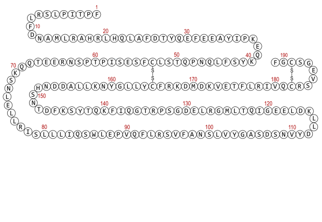
Reading the chain, the features that matter are sparse and well separated. Four cysteines form two bonds: Cys53 to Cys165, which clamps the long loop between the second and third helices against the bundle, and Cys182 to Cys189, which closes a six-residue loop near the C-terminus. Three methionines sit at positions 14, 125 and 170, three histidines at 18, 21 and 151, and the single tryptophan at position 86. His18 and His21 sit near one end of the first helix, and together with Glu174 near the C-terminus they form the zinc site, which is the clearest illustration that the folded protein brings distant parts of the sequence into one small cluster.[1,19,6]
The two receptor-binding surfaces are also discontinuous in sequence. The alanine scan that first mapped site 1 at residue resolution worked through three separate stretches, residues 2 to 19, 54 to 74 and 167 to 191, because those were the segments the earlier work had implicated; site 2 is a different face, centred on Asp116 and nearby residues. Neither surface can be recognised by reading the sequence, and neither can be reproduced by a fragment of it.[18,30]
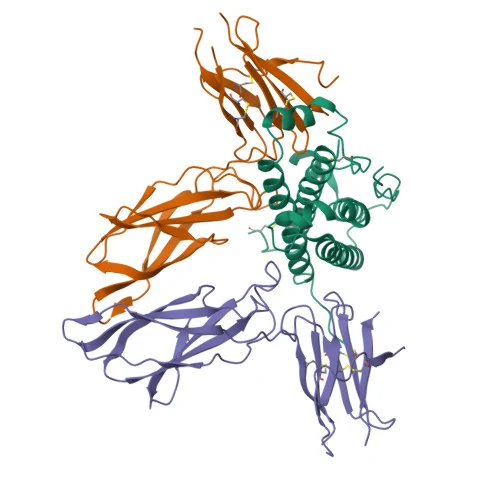
The 3D view shows the hormone as it sits in the 1:2 complex with two copies of the receptor extracellular domain, at 2.8 Å. Two things it does not show are worth stating. The last residue, Phe191, is outside the deposited construct, and residues 149 to 153 have no coordinates, so the loop that carries His151 is absent from the model. More broadly, it is a bound conformation in a crystal: no deposited structure of the free protein with this exact sequence was located, since the free-hormone entry titled as wild-type has a deposited sequence differing at twelve positions and the high-resolution free-hormone structure is a fifteen-substitution variant. In that variant the first turn of the third core helix is unwound, which its authors attribute to crystal packing rather than to the mutations, and the model again breaks between residues 128 and 155. The same loop goes missing in three independent crystals, which is itself the finding: that region is mobile.[31,2,32,33,34]
- F1
- P2
- T3
- I4
- P5
- L6
- S7
- R8
- L9
- F10
- D11
- N12
- A13
- M14
- L15
- R16
- A17
- H18
- R19
- L20
- H21
- Q22
- L23
- A24
- F25
- D26
- T27
- Y28
- Q29
- E30
- F31
- E32
- E33
- A34
- Y35
- I36
- P37
- K38
- E39
- Q40
- K41
- Y42
- S43
- F44
- L45
- Q46
- N47
- P48
- Q49
- T50
- S51
- L52
- C53
- F54
- S55
- E56
- S57
- I58
- P59
- T60
- P61
- S62
- N63
- R64
- E65
- E66
- T67
- Q68
- Q69
- K70
- S71
- N72
- L73
- E74
- L75
- L76
- R77
- I78
- S79
- L80
- L81
- L82
- I83
- Q84
- S85
- W86
- L87
- E88
- P89
- V90
- Q91
- F92
- L93
- R94
- S95
- V96
- F97
- A98
- N99
- S100
- L101
- V102
- Y103
- G104
- A105
- S106
- D107
- S108
- N109
- V110
- Y111
- D112
- L113
- L114
- K115
- D116
- L117
- E118
- E119
- G120
- I121
- Q122
- T123
- L124
- M125
- G126
- R127
- L128
- E129
- D130
- G131
- S132
- P133
- R134
- T135
- G136
- Q137
- I138
- F139
- K140
- Q141
- T142
- Y143
- S144
- K145
- F146
- D147
- T148
- N149
- S150
- H151
- N152
- D153
- D154
- A155
- L156
- L157
- K158
- N159
- Y160
- G161
- L162
- L163
- Y164
- C165
- F166
- R167
- K168
- D169
- M170
- D171
- K172
- V173
- E174
- T175
- F176
- L177
- R178
- I179
- V180
- Q181
- C182
- R183
- S184
- V185
- E186
- G187
- S188
- C189
- G190
- F191
Modifications
- Cys53 to Cys165 The large disulfide bond, which ties the long loop between the second and third core helices back to the body of the protein. Numbered 79 to 191 in the precursor numbering UniProt uses and 53 to 165 in the mature numbering used here and by the substance registry.[1,14]
- Cys182 to Cys189 The small C-terminal disulfide bond, enclosing a short loop of six residues. It is the chemically vulnerable one: a variant isolated from bacterially expressed material had this bridge replaced by a non-reducible thioether, with the change detected by peptide mapping, de novo sequencing and near-UV circular dichroism.[1,14,29]
- Whole chain No carbohydrate. The protein carries no N-linked or O-linked glycan, which is why a bacterially expressed chain can reproduce the natural sequence exactly and why the measured mass of intact material is compared directly against the sum over the sequence.[1,15]
All residues are the L enantiomers of the twenty proteinogenic amino acids, with no D-residues, no non-canonical residues and no cyclisation of the backbone. The only covalent cross-links are the two disulfide bonds, so the folded state is held almost entirely by non-covalent packing of the helix bundle. Separately from covalent structure, the protein binds zinc: two zinc ions associate per protein dimer in a cooperative fashion, cobalt(II) can substitute and gives a spectrum characteristic of tetrahedral coordination by oxygen and nitrogen ligands, and replacing His18, His21 or Glu174 with alanine weakened both metal binding and dimer formation. A zinc ion is present in the deposited growth hormone complex with the prolactin receptor extracellular domain.[19,20,1]
What studies on Somatropin show
Each card names the experimental system and what was measured. A result in one system is not evidence about another.
X-ray crystallography · Crystals of purified human growth hormone with the extracellular domain of the human growth hormone receptor
The complex contains one hormone per two receptor domains. The hormone is a four-helix bundle of unusual topology; each receptor domain comprises two subdomains with some resemblance to immunoglobulin folds but a different relative orientation, and both contribute residues to hormone binding. Both receptors donate essentially the same residues even though the two binding sites on the hormone have no structural similarity. There is also a substantial contact between the C-terminal subdomains of the two receptors, and the relative sizes of the interfaces support a sequential mechanism of assembly.[2]
- Material
- Wild-type hormone, residues 1 to 190 modelled, with two copies of the receptor extracellular domain (PDB 3HHR)
- Measured
- The structure of the complex at 2.8 Å, its stoichiometry, and the identity of the contact residues on both proteins
- Compared with
- Interfaces predicted by the earlier mutational mapping
- Limitation
- Isolated receptor extracellular domains in a crystal: no membrane, no transmembrane helix, no intracellular region, and no information about the free hormone in solution. Residues 149 to 153 are unmodelled and Phe191 is outside the construct.
de Vos AM et al., 1992, Science
Alanine-scanning mutagenesis with receptor binding measurement · Binding to the growth hormone receptor cloned from human liver
A cluster of about a dozen large side chains each lost more than fourfold affinity when replaced by alanine, defining site 1 at residue resolution. One residue behaved the opposite way: alanine at Glu174 raised affinity more than fourfold, so that side chain hinders binding in the wild-type protein. Many of the residues that promote binding differ in placental lactogens and prolactins, which do not bind this receptor tightly.[18]
- Material
- 62 single alanine substitutions covering every residue in positions 2 to 19, 54 to 74 and 167 to 191
- Measured
- Change in receptor binding affinity for each single mutant
- Compared with
- Wild-type hormone; folding checked against seven conformation-sensitive monoclonal antibodies
- Limitation
- Affinity for one receptor, measured before any structure existed. Alanine scanning reports the energetic contribution of a side chain and not contact geometry, and the antibody check establishes that the fold survived rather than that nothing moved locally.
Cunningham BC et al., 1989, Science
Solution biophysics: size-exclusion chromatography, calorimetry, fluorescence quenching · Purified hormone with the receptor extracellular binding protein
The hormone forms a 1:2 complex with the receptor extracellular domain, and binding is sequential: a first receptor binds site 1, then a second binds the adjacent but distinct site 2. Two overlapping surfaces on the receptor were implicated in binding the hormone.[3]
- Material
- Wild-type hormone plus engineered variants
- Measured
- Complex stoichiometry, the order of binding, and which hormone residues belong to which site
- Compared with
- Variants altered at each of the two candidate surfaces
- Limitation
- Isolated extracellular domains in solution. Receptor dimerisation is proposed to be relevant to signalling; the study measures assembly, not signalling.
Cunningham BC et al., 1991, Science
Alanine scan of the receptor side, read against the crystal structure · Receptor extracellular domain mutants binding the hormone
About 30 side chains from each protein are in contact, but a central hydrophobic region dominated by two tryptophans accounts for more than three quarters of the binding free energy. The surrounding contacts are generally hydrophilic and partially hydrated, so the interface resembles a cross section through a globular protein with a packed core and a wet rim. The functionally important receptor residues contact the functionally important hormone residues directly.[4]
- Material
- Individual alanine replacements of receptor contact residues
- Measured
- Contribution of each contact residue to binding free energy
- Compared with
- Wild-type receptor domain; the structural contact set from the crystal
- Limitation
- Energetics inferred from binding measurements on mutants rather than from structures of each mutant, and the scan is of the receptor, so it defines the hormone's partner surface rather than the hormone's own.
Clackson T et al., 1995, Science
X-ray crystallography at 2.6 Å with binding and kinetic analysis · Crystals of a 1:1 hormone-receptor complex, plus mutant binding kinetics
The 1:1 complex closely resembles the corresponding part of the 1:2 complex, so assembling the active complex does not require a major conformational change. A segment that makes receptor-receptor contact in the 1:2 complex is disordered here, which suggests its ordered conformation is stabilised by the second receptor. The hot spot is assembled cooperatively: several hydrophobic residues serve mainly to orient Trp104 and Trp169, and the directly energetic epitope may comprise as few as six side chains, mostly in alkyl-aromatic stacking. Replacing seven peripheral residues with alanine raised affinity up to tenfold, so the rim attenuates rather than adds to the central interaction.[16]
- Material
- G120R hormone, which can bind only one receptor, with the receptor extracellular domain (PDB 1A22)
- Measured
- Whether forming the complex changes either protein's conformation, and how the site 1 hot spot is assembled
- Compared with
- The equivalent portion of the wild-type 1:2 complex
- Limitation
- The hormone here is an engineered single substitution, G120R, chosen because it cannot bind a second receptor, so it is not somatropin and the site 2 surface is deliberately disabled.
Clackson T et al., 1998, Journal of Molecular Biology
Trimolecular surface plasmon resonance kinetics · Binding of a second receptor extracellular domain to a preassembled 1:1 hormone-receptor complex
Site 2 has no distinct hot spot. Its important residues are distributed over the whole binding surface rather than clustered, and pairwise alanine mutations showed significant negative cooperativity, including between residues more than 10 Å apart: substitutions that alone did little, or even improved binding, behaved differently when paired with the Asp116 substitution. The second interface is therefore both structurally and energetically malleable, in contrast to site 1.[17]
- Material
- 38 single and double alanine mutants in the hormone's site 2 surface
- Measured
- Association and dissociation kinetics for the second binding step
- Compared with
- Wild-type hormone; pairwise double mutants against the single mutants
- Limitation
- Kinetics on isolated extracellular domains with no cellular or signalling readout, and cooperativity is inferred from binding energies rather than observed structurally.
Walsh ST et al., 2003, Protein Science
Metal binding and protein association: size-exclusion chromatography, sedimentation equilibrium, radiolabelled zinc Scatchard analysis, cobalt substitution spectroscopy, denaturation · Purified hormone in solution
Zinc induced dimerisation, with two zinc ions per hormone dimer bound cooperatively. Cobalt(II) substituted and produced a visible spectrum characteristic of tetrahedral coordination by oxygen- and nitrogen-containing ligands. Alanine at His18, His21 or Glu174 weakened both metal binding and dimer formation, identifying those three side chains as the ligands. The zinc-bound dimer resisted chemical denaturation better than the monomer.[19]
- Material
- Wild-type hormone and His18, His21 and Glu174 alanine mutants
- Measured
- Zinc stoichiometry and cooperativity, metal coordination geometry, dimer formation, and resistance to guanidine hydrochloride denaturation
- Compared with
- Hormone without added zinc; cobalt(II) in place of zinc
- Limitation
- In vitro with metal added as an assay condition. The suggestion that this matters for storage in secretory granules is a proposal in the paper, not a measurement in it.
Cunningham BC et al., 1991, Science
Liquid chromatography with online tandem mass spectrometry · Recombinant human growth hormone material from three sources, described by the authors as innovator, follow-on and counterfeit
All three materials gave the expected sequence and disulfide pattern, yet differed measurably in chemistry: methionine oxidation followed the order Met14, then Met125, then Met170 in each case, with the follow-on material the most oxidised and the innovator material the least; deamidation at Asn149 was lowest in the follow-on and highest in the counterfeit; and backbone cleavage was site-specific, between Asn99 and Ser100 or between Glu30 and Phe31 depending on the source. The authors attribute the differences to manufacturing, formulation and storage rather than to sequence.[6]
- Material
- Intact protein digested for peptide mapping, with accurate mass and fragment-based sequence assignment
- Measured
- Complete sequence coverage with disulfide linkages, and the extent of oxidation, deamidation and chain cleavage at each site
- Compared with
- The three materials against each other; modified against unmodified peptide peak areas
- Limitation
- One sample per source, with the counterfeit material unidentified as to origin. The measurements are of chemical modification only and establish nothing about receptor binding or any functional property.
Jiang H et al., 2009, Biotechnology Progress
Mechanistic model of kinase activation from resonance energy transfer, engineered receptor constructs, atomistic simulation and domain docking · The growth hormone receptor treated as an archetypal cytokine receptor: fluorescence resonance energy transfer to report the position of the JAK2 binding motif in the receptor dimer, receptor extracellular domains replaced by Jun zippers to control the relative position of the transmembrane helices, atomistic modelling of those helix movements, and docking of the separately determined JAK2 kinase and inhibitory pseudokinase structures against an opposing pair
A complete mechanistic model for activation of receptor-bound JAK2, in which the activating event is a change in the relative position of the two transmembrane helices that separates the two JAK2 binding motifs and so breaks an inhibitory pairing between one receptor's kinase domain and the other's pseudokinase domain. This is the step the binding work on this page stops short of, and it is the reason simply bringing two receptors together is not sufficient.[24]
- Material
- Engineered receptor constructs, not the hormone: somatropin enters only as the physiological trigger the model has to account for
- Measured
- Position of the JAK2 binding motif within the receptor dimer, transmembrane helix movement, and the arrangement of kinase and pseudokinase domains
- Compared with
- Zipper constructs holding the transmembrane helices in defined relative positions, read against the arrangement a ligand produces
- Limitation
- The molecule under test is the receptor and not somatropin, which is absent from the constructs that carry the argument, and the kinase arrangement is docked from structures determined separately rather than observed in one assembly. No deposited structure of a hormone-bound, membrane-spanning receptor with its kinase was located, so the hormone's own contribution is still attributed rather than observed: it is the asymmetric placement of its two binding sites.
Brooks AJ et al., 2014, Science
How somatropin is studied at its own receptor and at the prolactin receptor
The molecular account is unusually complete for the binding step and deliberately incomplete after it. One hormone molecule recruits two copies of the receptor extracellular domain in order, site 1 first and then site 2, and the crystal structures of the 1:2 and 1:1 complexes are close enough that assembly requires no large conformational change in either protein. The hormone's two binding faces are not variations on one surface: they share no structural similarity, they are read by the same set of receptor residues, and they are energetically quite different, with a compact tryptophan-dominated hot spot at site 1 and a diffuse, malleable surface at site 2.[3,2,16,4,17]
The two sites are not independent. Mutagenesis across 81 single and 32 double positions showed that changes at site 1 reorganise site 2 both conformationally and energetically, focused on the contact between hormone Asp116 and receptor Trp169, and in that work the reorganisation happened without a loss of overall affinity. The crystal structure of a fifteen-substitution site 1 variant with two receptor domains makes the same point structurally: only one of ten hydrogen bonds in site 1 survived the substitutions, parts of the hormone moved by up to 9 Å, and site 2 was extensively rebuilt although no mutations were made there. A separate experiment, deleting a tryptophan from the receptor to create a cavity and selecting a hormone pentamutant that fills it, produced large rearrangements at interface positions distant from any mutation. The interface is plastic rather than lock-and-key.[30,35,36]
The same molecule also binds a second receptor, and the comparison is informative about specificity. Growth hormone binds the prolactin receptor while prolactin does not bind the growth hormone receptor, and mutagenesis had shown the two receptor-binding sites on the hormone overlap, which the crystal structure of the hormone with the prolactin receptor extracellular domain then explained as one hormone surface accommodating two different receptor surfaces. That interaction is metal-dependent in a way the hormone's own receptor is not: added zinc raised affinity for the prolactin receptor binding domain about 8000-fold, with one zinc per complex and His18, His21 and Glu174 on the hormone plus a histidine on the receptor implicated as its ligands, while zinc was required neither for growth hormone at its own receptor nor for prolactin at the prolactin receptor. A later kinetic study found a further asymmetry at that shared receptor: prolactin's dissociation rate changed 500-fold between pH 8.3 and 5.8 whereas growth hormone binding to the same receptor domain showed no significant pH dependence.[37,20,38]

What is still unknown about Somatropin
There is no deposited structure of the free protein with the verified sequence. The entry titled as the wild-type free hormone carries a deposited sequence differing from the mature chain at twelve positions, a discrepancy no record consulted here explains and whose primary citation has no PubMed identifier; the highest-resolution free-hormone structure is a fifteen-substitution affinity-matured variant. Everything structural about somatropin itself therefore comes from complexes, and three independent crystals leave the loop around residues 149 to 153 unmodelled, so that region's conformation is unknown rather than known to be flexible in a described way.[32,33,31,34]
Two of the most quoted facts about this molecule rest on a single record each, and one of them is internally inconsistent. The FDA substance registry states a molecular formula that disagrees with a sum over its own stated sequence by two hydrogens and one oxygen, while its molecular weight matches the sum; the summed formula is printed here and the disagreement is recorded rather than resolved. Separately, three of the nine identifiers on this page (DrugBank, IUPHAR/BPS and the ChEBI class term) are cross-references read from that one registry record rather than from the databases themselves, and the ChEBI term in particular resolves to a general class, not to this 191-residue protein. The ChEMBL identifier was a fourth until that record was read at source, where it resolves to somatropin typed as a protein.[14,1,39,40]
Covalent identity and chemical identity are not the same question, and the literature on this protein is unusually explicit about the gap. Material from three sources, all nominally the same protein with the same sequence and the same disulfide pattern, differed measurably in methionine oxidation, in deamidation at Asn149 and in where the backbone had cleaved. A variant isolated from bacterially expressed preparations had its small C-terminal disulfide replaced by a non-reducible thioether, which the authors note falls in a region earlier mutagenesis had implicated in receptor binding, although the functional consequence was discussed rather than measured. Routine disulfide assignment is now done analytically for exactly this reason, because incorrect folding and disulfide scrambling are not detectable from a sequence read.[6,29,41]
Finally, the step this page cannot describe at molecular resolution is the one between binding and signalling, and the hormone's role in it is inferred rather than observed. Comparing liganded and unliganded receptor extracellular domains found no substantial conformational change on binding, and the full-length receptor could be activated with no ligand at all by inserting a defined number of alanine residues into its transmembrane segment, which argues that bringing two receptors together is not by itself the activating event. The later model, in which activation separates the two JAK2-binding motifs through a change from a parallel transmembrane helix pair to a left-handed crossover, was assembled from engineered constructs, resonance energy transfer, atomistic simulation and docking of separately determined domains. No deposited structure of a hormone-bound, membrane-spanning receptor with its kinase was located, and the hormone's contribution is attributed to the asymmetric placement of its two binding sites.[42,24]
Key papers on Somatropin
- Human growth hormone and extracellular domain of its receptor: crystal structure of the complex.
de Vos AM, Ultsch M, Kossiakoff AA (1992), Science · X-ray crystallography
The structure that defined the molecule: the four-helix bundle, the 1:2 stoichiometry and the two dissimilar binding faces, all in one paper.
- High-resolution epitope mapping of hGH-receptor interactions by alanine-scanning mutagenesis.
Cunningham BC, Wells JA (1989), Science · Alanine-scanning mutagenesis
The residue-level map of site 1, and a model of how to read a binding surface before any structure exists. It also found the one residue that hinders binding.
- A hot spot of binding energy in a hormone-receptor interface.
Clackson T, Wells JA (1995), Science · Interface energetics
The clearest statement of why a structural contact map is not an energy map: thirty contacts, two tryptophans, three quarters of the binding energy.
- Site2 binding energetics of the regulatory step of growth hormone-induced receptor homodimerization.
Walsh ST, Jevitts LM, Sylvester JE, Kossiakoff AA (2003), Protein Science · Binding kinetics of alanine mutants
The counterpart for the second site, and the reason site 1 and site 2 should never be described in the same terms.
- Dimerization of human growth hormone by zinc.
Cunningham BC, Mulkerrin MG, Wells JA (1991), Science · Metal binding and protein association
The metal chemistry: stoichiometry, coordination geometry, the three side chains involved, and the effect on stability.
- Somatropin and its variants: structural characterization and methods of analysis.
Bayol A, Bristow A, Charton E, Girard M, Jongen P (2004), Pharmeuropa Bio · Review of structural characterisation and analytical methods
Read for identity rather than for mechanism: what somatropin is defined as, how it came to be characterised physicochemically rather than by bioassay, and why the methionyl form is a separate substance.
- Mass spectrometric analysis of innovator, counterfeit, and follow-on recombinant human growth hormone.
Jiang H, Wu SL, Karger BL, Hancock WS (2009), Biotechnology Progress · Mass spectrometric characterisation
The demonstration that the same sequence is not the same material, with the modification sites named.
References
- 1.UniProt Consortium. UniProtKB entry P01241 (SOMA_HUMAN), Somatotropin, Homo sapiens. UniProtKB. source · database record
- 2.de Vos AM, Ultsch M, Kossiakoff AA. (1992). Human growth hormone and extracellular domain of its receptor: crystal structure of the complex. Science. 255:306-12. doi:10.1126/science.1549776 · abstract only
- 3.Cunningham BC, Ultsch M, De Vos AM, Mulkerrin MG, Clauser KR, Wells JA. (1991). Dimerization of the extracellular domain of the human growth hormone receptor by a single hormone molecule. Science. 254:821-5. doi:10.1126/science.1948064 · abstract only
- 4.Clackson T, Wells JA. (1995). A hot spot of binding energy in a hormone-receptor interface. Science. 267:383-6. doi:10.1126/science.7529940 · abstract only
- 5.Goeddel DV, Heyneker HL, Hozumi T, Arentzen R, Itakura K, Yansura DG, Ross MJ, Miozzari G, Crea R, Seeburg PH. (1979). Direct expression in Escherichia coli of a DNA sequence coding for human growth hormone. Nature. 281:544-8. doi:10.1038/281544a0 · abstract only
- 6.Jiang H, Wu SL, Karger BL, Hancock WS. (2009). Mass spectrometric analysis of innovator, counterfeit, and follow-on recombinant human growth hormone. Biotechnology Progress. 25:207-18. doi:10.1002/btpr.72 · abstract only
- 7.U.S. Food and Drug Administration. (2026). Drugs@FDA: FDA-Approved Drugs. Drugs@FDA database. source · database record
- 8.U.S. National Library of Medicine. (2026). DailyMed: somatropin (Sandoz Inc.) prescribing information, Indications and Usage. DailyMed. source · full text read
- 9.European Medicines Agency. (2026). Medicines: search for human medicines (centrally authorised medicines). European Medicines Agency medicines database. source · database record
- 10.Health Canada. (2026). Drug Product Database online query. Health Canada Drug Product Database. source · database record
- 11.United States Congress. (2026). 21 U.S. Code § 333: Penalties, subsection (e), Prohibited distribution of human growth hormone. United States Code. source · full text read
- 12.World Anti-Doping Agency. (2025). World Anti-Doping Code International Standard: Prohibited List 2026. World Anti-Doping Agency. source · full text read
- 13.US Food and Drug Administration. (2026). Certain Bulk Drug Substances for Use in Compounding that May Present Significant Safety Risks. FDA human drug compounding. source · full text read
- 14.National Center for Advancing Translational Sciences. Global Substance Registration System record for SOMATROPIN, UNII NQX9KB6PCL. FDA Global Substance Registration System. source · database record
- 15.Bayol A, Bristow A, Charton E, Girard M, Jongen P. (2004). Somatropin and its variants: structural characterization and methods of analysis. Pharmeuropa Bio. 2004:35-45. PMID 15659284 · abstract only
- 16.Clackson T, Ultsch MH, Wells JA, de Vos AM. (1998). Structural and functional analysis of the 1:1 growth hormone:receptor complex reveals the molecular basis for receptor affinity. Journal of Molecular Biology. 277:1111-28. doi:10.1006/jmbi.1998.1669 · abstract only
- 17.Walsh ST, Jevitts LM, Sylvester JE, Kossiakoff AA. (2003). Site2 binding energetics of the regulatory step of growth hormone-induced receptor homodimerization. Protein Science. 12:1960-70. doi:10.1110/ps.03133903 · abstract only
- 18.Cunningham BC, Wells JA. (1989). High-resolution epitope mapping of hGH-receptor interactions by alanine-scanning mutagenesis. Science. 244:1081-5. doi:10.1126/science.2471267 · abstract only
- 19.Cunningham BC, Mulkerrin MG, Wells JA. (1991). Dimerization of human growth hormone by zinc. Science. 253:545-8. doi:10.1126/science.1907025 · abstract only
- 20.Cunningham BC, Bass S, Fuh G, Wells JA. (1990). Zinc mediation of the binding of human growth hormone to the human prolactin receptor. Science. 250:1709-12. doi:10.1126/science.2270485 · abstract only
- 21.Li CH, Dixon JS. (1971). Human pituitary growth hormone. 32. The primary structure of the hormone: revision. Archives of Biochemistry and Biophysics. 146:233-6. doi:10.1016/s0003-9861(71)80060-7 · abstract only
- 22.Niall HD, Hogan ML, Sauer R, Rosenblum IY, Greenwood FC. (1971). Sequences of pituitary and placental lactogenic and growth hormones: evolution from a primordial peptide by gene reduplication. Proceedings of the National Academy of Sciences of the United States of America. 68:866-70. doi:10.1073/pnas.68.4.866 · abstract only
- 23.Dieguez C, López M, Casanueva F. (2025). Hypothalamic GHRH. Reviews in Endocrine and Metabolic Disorders. 26:297-303. doi:10.1007/s11154-025-09951-y · abstract only
- 24.Brooks AJ, Dai W, O'Mara ML, Abankwa D, Chhabra Y, Pelekanos RA, Gardon O, Tunny KA, Blucher KM, Morton CJ, Parker MW, Sierecki E, Gambin Y, Gomez GA, Alexandrov K, Wilson IA, Doxastakis M, Mark AE, Waters MJ. (2014). Mechanism of activation of protein kinase JAK2 by the growth hormone receptor. Science. 344:1249783. doi:10.1126/science.1249783 · abstract only
- 25.UniProt Consortium. UniProtKB entry P10912 (GHR_HUMAN), Growth hormone receptor, Homo sapiens. UniProtKB. source · database record
- 26.Arsene C, Schulze D, Röthke A, Thevis M, Henrion A. (2018). Growth hormone isoform-differential mass spectrometry for doping control purposes. Drug Testing and Analysis. 10:938-946. doi:10.1002/dta.2350 · abstract only
- 27.Vickers MH, Gilmour S, Gertler A, Breier BH, Tunny K, Waters MJ, Gluckman PD. (2009). 20-kDa placental hGH-V has diminished diabetogenic and lactogenic activities compared with 22-kDa hGH-N while retaining antilipogenic activity. American Journal of Physiology. Endocrinology and Metabolism. 297:E629-37. doi:10.1152/ajpendo.00221.2009 · abstract only
- 28.HUGO Gene Nomenclature Committee. HGNC:4261, GH1, growth hormone 1. HGNC. source · database record
- 29.Datola A, Richert S, Bierau H, Agugiaro D, Izzo A, Rossi M, Cregut D, Diemer H, Schaeffer C, Van Dorsselaer A, Giartosio CE, Jone C. (2007). Characterisation of a novel growth hormone variant comprising a thioether link between Cys182 and Cys189. ChemMedChem. 2:1181-9. doi:10.1002/cmdc.200700042 · abstract only
- 30.Walsh ST, Sylvester JE, Kossiakoff AA. (2004). The high- and low-affinity receptor binding sites of growth hormone are allosterically coupled. Proceedings of the National Academy of Sciences of the United States of America. 101:17078-83. doi:10.1073/pnas.0403336101 · abstract only
- 31.de Vos AM, Ultsch M, Kossiakoff AA. (1993). PDB 3HHR: human growth hormone and extracellular domain of its receptor, crystal structure of the complex. RCSB Protein Data Bank. doi:10.2210/pdb3hhr/pdb · database record
- 32.Chantalat L, Jones ND, Korber F, Navaza J, Pavlovsky AG. (1995). PDB 1HGU: the crystal structure of wild-type growth hormone at 2.5 angstrom resolution. RCSB Protein Data Bank. doi:10.2210/pdb1hgu/pdb · database record
- 33.Ultsch MH, Somers W, Kossiakoff AA, de Vos AM. (1994). The crystal structure of affinity-matured human growth hormone at 2 A resolution. Journal of Molecular Biology. 236:286-99. doi:10.1006/jmbi.1994.1135 · abstract only
- 34.Sundström M, Lundqvist T, Rödin J, Giebel LB, Milligan D, Norstedt G. (1996). PDB 1HWG: 1:2 complex of human growth hormone with its soluble binding protein, and PDB 1HWH, the 1:1 complex of the G120R mutant. RCSB Protein Data Bank. doi:10.2210/pdb1hwg/pdb · database record
- 35.Schiffer C, Ultsch M, Walsh S, Somers W, de Vos AM, Kossiakoff A. (2002). Structure of a phage display-derived variant of human growth hormone complexed to two copies of the extracellular domain of its receptor: evidence for strong structural coupling between receptor binding sites. Journal of Molecular Biology. 316:277-89. doi:10.1006/jmbi.2001.5348 · abstract only
- 36.Atwell S, Ultsch M, De Vos AM, Wells JA. (1997). Structural plasticity in a remodeled protein-protein interface. Science. 278:1125-8. doi:10.1126/science.278.5340.1125 · abstract only
- 37.Somers W, Ultsch M, De Vos AM, Kossiakoff AA. (1994). The X-ray structure of a growth hormone-prolactin receptor complex. Nature. 372:478-81. doi:10.1038/372478a0 · abstract only
- 38.Keeler C, Jablonski EM, Albert YB, Taylor BD, Myszka DG, Clevenger CV, Hodsdon ME. (2007). The kinetics of binding human prolactin, but not growth hormone, to the prolactin receptor vary over a physiologic pH range. Biochemistry. 46:2398-410. doi:10.1021/bi061958v · abstract only
- 39.ChEBI curators. ChEBI:37845, growth hormone. Chemical Entities of Biological Interest, read through the EBI Ontology Lookup Service. source · database record
- 40.ChEMBL curators. ChEMBL molecule record CHEMBL1201621, SOMATROPIN. ChEMBL, European Bioinformatics Institute. source · database record
- 41.Wu SL, Jiang H, Lu Q, Dai S, Hancock WS, Karger BL. (2009). Mass spectrometric determination of disulfide linkages in recombinant therapeutic proteins using online LC-MS with electron-transfer dissociation. Analytical Chemistry. 81:112-22. doi:10.1021/ac801560k · abstract only
- 42.Brown RJ, Adams JJ, Pelekanos RA, Wan Y, McKinstry WJ, Palethorpe K, Seeber RM, Monks TA, Eidne KA, Parker MW, Waters MJ. (2005). Model for growth hormone receptor activation based on subunit rotation within a receptor dimer. Nature Structural and Molecular Biology. 12:814-21. doi:10.1038/nsmb977 · abstract only
Related entries
- IGF-1 LR3IGF-1 LR3 is an engineered 83-residue analogue of human insulin-like growth factor I that binds the IGF binding proteins poorly. Its only established use is as a reagent in serum-free cell culture.
- SermorelinSermorelin is a synthetic 29-residue fragment of growth hormone-releasing hormone, the hypothalamic peptide that signals the pituitary, ending in an arginine amide.
- TesamorelinTesamorelin is the full 44-residue human growth hormone-releasing hormone sequence, C-terminally amidated as the natural hormone is. It carries one added group: a trans-3-hexenoyl acyl chain on the amine of the N-terminal tyrosine.
- CJC-1295 with DACCJC-1295 with DAC is a 30-residue analogue of the first 29 residues of human growth hormone-releasing hormone, carrying four changes and an added lysine. That C-terminal lysine's side chain bears a maleimide, which reacts with a free protein thiol, so the peptide ends up covalently attached to a plasma protein rather than circulating alone; DAC, drug affinity complex, is the name for that arm.
- IpamorelinIpamorelin is a synthetic pentapeptide amide that activates the growth hormone secretagogue receptor, reported in 1998. It is known for not raising corticotropin or cortisol in the species tested.
