DSIP in brief
DSIP is a linear, unmodified nonapeptide, Trp-Ala-Gly-Gly-Asp-Ala-Ser-Gly-Glu (C35H48N10O15, about 848.8 g/mol), with the International Nonproprietary Name emideltide. It was sequenced in 1977 and 1978 from rabbit cerebral venous blood and named for an EEG change measured in rabbits. In almost fifty years no receptor, gene or protein precursor has been identified, and ChEMBL holds no bioactivity measurement for it. Its best-measured properties are physical: saturable transport across the blood-brain barrier in perfused guinea pig brain, and a plasma half-life of minutes in several species. Human evidence exists but is thin and contradictory: three double-blind polysomnography studies in chronic insomnia reached opposite conclusions, and the largest human dataset is an uncontrolled series. Much of the literature under the name concerns other molecules, including the cross-reacting protein GILZ, so a reader should check which molecule a study actually tested.[1,2,3,4,5,6,7,8,9]
DSIP regulatory status
No FDA, EMA or Health Canada marketing authorisation was found for emideltide in Drugs@FDA (queried through openFDA), the EMA medicines database or the Health Canada Drug Product Database, and a ClinicalTrials.gov search for emideltide returned no registered trial (all searched November 2026). FDA lists emideltide (DSIP) among bulk drug substances nominated for compounding whose nominations were withdrawn (page current as of 22 April 2026), stating that compounded drugs containing it may pose a risk of immunogenicity for certain routes of administration, may have complexities with peptide-related impurities and characterisation of the active ingredient, and that the agency has not identified safety-related information sufficient to know whether it would cause harm if administered to humans. The 2026 WADA Prohibited List does not name emideltide; its S0 class prohibits at all times any pharmacological substance not addressed elsewhere on the List and with no current approval by a governmental regulatory health authority for human therapeutic use.[10,11,12,13,14,15]
DSIP at a glance
- A linear nine-residue peptide, WAGGDASGE, all L, with free termini and no modification of any kind, carrying the International Nonproprietary Name emideltide. Its registry identity is consistent across PubChem, ChEBI, ChEMBL and UniProt.[1,16,17,18]
- The sequence was determined in 1977 and 1978 by amino-acid analysis and sequencing of material recovered from the cerebral venous blood of rabbits during thalamic stimulation, and confirmed by synthesising the nonapeptide alongside five candidate fragments, two substituted analogues and a related tripeptide.[19,2]
- No receptor for this peptide has ever been identified, and no gene or protein precursor has been isolated. ChEMBL holds no bioactivity measurement for it. A 2006 review in the Journal of Neurochemistry describes the molecule as a still unresolved riddle on exactly those grounds.[4,16,20]
- Human evidence exists and it is thin. Three double-blind polysomnography studies in chronic insomnia reached opposite conclusions: two, one crossover and one parallel-group, found the sleep changes weak or of little clinical significance, while a third reported substantial improvement. The largest human dataset, 107 inpatients in alcohol or opiate withdrawal, was open and uncontrolled with the outcome judged by clinical impression.[5,6,21,7]
- Its best-measured molecular property is transport across the blood-brain barrier, and it is saturable: in a vascularly perfused guinea pig forebrain the unidirectional transfer constant fell to the range measured for mannitol when excess unlabelled peptide was present, and free L-tryptophan inhibited it too, so the transporter responds to something about the N-terminal residue. It is also short-lived, with a measured plasma half-life of a few minutes in dogs, a monkey and rats. Two earlier studies had argued for barrier passage less directly, one from a central electrophysiological change after peripheral administration in rabbits and one from chromatographically identified peptide appearing in canine cerebrospinal fluid.[22,23,3,8]
- Much of the literature filed under this name is about other molecules: a transcriptional regulator (GILZ) that acquired the name through antibody cross-reactivity, an unrelated 77-residue porcine brain peptide isolated because it cross-reacted, and the Ser7-phosphorylated analogue, which behaves measurably differently from the parent peptide.[24,9,25]
What is DSIP?
DSIP is a synthetic nonapeptide. Its sequence was reported in 1977 by Schoenenberger and Monnier, who recovered the peptide from an extracorporeal dialysate of cerebral venous blood taken from rabbits during electrical stimulation of the intralaminar thalamic area, and named it for the electroencephalographic change they measured: enhancement of delta and spindle activity. The companion 1978 paper gives the amino-acid analysis and the sequence determination. Both papers tested the sequence by synthesis, infusing the nonapeptide alongside five candidate fragments spanning residues 1 to 8, 2 to 9, 2 to 8, 1 to 4 and 5 to 9, two substituted nonapeptide analogues (the 1978 paper records one with a single residue exchanged and one with two) and a Trp-Ser-Glu tripeptide, under double-blind conditions in rabbits. Of the nine synthetic peptides only the complete nonapeptide changed the measured EEG endpoint. The experiment did not identify any molecular target, and nothing in it establishes a mechanism.[19,2]
As a chemical entity it is about as plain as a peptide gets: a linear chain of nine residues, three of them glycine, with a single aromatic side chain at the N-terminus, an acidic residue at position 5 and another at position 9, a serine hydroxyl at position 7 and no modification, bridge or metal anywhere. It belongs to no recognised peptide family. The 2006 review makes that point explicitly, noting that its structure differs from any other known representative of the various peptide families, which is one reason no receptor search has had an obvious place to start.[1,4]
What distinguishes this entry from most peptides of comparable age is how little of its literature is actually about it. Three substitutions have to be kept apart. Antisera raised against the nonapeptide detect large immunoreactive forms, and when one of those was purified from porcine brain and sequenced in 1993 it turned out to be a 77-residue leucine-zipper peptide with no sequence relationship to WAGGDASGE. Separately, the gene product called the delta sleep-inducing peptide immunoreactor is the same protein as glucocorticoid-induced leucine zipper, GILZ, a transcriptional regulator, so a search on the compound name returns a substantial modern literature about a protein rather than a peptide. And the Ser7-phosphorylated analogue, widely studied beside the parent molecule, is a separate compound whose measured behaviour differs from it. The tissue-distribution literature inherits all of this: immunohistochemical mapping, such as a preliminary survey of seven human hypothalami from the first postnatal year, locates immunoreactivity rather than an identified molecule.[9,24,25,26]
The catalogue rows behind this entry list it simply as DSIP, with no sequence, salt form or analytical claim attached. Nothing on a label of that kind distinguishes the alpha-aspartyl peptide described here from its beta-aspartyl isomer, which has the same formula and the same nominal mass and which the original characterisation reports as inactive on the endpoint the peptide was named for.[2,1]
Not to be confused with
- Glucocorticoid-induced leucine zipper (GILZ), also indexed as the delta sleep-inducing peptide immunoreactor
- A transcriptional regulator protein that acquired this peptide's name through antibody cross-reactivity, not through any chemical relationship. A review of adipose circadian biology states the equivalence of the two names outright. A reader searching the compound's name therefore meets a large literature about a protein with a different sequence, a different size and a different biology, and none of it describes the nonapeptide.[24]
- DIP, the 77-residue porcine brain peptide
- Purified from an acid extract of porcine brain precisely because antisera raised against the nonapeptide detected large immunoreactive forms. Sequencing showed it is unrelated to WAGGDASGE: it carries a putative leucine zipper, a proline and glutamate rich domain, three potential phosphorylation sites and an acetylated N-terminus. It is also the only molecule in this family with a deposited experimental structure, which makes mistaking it for the nonapeptide an easy error to commit.[9,29]
- The Ser7-phosphorylated analogue (P-DSIP)
- A different molecule, consistently measured as behaving differently. It degraded more slowly than the parent peptide in blood, acted at a lower assay concentration on pineal N-acetyltransferase, had a faster onset and offset on a rat thermoregulatory endpoint, and was not neutralised by an antiserum that abolished the parent peptide's effect in the same experiment. Results reported for it are not results for this entry's molecule.[33,31,25,32]
- Structural analogues such as KND
- Several of the measured effects most often attributed to this peptide were obtained with modified sequences rather than with WAGGDASGE. In a 2021 pair of acute ischaemia models the analogue KND, not the nonapeptide, carried the reported reductions in infarct size, and a 2006 review notes that certain synthetic analogues changed slow-wave sleep measures in rabbits and rats where the nonapeptide itself did not.[46,4]
- The beta-aspartyl isomer
- The same nine residues joined through the beta-carboxyl of aspartate rather than its alpha-carboxyl: identical formula, identical mass, a different molecule. The 1978 characterisation reports that only the pure alpha-aspartyl peptide changed the measured EEG endpoint and that the beta isomer did not, which makes the aspartate linkage an analytical criterion for this material rather than a detail.[2]
- Drosophila DIP-alpha and related IgSF proteins
- Insect cell-surface proteins whose abbreviation collides with DIP in literature searches. No relationship of any kind to this peptide; listed because they dominate recent search results for the abbreviation.[29]
DSIP structure and sequence
Reading the chain from the N-terminus: tryptophan, alanine, glycine, glycine, aspartate, alanine, serine, glycine, glutamate. The registry's systematic name, L-tryptophyl-L-alanylglycylglycyl-L-alpha-aspartyl-L-alanyl-L-serylglycyl-L-glutamic acid, records the one connectivity detail that is easy to get wrong: aspartate at position 5 is joined into the backbone through its alpha-carboxyl, leaving its beta-carboxyl as a free side chain. That distinction is not pedantry here. The 1978 characterisation separated the alpha-aspartyl peptide from its beta isomer and reports that only the former changed the EEG endpoint, so the linkage is part of what identifies the material.[1,2]
Three of the nine residues are glycine, which removes side chains and backbone constraint at those positions, and there is no proline, no cysteine and nothing to cyclise. The predictable consequence is conformational mobility, and that is what the measurements show. The one experimental conformational study, which compared this peptide with neuromedin B using steady-state and time-resolved tryptophan fluorescence, circular dichroism and molecular dynamics, found both peptides in energetically equivalent conformations in buffer. On moving to a lipid phase neuromedin B's tryptophan emission shifted to the blue and gained intensity, consistent with the indole becoming buried in a helix-like structure, while this peptide's fluorescence was almost unchanged; circular dichroism and the simulation agreed, describing a globule-like state with the indole ring surface-exposed. The authors concluded that its interaction with the lipid phase is not driven by an amphipathic structure, and suggested hydrophobicity-driven diffusion across the bilayer instead.[27]
Charge is the other property with experimental support. Two side-chain carboxylates plus the C-terminal carboxylate against a single N-terminal amine give a net negative charge at neutral pH, and an entrapment study is consistent with it: essentially the whole load was taken up by a cationic macroporous matrix while 2 to 6 percent adsorbed onto an anionic one, and release occurred only in salt solutions rather than in water or ethanol, which the authors attribute to ionic strength. There is nothing to show in three dimensions. No experimental structure of this peptide has been deposited, and PubChem generated no conformer for it, so the structure panel on this page is empty by fact rather than by omission; the deposited NMR structure that carries the name belongs to the unrelated 77-residue peptide.[28,1,29,27]
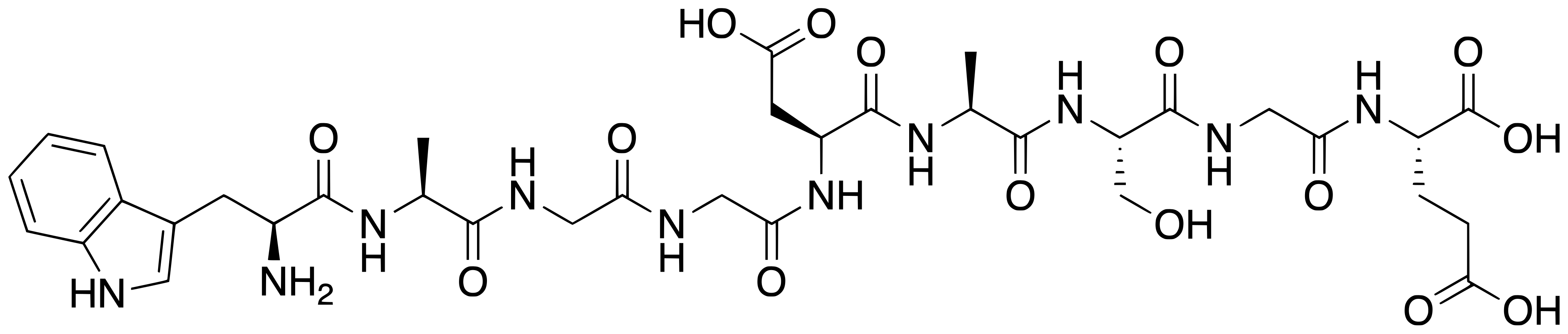
Six stereocentres, at Trp1, Ala2, Asp5, Ala6, Ser7 and Glu9; the three glycines are achiral. All are L in the registry record, expressed in the isomeric SMILES and in the InChIKey ZRZROXNBKJAOKB-GFVHOAGBSA-N. There is no cysteine and therefore no disulfide, no cyclisation, no lipidation, no PEG, no glycosylation and no metal coordination. At neutral pH the molecule is expected to carry one ammonium group against three carboxylates, two of them side chains, giving a net negative charge; an entrapment study is consistent with that, since essentially all of the peptide loaded onto a positively charged macroporous matrix while only 2 to 6 percent adsorbed to a negatively charged one.[1,28]
What studies on DSIP show
Each card names the experimental system and what was measured. A result in one system is not evidence about another.
Blood-brain barrier transport, multiple-time brain uptake analysis in a vascularly perfused preparation · Ipsilateral in situ perfused guinea pig forebrain; parietal cortex, caudate nucleus and hippocampus
Transfer constants were 0.93, 1.33 and 1.66 microlitres per minute per gram in cortex, caudate and hippocampus. Excess unlabelled peptide reduced them to 0.23 to 0.38, which is the mannitol range, establishing that uptake is saturable rather than purely diffusional. The rapidly equilibrating volume exceeded mannitol's and collapsed to it under competition, which the authors read as binding at sites on the capillary membrane. Free L-tryptophan inhibited transfer without affecting that volume; vasopressin inhibited only weakly and at a far higher concentration.[3]
- Material
- Radioiodinated nonapeptide, with unlabelled nonapeptide, free L-tryptophan and [Arg8]-vasopressin as competitors and mannitol as the diffusional reference
- Measured
- Regional unidirectional transfer constants across the luminal interface, and the rapidly equilibrating brain volume
- Compared with
- Mannitol transfer constants, and the same tissue under competition
- Limitation
- A perfused preparation, not an intact animal. Inhibition by free tryptophan indicates the transporter recognises something about the N-terminal residue, so saturability here is evidence about a transport mechanism and not about a receptor for the peptide. No affinity constant for any protein was measured.
Zlokovic BV et al., 1989, Peptides
Spectroscopy (tryptophan fluorescence, decay-associated spectra, circular dichroism) with molecular dynamics · Aqueous buffer compared against a model membrane lipid phase
Both peptides occupy energetically equivalent conformations in solution. In lipid, neuromedin B's emission blue-shifted with a rise in the second lifetime component, consistent with burial of its indole in a helix-like structure, while this peptide's emission was essentially unchanged. Circular dichroism and the simulation agree on a globule-like state with the indole surface-exposed, and the authors conclude its membrane interaction is not amphipathic-structure driven.[27]
- Material
- Synthetic nonapeptide; neuromedin B as the comparator peptide in the same experiments
- Measured
- Steady-state and time-resolved tryptophan emission, decay-associated spectra, circular dichroism signal, and simulated conformational ensembles
- Compared with
- Neuromedin B, and the same peptide in buffer
- Limitation
- A model lipid phase, not a cell membrane. Fluorescence and circular dichroism report ensemble averages, not a structure, and the molecular dynamics component is a calculation. No protein partner is identified anywhere in the work.
Polverini E et al., 1998, Archives of Biochemistry and Biophysics
Peptide isolation with amino-acid analysis, sequencing, mass spectrometry and radioimmunoassay · Acid extract of porcine brain
The purified species is a 77-residue peptide, named DIP, with an acetylated N-terminus, a putative leucine-zipper motif, a proline and glutamate rich domain and three potential phosphorylation sites. Its sequence bears no relation to WAGGDASGE, although the antiserum recognises it. Its N-terminal region resembles a protein induced by transforming growth factor beta 1 in mouse osteoblastic cells, and it is structurally similar to baculoviral p10.[9]
- Material
- The large immunoreactive species detected by an antiserum raised against the synthetic rabbit nonapeptide
- Measured
- Sequence, mass and immunoreactivity of the purified material
- Compared with
- The nonapeptide's own sequence
- Limitation
- One species and one tissue extract. It identifies a cross-reactive molecule, which does not prove that all immunoreactivity attributed to the nonapeptide comes from this peptide. The function of the isolated peptide is left open by the authors.
Sillard R et al., 1993, European Journal of Biochemistry
Agonist-evoked current recording in neurons, plus radiolabelled calcium uptake into synaptosomes · Rat cortical, hippocampal and cerebellar neurons; rat cortical synaptosomes
GABA-activated currents in hippocampal and cerebellar neurons were potentiated concentration-dependently, and NMDA-activated potentiation in cortical and hippocampal neurons was blocked. Calcium uptake into cortical synaptosomes stimulated by glutamate or NMDA changed, which the authors read as modulation of presynaptic NMDA receptors.[30]
- Material
- The nonapeptide applied to the preparation
- Measured
- GABA-activated and NMDA-activated currents; 45Ca2+ uptake into synaptosomes after stimulation with glutamate or NMDA
- Compared with
- The same preparation with GABA, glutamate or NMDA alone
- Limitation
- Modulation of a current is not binding: no site, affinity or subunit dependence is established, and an indirect action on either receptor is not excluded. A short report, with no concentrations given in the abstract and no independent replication located.
Grigor'ev VV et al., 2006, Bulletin of Experimental Biology and Medicine
Enzyme activity assay with receptor-antagonist dissection · Rat pineal gland in vitro
Both peptides increased norepinephrine-stimulated N-acetyltransferase activity, the phosphorylated analogue at a lower assay concentration. Prazosin abolished the enhancement while propranolol did not, which places the effect at the alpha-1 adrenergic receptor rather than at the beta receptor. The same group had separately found the peptide reduced the nocturnal rise of the same enzyme in the intact animal, so the direction of the change depends on the preparation.[31]
- Material
- The nonapeptide at assay concentrations of 20 to 300 nM, and its Ser7-phosphorylated analogue, against norepinephrine as the stimulus
- Measured
- N-acetyltransferase activity in the gland
- Compared with
- Norepinephrine alone, and the same stimulus with the alpha-1 antagonist prazosin or the beta antagonist propranolol
- Limitation
- Modulating a receptor-driven enzyme response is not binding at that receptor, and the mechanism is offered as a conclusion rather than measured. One species, isolated gland, and the in vivo and in vitro directions disagree. A 1986 review of the field had already stated that this adrenergic mechanism remained to be established.
Graf MV et al., 1987, Journal of Neurochemistry
In vitro kinase assay · Purified casein kinase II with the peptide as substrate
The peptide is a substrate for casein kinase II, with an apparent Km reported as 20 mM and a Vmax of 90.9 nmol per minute per milligram of protein. Both ATP and GTP served as phosphoryl donors, heparin inhibited and spermine enhanced the reaction. The work was framed as a candidate route to the Ser7-phosphorylated form that immunochemical studies had reported.[32]
- Material
- The unmodified nonapeptide
- Measured
- Phosphorylation rate, apparent Km and Vmax
- Compared with
- Phosphoryl donor variation (ATP against GTP), with heparin and spermine as modifiers
- Limitation
- A millimolar Km is orders of magnitude above any plausible tissue concentration of a free nonapeptide, so this establishes chemical competence as a substrate rather than a physiological pathway. The phosphorylation site is not determined here; it is carried over from earlier immunochemical work.
Nakamura A et al., 1991, Peptides
Stability and binding study in blood, with gel filtration · Human and rat blood, plasma and serum, in vitro at varied temperature and time
Incubation released products eluting like free tryptophan, dependent on temperature, time and species, consistent with degradation beginning at the N-terminal residue. The labelled peptides degraded more slowly and formed complexes that excess unlabelled material did not displace, which the authors interpret as non-specific binding or aggregation.[33]
- Material
- The nonapeptide; radioiodinated N-Tyr-nonapeptide; radioiodinated N-Tyr-phosphorylated analogue
- Measured
- Appearance of degradation products by gel-filtration retention time, and formation of high-molecular-weight complexes
- Compared with
- The two labelled analogues against the parent peptide, and excess unlabelled peptide as a displacement control
- Limitation
- Retention time is weak evidence of a product's identity, and no cleavage site was mapped. The non-displaceable complex is the instructive negative result: an apparent binding signal in this system is not evidence of a receptor.
Graf MV et al., 1987, Peptides
Sequence homology search of gene and protein databases, with follow-up peptide synthesis · Mammalian sequence databases including human; synthesised peptide screened in animal tests
The closest homologous site found in mammalian proteins lies in JmjC-domain-containing histone demethylases of the JMJD1B type. The authors synthesised that nonapeptide, screened it preliminarily, and propose those proteins as possible precursors of endogenous peptides with similar activity.[20]
- Material
- WKGGNASGE, the closest mammalian match, differing from WAGGDASGE at positions 2 and 5
- Measured
- Presence and identity of homologous sequence sites in known proteins
- Compared with
- The nonapeptide's own sequence
- Limitation
- Homology at a two-residue remove is a hypothesis about a precursor, not evidence of processing. Nothing here demonstrates that WAGGDASGE is produced from these proteins in any tissue, and the authors describe the biological screening as preliminary.
Mikhaleva II et al., 2011, Peptides
Placebo-controlled, double-blind polysomnography study in people · 14 middle-aged patients with severe chronic insomnia; polysomnograms at a placebo baseline night, at the beginning and the end of a seven-night treatment period and on one placebo night afterwards, with daytime psychological state and mental performance tested before and after
The author reports that night sleep improved from the first exposure and further with repetition, that the improvement persisted into the first placebo night afterwards, that the efficiency of night sleep and of daytime rest reached the levels of normal controls, and that daytime alertness and performance rose significantly.[6]
- Material
- The nonapeptide
- Measured
- Night-sleep efficiency and structure by polysomnography, daytime rest, alertness and mental performance
- Compared with
- Placebo, double-blind
- Limitation
- Fourteen patients, one author, one centre, and read here as an abstract only, so no figure and no statistic is in hand. The conclusion of efficacy is the author's. It has not been replicated: two independent double-blind studies in the same population, one crossover and one parallel-group, measured changes in the same direction and concluded they were weak or of little clinical significance.
Schneider-Helmert D, 1987, European Neurology
Double-blind, matched-pairs, parallel-group polysomnography study in people · 16 patients with chronic insomnia sleeping five consecutive nights in a laboratory, the first for adaptation and the second for baseline, with measurements on the last three
Objective sleep quality was better than placebo, with higher sleep efficiency and shorter sleep latency, and one measure of subjective tiredness fell within the treated group. The authors report that the statistically significant effects were weak and could in part be due to an incidental change in the placebo group, that no other measure including subjective sleep quality changed at all, and they conclude that short-term treatment of chronic insomnia with the peptide is not likely to be of major therapeutic significance.[5]
- Material
- The nonapeptide
- Measured
- Sleep structure, objective sleep quality by polysomnography, subjective sleep quality and subjective tiredness
- Compared with
- A glucose solution as placebo, in matched pairs
- Limitation
- Sixteen patients split across two parallel groups, so the comparison rests on matched pairs rather than within-subject change, and the authors attribute part of the difference to movement in the placebo arm. Three measurement nights, and no molecular endpoint of any kind.
Bes F et al., 1992, Neuropsychobiology
Double-blind crossover polysomnography study in people · Patients with chronic insomnia recorded by polysomnography across four nights in a double-blind crossover design
Awakenings, NREM sleep latency, total waking time and waking time after sleep onset all fell under the peptide, but none of them differed significantly from baseline or from the double-blind placebo nights. Total sleep time and NREM sleep time rose, the rise carried by stage 2, with stage 1, slow-wave sleep and REM sleep unchanged. The NREM and stage 2 differences from placebo were significant, but the same differences were already present in the baseline values, and the authors conclude that the sleep improvement is of little clinical significance.[21]
- Material
- The nonapeptide
- Measured
- Nocturnal awakenings, NREM sleep latency, total waking time, waking time after sleep onset, total sleep time, NREM sleep time and the individual sleep stages
- Compared with
- Placebo nights and each patient's own baseline
- Limitation
- The significant comparisons are confounded by pre-existing baseline differences, which the authors state themselves. Four nights, a single centre, a sample size the abstract does not give, and abstract-level reading only.
Monti JM et al., 1987, International Journal of Clinical Pharmacology Research
Open, uncontrolled clinical series in people · 107 inpatients with symptoms of alcohol withdrawal (47) or opiate withdrawal (60), with the effect judged by clinical evaluation by the treating physician and the nursing staff
About 13 per cent of the alcohol group and 22 per cent of the opiate group did not meet the requirements for evaluation. Among those evaluated, clinical symptoms and signs disappeared or improved markedly and rapidly in 97 per cent of opiate and 87 per cent of alcohol cases; anxiety resolved more slowly and the opiate cases ran a longer course. The authors' stated rationale came from earlier animal work in which the peptide, like morphine, alcohol and pentobarbital, produced slow-wave sleep with spindles when applied to a brainstem and thalamic region, an effect naloxone reversed, from which they postulated agonist activity at opiate receptors.[7]
- Material
- The nonapeptide
- Measured
- Clinical symptoms and signs of withdrawal, rated by physician and nursing staff
- Compared with
- None: no placebo, no randomisation, no blinding
- Limitation
- The largest human dataset for this compound and the weakest in design: no control group, no blinding, no randomisation, and an unblinded clinical impression as the endpoint in a condition that resolves by itself over days. Roughly one in five enrolled patients was dropped from evaluation after enrolment. The opiate-receptor rationale was a hypothesis the series did not test, and no receptor for this peptide has been identified since.
Dick P et al., 1984, European Neurology
Why this peptide still has no mechanism, and what has been measured instead
There is no identified receptor for this peptide, and that is the central fact about its pharmacology rather than a gap at its edges. No radioligand binding study, saturation isotherm or dissociation constant was found for it, ChEMBL records no bioactivity measurement at all, and the 2006 review states that no gene, protein precursor or candidate receptor has been isolated. The nearest thing to a molecular partner anywhere in the literature is the blood-brain barrier transport site inferred from competition in perfused guinea pig forebrain, and even there the inhibition by free L-tryptophan suggests a transporter reading the N-terminal residue rather than a receptor reading the peptide. The field has known this for decades: a 1986 review of the literature concluded that even the proposed modulation of adrenergic transmission remained to be established.[4,16,3,34]
What exists instead is a set of measurements in which the peptide changes some other system's response. It potentiated GABA-activated currents and blocked NMDA-activated potentiation in rat neurons; applied locally to neurons of cortex, hippocampus, thalamus and hypothalamus it mostly raised firing rates and prevented the rise produced by glutamate, and after the non-competitive NMDA antagonist MK-801 fewer neurons responded to it. In isolated rat pineal gland it increased norepinephrine-stimulated N-acetyltransferase activity in a way prazosin abolished and propranolol did not. In isolated median eminence it reduced somatostatin release, blocked by the dopamine antagonist pimozide, and in cultured rat pituitary cells it changed growth hormone release with a maximum at an assay concentration of 0.1 nM and no significant effect at micromolar concentrations. Further down the scale of organisation, isolated rat brain mitochondria showed a higher rate of phosphorylating respiration and of ADP phosphorylation with uncoupled respiration unchanged, so the respiratory control ratio rose. Each result is a measured endpoint in a named preparation. None of them locates the peptide on a protein, and the mitochondrial measurement has the additional problem that no route into an intact organelle is established for a charged nonapeptide.[30,35,31,36,37,38]
Two features recur across that list and both argue for caution. The concentration-response relations are repeatedly non-monotonic, described as inverted bell-shaped in the pituitary and thermoregulatory work and as U-shaped as early as the 1984 reviews, and the effect sizes are modest, around 50 percent over basal at the most effective concentration in the pituitary experiment. A bell-shaped curve of that amplitude is as consistent with an indirect or non-specific action as with a receptor-mediated one. Separately, the molecule is short-lived and easy to cut, and here there are measured figures. An enzyme immunoassay study in anaesthetised dogs reported a mean metabolic clearance rate of 30.7 plus or minus 2.5 mL per kg per minute and a mean plasma half-life of 4.0 plus or minus 0.7 minutes, with 2.9 minutes measured in a monkey and about 2.0 minutes in rats; a second dog study from the same group, of entry into cerebrospinal fluid, found that entry correlated with plasma concentration, with plasma half-life and with lipophilicity but not with plasma protein binding or molecular weight, the three correlating factors combining into a single parameter at r = 0.813. The cutting machinery is partly identified: an aminopeptidase of roughly 80 kDa purified from human cerebrospinal fluid degrades the peptide, is blocked by amastatin, bestatin and EDTA, and also degrades enkephalins, while in blood the first products elute like free tryptophan and the phosphorylated analogue degrades more slowly. The figure of about fifteen minutes that secondary pages quote as an in vitro half-life is attributed to the 1984 characterisation review, which was read here at abstract level and states no such number; the measured animal figures above are shorter. Recent work reflects all of this by treating the sequence as cargo rather than as a ligand, fusing it to a barrier-crossing peptide and secreting the construct from Pichia pastoris.[37,25,39,40,33,41,8,42,43]
What is still unknown about DSIP
The chemistry is settled; the biology is not. Formula, mass, sequence, stereochemistry and the alpha-aspartyl linkage agree across the registry records and the original characterisation, and the molecule has an International Nonproprietary Name. Against that, no gene, no precursor protein and no receptor has been isolated in almost fifty years. The best available account of an endogenous origin is a sequence homology: a JMJD1B-type histone demethylase contains WKGGNASGE, two residues different, and the authors who found it propose those proteins as candidate precursors without claiming to have shown processing. A suggestion that the sequence is of bacterial origin circulates on the leading general-reference page for this peptide; it is not stated as a hypothesis here because a PubMed search located no primary sequence-comparison study behind it, and the citation attached to it there is a book chapter on sleep and immunity in Drosophila.[1,2,4,20,18]
A large part of the historical evidence that the peptide occurs naturally rests on antibodies, and that evidence was undercut by the isolation of a cross-reactive molecule. Radioimmunoassay and immunohistochemistry were the main tools for locating the material in brain and peripheral tissues, as the 1984 review records, and in 1993 a large immunoreactive species purified from porcine brain proved to be an unrelated 77-residue leucine-zipper peptide. Everything that literature reports is therefore an immunoreactivity measurement rather than a measurement of the nonapeptide: a preliminary survey of seven human hypothalami mapped immunoreactive cell bodies and fibres through the anterior and mediobasal hypothalamus and is honest about being preliminary, and a plasma study of four groups of infants found immunoreactivity higher in healthy full-term infants (median 1885 pmol/L) than in preterm infants (1595) or in infants with long mean apnoea durations (1542), with no difference by age across the first year. The 2006 review turns this into a positive hypothesis, arguing that a different endogenous peptide accounts for both the immunoreactivity and much of the attributed activity, and noting that immunoreactivity concentrates in neurosecretory hypothalamic nuclei not specifically associated with sleep regulation.[39,9,26,4,44]
The human literature is the part a reader is most likely to arrive with and the part that needs reading most carefully, because its three controlled studies disagree and its largest dataset is uncontrolled. Two independent double-blind polysomnography studies in chronic insomnia, one a crossover and one a matched-pairs parallel-group design, each measured sleep changes in the expected direction and each concluded that they were weak or of little clinical significance, one of them attributing part of its own significant result to an incidental shift in the placebo group; a third, by a single author in 14 patients, reported substantial improvement sustained into a post-treatment placebo night and concluded the opposite. Nothing larger or more recent settles it, and no registered trial exists. On the uncontrolled side, a series of 107 inpatients in alcohol or opiate withdrawal and a pilot in 7 patients with chronic pain episodes both report high proportions improving, and both judge the outcome by unblinded clinical assessment in conditions that change on their own, with about one in five of the withdrawal patients excluded from evaluation after enrolment. Those are reports of what was observed, not evidence of an effect.[5,6,21,7,45,13]
A recurring and awkward pattern is that modified sequences often perform better than the molecule itself. The 2006 review observes that certain synthetic analogues changed slow-wave sleep measures in rabbits and rats where the nonapeptide did not. In a 2021 pair of acute ischaemia models the analogue KND, not the nonapeptide, carried the reported reductions in infarct area and volume, and in a separate rat focal-stroke study of the nonapeptide itself the infarct volume difference from vehicle was not significant while a motor-performance measure differed. The Ser7-phosphorylated analogue likewise degrades more slowly, acts at lower assay concentrations and is not neutralised by an antiserum that abolishes the parent peptide's effect. A reader meeting a claim about this compound should check which molecule was actually tested.[4,46,47,33,31,25]
Structurally there is nothing to resolve against. No experimental structure of the nonapeptide has been deposited, PubChem generated no computed conformer, and the PDB's sequence search cannot even accept a nine-residue query. The available conformational description is an ensemble inferred from fluorescence, circular dichroism and simulation, which suits a flexible peptide with three glycines but cannot be checked against a measured structure. Until some protein partner is identified there is also nothing to solve a complex with, so the structural gap and the receptor gap are the same gap seen twice.[1,29,27,4]
Key papers on DSIP
- Delta sleep-inducing peptide (DSIP): a still unresolved riddle.
Kovalzon VM, Strekalova TV (2006), Journal of Neurochemistry · review
The orientation to read first. A 2006 review that sets out exactly why nearly fifty years of work has produced no receptor, no gene and no precursor, and argues that a different endogenous peptide may account for the immunoreactivity.
- The delta EEG (sleep)-inducing peptide (DSIP). XI. Amino-acid analysis, sequence, synthesis and activity of the nonapeptide.
Schoenenberger GA, Maier PF, Tobler HJ, Wilson K, Monnier M (1978), Pflugers Archiv: European Journal of Physiology · primary-study
The 1978 paper that establishes the sequence by amino-acid analysis and sequencing and confirms it by synthesis, alongside candidate fragments and analogues. This is where the identity on this page comes from.
- Saturable mechanism for delta sleep-inducing peptide (DSIP) at the blood-brain barrier of the vascularly perfused guinea pig brain.
Zlokovic BV, Susic VT, Davson H, Begley DJ, Jankov RM, Mitrovic DM, Lipovac MN (1989), Peptides · primary-study
The best-measured molecular property: saturable transport across the blood-brain barrier in a vascularly perfused preparation, with the competition and tryptophan-inhibition controls that make it a mechanism rather than a correlation.
- A novel 77-residue peptide from porcine brain contains a leucine-zipper motif and is recognized by an antiserum to delta-sleep-inducing peptide.
Sillard R, Schulz-Knappe P, Vogel P, Raida M, Bensch KW, Forssmann WG, Mutt V (1993), European Journal of Biochemistry · primary-study
The paper that breaks the antibody evidence. A large immunoreactive species purified from porcine brain turns out to be an unrelated 77-residue leucine-zipper peptide, which is why immunohistochemical maps under this name have to be read as immunoreactivity.
- Effects of delta sleep-inducing peptide on sleep of chronic insomniac patients. A double-blind study.
Bes F, Hofman W, Schuur J, Van Boxtel C (1992), Neuropsychobiology · primary-study
The clearest of the human studies to read, because it is candid about its own weakness: a double-blind polysomnography study that found statistically significant sleep changes and still concluded they were weak and partly attributable to the placebo group.
- JmjC-domain-containing histone demethylases of the JMJD1B type as putative precursors of endogenous DSIP.
Mikhaleva II, Prudchenko IA, Ivanov VT, Voitenkov VB (2011), Peptides · primary-study
The only published attempt at the origin question: a database search finding a two-residue-different sequence in the JMJD1B family of histone demethylases, proposed as candidate precursors without processing being shown.
References
- 1.National Center for Biotechnology Information. PubChem Compound Summary for CID 68816, Emideltide. PubChem. source · database record
- 2.Schoenenberger GA, Maier PF, Tobler HJ, Wilson K, Monnier M. (1978). The delta EEG (sleep)-inducing peptide (DSIP). XI. Amino-acid analysis, sequence, synthesis and activity of the nonapeptide. Pflugers Archiv: European Journal of Physiology. 376:119-129. doi:10.1007/BF00581575 · abstract only
- 3.Zlokovic BV, Susic VT, Davson H, Begley DJ, Jankov RM, Mitrovic DM, Lipovac MN. (1989). Saturable mechanism for delta sleep-inducing peptide (DSIP) at the blood-brain barrier of the vascularly perfused guinea pig brain. Peptides. 10:249-254. doi:10.1016/0196-9781(89)90026-0 · abstract only
- 4.Kovalzon VM, Strekalova TV. (2006). Delta sleep-inducing peptide (DSIP): a still unresolved riddle. Journal of Neurochemistry. 97:303-309. doi:10.1111/j.1471-4159.2006.03693.x · abstract only
- 5.Bes F, Hofman W, Schuur J, Van Boxtel C. (1992). Effects of delta sleep-inducing peptide on sleep of chronic insomniac patients. A double-blind study. Neuropsychobiology. 26:193-197. doi:10.1159/000118919 · abstract only
- 6.Schneider-Helmert D. (1987). Effects of delta-sleep-inducing peptide on 24-hour sleep-wake behaviour in severe chronic insomnia. European Neurology. 27:120-129. doi:10.1159/000116143 · abstract only
- 7.Dick P, Costa C, Fayolle K, Grandjean ME, Khoshbeen A, Tissot R. (1984). DSIP in the treatment of withdrawal syndromes from alcohol and opiates. European Neurology. 23:364-371. doi:10.1159/000115715 · abstract only
- 8.Kato N, Honda Y, Ebihara S, Naruse H, Takahashi Y. (1984). Development of an enzyme immunoassay for delta sleep-inducing peptide (DSIP) and its use in the determination of the metabolic clearance rate of DSIP administered to dogs. Neuroendocrinology. 39:39-44. doi:10.1159/000123952 · abstract only
- 9.Sillard R, Schulz-Knappe P, Vogel P, Raida M, Bensch KW, Forssmann WG, Mutt V. (1993). A novel 77-residue peptide from porcine brain contains a leucine-zipper motif and is recognized by an antiserum to delta-sleep-inducing peptide. European Journal of Biochemistry. 216:429-436. doi:10.1111/j.1432-1033.1993.tb18160.x · abstract only
- 10.U.S. Food and Drug Administration. (2026). Drugs@FDA data, openFDA drugsfda endpoint, active-ingredient search for EMIDELTIDE (searched November 2026). openFDA. source · database record
- 11.European Medicines Agency. (2026). Medicines data: all centrally processed human and veterinary medicines (JSON report), searched for emideltide and delta sleep-inducing peptide, retrieved November 2026. European Medicines Agency. source · database record
- 12.Health Canada. (2026). Drug Product Database, active ingredient search for emideltide and delta (searched November 2026). Health Canada Drug Product Database. source · database record
- 13.ClinicalTrials.gov. (2026). Registry search for emideltide (searched November 2026), no records returned. ClinicalTrials.gov. source · database record
- 14.U.S. Food and Drug Administration. (2026). Certain Bulk Drug Substances for Use in Compounding that May Present Significant Safety Risks (Category 2 of the bulk substances nominated under sections 503A or 503B), content current as of 04/22/2026. U.S. Food and Drug Administration. source · full text read
- 15.World Anti-Doping Agency. (2025). World Anti-Doping Code International Standard: Prohibited List 2026. World Anti-Doping Agency. source · full text read
- 16.European Molecular Biology Laboratory, European Bioinformatics Institute. ChEMBL molecule record CHEMBL2104403, EMIDELTIDE. ChEMBL. source · database record
- 17.European Bioinformatics Institute. (2026). ChEBI entry CHEBI:750819, emideltide. Chemical Entities of Biological Interest (ChEBI). source · database record
- 18.UniProt Consortium. (2026). UniProtKB entry P01158 (DSIP_RABIT), Delta sleep-inducing peptide, Oryctolagus cuniculus. UniProt. source · database record
- 19.Schoenenberger GA, Monnier M. (1977). Characterization of a delta-electroencephalogram (-sleep)-inducing peptide. Proceedings of the National Academy of Sciences of the United States of America. 74:1282-1286. doi:10.1073/pnas.74.3.1282 · abstract only
- 20.Mikhaleva II, Prudchenko IA, Ivanov VT, Voitenkov VB. (2011). JmjC-domain-containing histone demethylases of the JMJD1B type as putative precursors of endogenous DSIP. Peptides. 32:826-831. doi:10.1016/j.peptides.2011.01.006 · abstract only
- 21.Monti JM, Debellis J, Alterwain P, Pellejero T, Monti D. (1987). Study of delta sleep-inducing peptide efficacy in improving sleep on short-term administration to chronic insomniacs. International Journal of Clinical Pharmacology Research. 7:105-110. PMID 3583493 · abstract only
- 22.Monnier M, Dudler L, Gächter R, Schoenenberger GA. (1977). Transport of the synthetic peptide DSIP through the blood-brain barrier in rabbit. Experientia. 33:1609-1610. doi:10.1007/BF01934026 · abstract only
- 23.Banks WA, Kastin AJ, Coy DH. (1982). Delta sleep-inducing peptide crosses the blood-brain-barrier in dogs: some correlations with protein binding. Pharmacology, Biochemistry, and Behavior. 17:1009-1014. doi:10.1016/0091-3057(82)90486-5 · abstract only
- 24.Gimble JM, Ptitsyn AA, Goh BC, Hebert T, Yu G, Wu X, Zvonic S, Shi XM, Floyd ZE. (2009). Delta sleep-inducing peptide and glucocorticoid-induced leucine zipper: potential links between circadian mechanisms and obesity?. Obesity Reviews. 10 Suppl 2:46-51. doi:10.1111/j.1467-789X.2009.00661.x · abstract only
- 25.Tsunashima K, Masui A, Kato N. (1990). The effect of delta sleep-inducing peptide (DSIP) and phosphorylated DSIP (P-DSIP) on the apomorphine-induced hypothermia in rats. Brain Research. 510:171-174. doi:10.1016/0006-8993(90)90748-z · abstract only
- 26.Najimi M, Bennis M, Moyse E, Kopp N, Chigr F. (2001). Immunohistochemical distribution of DSIP immunoreactivity in the human hypothalamus during the first postnatal year. A preliminary report. Folia Biologica (Praha). 47:66-70. doi:10.14712/fb2001047020066 · abstract only
- 27.Polverini E, Casadio R, Neyroz P, Masotti L. (1998). Conformational changes of neuromedin B and delta sleep-inducing peptide induced by their interaction with lipid membranes as revealed by spectroscopic techniques and molecular dynamics simulation. Archives of Biochemistry and Biophysics. 349:225-235. doi:10.1006/abbi.1997.0395 · abstract only
- 28.Sukhanova TV, Artyukhov AA, Gurevich YM, Semenikhina MA, Prudchenko IA, Shtilman MI, Markvicheva EA. (2014). Delta-sleep inducing peptide entrapment in the charged macroporous matrices. Materials Science and Engineering C. 42:461-465. doi:10.1016/j.msec.2014.05.059 · abstract only
- 29.RCSB Protein Data Bank. PDB entry 1DIP: The solution structure of porcine delta-sleep-inducing peptide immunoreactive peptide, NMR, 10 structures. Protein Data Bank. source · database record
- 30.Grigor'ev VV, Ivanova TA, Kustova EA, Petrova LN, Serkova TP, Bachurin SO. (2006). Effects of delta sleep-inducing peptide on pre- and postsynaptic glutamate and postsynaptic GABA receptors in neurons of the cortex, hippocampus, and cerebellum in rats. Bulletin of Experimental Biology and Medicine. 142:186-188. doi:10.1007/s10517-006-0323-9 · abstract only
- 31.Graf MV, Schoenenberger GA. (1987). Delta sleep-inducing peptide modulates the stimulation of rat pineal N-acetyltransferase activity by involving the alpha 1-adrenergic receptor. Journal of Neurochemistry. 48:1252-1257. doi:10.1111/j.1471-4159.1987.tb05654.x · abstract only
- 32.Nakamura A, Shiomi H. (1991). Phosphorylation of delta sleep-inducing peptide (DSIP) by casein kinase II in vitro. Peptides. 12:1375-1377. doi:10.1016/0196-9781(91)90222-b · abstract only
- 33.Graf MV, Saegesser B, Schoenenberger GA. (1987). Degradation and aggregation of delta sleep-inducing peptide (DSIP) and two analogs in plasma and serum. Peptides. 8:599-603. doi:10.1016/0196-9781(87)90031-3 · abstract only
- 34.Graf MV, Kastin AJ. (1986). Delta-sleep-inducing peptide (DSIP): an update. Peptides. 7:1165-1187. doi:10.1016/0196-9781(86)90148-8 · abstract only
- 35.Sudakov KV, Umriukhin PE, Rayevsky KS. (2004). Delta-sleep inducing peptide and neuronal activity after glutamate microiontophoresis: the role of NMDA-receptors. Pathophysiology. 11:81-86. doi:10.1016/j.pathophys.2004.03.003 · abstract only
- 36.Iyer KS, McCann SM. (1987). Delta sleep inducing peptide inhibits somatostatin release via a dopaminergic mechanism. Neuroendocrinology. 46:93-95. doi:10.1159/000124802 · abstract only
- 37.Iyer KS, McCann SM. (1987). Delta sleep-inducing peptide (DSIP) stimulates growth hormone (GH) release in the rat by hypothalamic and pituitary actions. Peptides. 8:45-48. doi:10.1016/0196-9781(87)90163-x · abstract only
- 38.Khvatova EM, Samartzev VN, Zagoskin PP, Prudchenko IA, Mikhaleva II. (2003). Delta sleep inducing peptide (DSIP): effect on respiration activity in rat brain mitochondria and stress protective potency under experimental hypoxia. Peptides. 24:307-311. doi:10.1016/s0196-9781(03)00040-8 · abstract only
- 39.Graf MV, Kastin AJ. (1984). Delta-sleep-inducing peptide (DSIP): a review. Neuroscience and Biobehavioral Reviews. 8:83-93. doi:10.1016/0149-7634(84)90022-8 · abstract only
- 40.Nyberg F, Thörnwall M, Hetta J. (1990). Aminopeptidase in human CSF which degrades delta-sleep inducing peptide (DSIP). Biochemical and Biophysical Research Communications. 167:1256-1262. doi:10.1016/0006-291x(90)90659-b · abstract only
- 41.Mu X, Qu L, Yin L, Wang L, Liu X, Liu D. (2024). Pichia pastoris secreted peptides crossing the blood-brain barrier and DSIP fusion peptide efficacy in PCPA-induced insomnia mouse models. Frontiers in Pharmacology. 15:1439536. doi:10.3389/fphar.2024.1439536 · abstract only
- 42.Schoenenberger GA. (1984). Characterization, properties and multivariate functions of delta-sleep-inducing peptide (DSIP). European Neurology. 23:321-345. doi:10.1159/000115711 · abstract only
- 43.Banks WA, Kastin AJ, Coy DH, Angulo E. (1986). Entry of DSIP peptides into dog CSF: role of physicochemical and pharmacokinetic parameters. Brain Research Bulletin. 17:155-158. doi:10.1016/0361-9230(86)90111-5 · abstract only
- 44.Scholle S, Zwacka G, Ekman R, Glaser S. (1992). Plasma levels of DSIP in infants in the first year of life and SIDS risk. Regulatory Peptides. 39:29-33. doi:10.1016/0167-0115(92)90005-f · abstract only
- 45.Larbig W, Gerber WD, Kluck M, Schoenenberger GA. (1984). Therapeutic effects of delta-sleep-inducing peptide (DSIP) in patients with chronic, pronounced pain episodes. A clinical pilot study. European Neurology. 23:372-385. doi:10.1159/000115716 · abstract only
- 46.Tukhovskaya EA, Shaykhutdinova ER, Ismailova AM, Slashcheva GA, Prudchenko IA, Mikhaleva II, Khokhlova ON, Murashev AN, Ivanov VT. (2021). DSIP-Like KND Peptide Reduces Brain Infarction in C57Bl/6 and Reduces Myocardial Infarction in SD Rats When Administered during Reperfusion. Biomedicines. 9:407. doi:10.3390/biomedicines9040407 · abstract only
- 47.Tukhovskaya EA, Ismailova AM, Shaykhutdinova ER, Slashcheva GA, Prudchenko IA, Mikhaleva II, Khokhlova ON, Murashev AN, Ivanov VT. (2021). Delta Sleep-Inducing Peptide Recovers Motor Function in SD Rats after Focal Stroke. Molecules. 26:5173. doi:10.3390/molecules26175173 · abstract only
- 48.Akhrem AA, Galaktionov SG, Golubovich VP, Kirnarskiĭ LI. (1982). [Theoretical conformational analysis of delta-sleep inducing peptide]. Biofizika. 27:324-325. PMID 6896159 · abstract only
Related entries
- SemaxSemax is a synthetic corticotropin-fragment peptide designed in Moscow in the 1980s and in Russian medical use since. It is made by joining residues 4 to 7 of adrenocorticotropic hormone to a Pro-Gly-Pro tail.
- SelankSelank is a synthetic tuftsin analogue from the Russian regulatory-peptide programme, a heptapeptide with no identified molecular target. It has been studied mainly in rodent anxiety-related behaviour, transcript arrays and enzyme assays.
- Epithalon (epitalon)Epithalon is a four-residue synthetic peptide (Ala-Glu-Asp-Gly) modelled on the amino-acid composition of a bovine pineal extract. It has been studied mainly for its effect on telomere length in cell culture.
