Selank in brief
Selank is a synthetic heptapeptide, Thr-Lys-Pro-Arg-Pro-Gly-Pro, built by appending Pro-Gly-Pro to the blood tetrapeptide tuftsin; it carries no other modification. It is registered as a medicine in Russia and is not approved elsewhere. Almost all of its experimental record comes from one Moscow research network, and most of it is behavioural, transcript-level or enzyme work rather than receptor pharmacology. No molecular target has been identified: the GABAergic description rests on a binding shift in isolated brain membranes, with no affinity or potency figure at any named receptor. Plasma cuts the chain into fragments, and one of them, Gly-Pro, reproduced most of the spleen transcript changes credited to the parent.[1,2,3,4,5,6]
Selank regulatory status
Selank is registered as a medicine in the Russian Federation. No FDA, EMA or Health Canada marketing authorisation was found in Drugs@FDA, the EMA medicines database or the Health Canada Drug Product Database (searched November 2026), ClinicalTrials.gov holds no registered study with it as an intervention, and it is not named on the WADA 2026 Prohibited List. The FDA's compounding safety-risk page lists selank acetate among bulk substances previously in category 2 whose nominations were withdrawn by the nominators, and keeps the agency's stated concern: a risk of immunogenicity due to the potential for aggregation and peptide-related impurities, and a lack of important information on any safety issues in humans (page current as of 22 April 2026).[3,7,8,9,10,11,12]
Selank at a glance
- Status: registered as a medicine in the Russian Federation and approved nowhere else. No FDA, EMA or Health Canada marketing authorisation was found, and the acetate appears on the FDA's compounding safety-risk page among bulk substances whose nominations were withdrawn, with the agency's stated concern a risk of immunogenicity from aggregation and peptide-related impurities together with a lack of important information on safety issues in humans.[3,7,8,9,12]
- It is tuftsin, a tetrapeptide found in mammalian blood, with the tripeptide Pro-Gly-Pro appended, giving the all-L heptapeptide Thr-Lys-Pro-Arg-Pro-Gly-Pro with free termini. The stated purpose of that appendage in the originating group's own account is to make a chimeric peptide more resistant to degradation than an unextended regulatory peptide.[13,2,1]
- What the literature actually measures, counted: of the 45 works counted for this entry, three used human participants (a randomised comparison in 62 patients with generalised anxiety disorder or neurasthenia, a cytokine study in the same patient population, and a placebo-controlled resting-state functional MRI study in 52 healthy volunteers). The rest is rodent behaviour, rodent tissue, transcript arrays, cell culture and cell-free enzyme or binding assays, and 36 of the 45 were readable only as abstracts.[14,15,16,17]
- Labelled-peptide work identifies the main plasma degradation products as TKPRP, TKP, Arg-Pro and Gly-Pro, and one of those fragments, Gly-Pro, reproduced most of the spleen transcript changes attributed to the intact heptapeptide.[5,6]
- No receptor has been identified. The molecular-level reports are a reported shift in tritiated GABA binding to isolated brain cell membranes, with the abstract naming no species, inhibition of plasma enkephalin hydrolysis at micromolar assay concentrations, and a failure to displace tritiated spiperone from rat brain membrane dopamine D2 sites.[4,18,19]
- There is no deposited structure and no computed 3D conformer, and almost the whole experimental record comes from one Moscow research network.[20,1,17]
What is Selank?
Selank is a seven-residue synthetic peptide from the Soviet and Russian regulatory-peptide programme centred on the Institute of Molecular Genetics in Moscow. Its design is explicit and additive: take tuftsin, the tetrapeptide Thr-Lys-Pro-Arg that Najjar and Nishioka isolated from mammalian blood in 1970 and named after Tufts University, and attach Pro-Gly-Pro to its C-terminus. The group that made it described the general method as building chimeric peptides whose glyproline tail (a glyproline is a peptide built from glycine and proline, here Pro-Gly-Pro) gives the whole molecule a stability they compare with ordinary pharmaceutical preparations. The same recipe applied to a corticotropin fragment gives Semax, which is why the two appear together so often. This molecule was one of a series: an early comparison measured ten peptides of the tuftsin family beside it, and the group attributed the differences between them to their sequences and to their degradation fragments.[13,2,21,22]
The earliest record found for the molecule calls it TP-7, and it appears under that name in work from outside the originating group as well, described plainly as tuftsin with three natural L-amino acids attached. Selanc is the other spelling in circulation, carried on the FDA substance register and used in one of the labelled-distribution papers. All of these names denote the same sequence; the supplier catalogue this library follows lists it simply as Selank.[23,24,25,26]
Side by side with Semax, the differences are easy to state and worth stating, because the two are constantly measured together. Parent fragment: tuftsin, the blood tetrapeptide Thr-Lys-Pro-Arg, against corticotropin 4 to 10 for Semax. Length: seven residues each, both ending in the same appended Pro-Gly-Pro, so the first four residues are where they differ and they differ completely. Composition: this peptide is Thr-Lys-Pro-Arg-Pro-Gly-Pro, with two basic side chains, three prolines and no aromatic residue, no histidine and no sulfur; Semax is Met-Glu-His-Phe-Pro-Gly-Pro, which brings a methionine, a histidine and a phenylalanine. Main assay types: for this peptide, rodent anxiety-related behaviour, transcript arrays, membrane radioligand binding and serum peptidase assays; for Semax, rodent ischaemia and learning models alongside the same transcript and peptidase work. Both have a human resting-state functional MRI study, from the same group and the same cohort. Neither has an identified receptor, and the shared C-terminal tripeptide has reported activity of its own, so a result obtained with one is not evidence about the other.[21,16,27,13,2]
The literature is unusually concentrated. Of 137 PubMed records returned for the name, the great majority come from one network of Moscow institutes, and a search of recent years returns reviews rather than new experiments. Independent work exists but is sparse: a Lublin group's behavioural study of TP-7, a Belgian control laboratory's identification of the peptide in preparations it examined, an Aachen electrophysiology group's channel screen that happened to include the sequence, and one review from outside Russia that calls the compound poorly studied. It is registered as a medicine in Russia and is not approved elsewhere.[24,28,29,17,3]
Not to be confused with
- Tuftsin, the tetrapeptide it is built from
- Tuftsin is Thr-Lys-Pro-Arg, isolated from mammalian blood at Tufts University in 1970, which is where its name comes from. This heptapeptide keeps those four residues and adds Pro-Gly-Pro, so the tetrapeptide's free C-terminal carboxylate is no longer free. That matters, because the best-characterised molecular interaction of tuftsin is binding to neuropilin-1 through a sequence resembling the one encoded by exon 8 of vascular endothelial growth factor, which is a basic C-terminal tail. Extending that tail is not a conservative change, and nothing in the neuropilin work tested the heptapeptide. The two have also been compared directly: in rats whose serotonin synthesis had been inhibited, the heptapeptide raised brain stem serotonin turnover 30 minutes after exposure while the tetrapeptide produced no brain stem change and lowered turnover in neocortex. Tuftsin has no entry of its own in this library, so what is known about the parent tetrapeptide is collected here.[13,30,50]
- Semax
- Met-Glu-His-Phe-Pro-Gly-Pro, a different heptapeptide from the same Moscow programme that also ends in Pro-Gly-Pro. The shared C-terminus is a design element rather than a shared identity: the first four residues differ completely, and Semax is built on a corticotropin fragment where this peptide is built on tuftsin. They are measured side by side often enough that results are easily transferred from one to the other, including in one plasma enzyme assay, one human imaging study and one analysis of preparations examined by a national control laboratory in which both were identified. Their side chains differ in what could coordinate a metal: Semax begins with methionine and carries a histidine, while position 1 here is threonine, there is no histidine, and there is no sulfur atom anywhere in the molecule, so the side-chain donors that peptide metal complexes are usually built on are absent. No metal-coordination study of this heptapeptide was found.[21,16,28]
- Its own degradation fragments, in particular Gly-Pro and Pro-Gly-Pro
- The major products recovered from plasma after the labelled heptapeptide are the pentapeptide TKPRP, the tripeptide TKP and the dipeptides Arg-Pro and Gly-Pro. Those fragments are not inert stand-ins: in mouse spleen the dipeptide Gly-Pro reproduced most of the transcript changes attributed to the parent, including a roughly threefold fall in complement C3 mRNA within 30 minutes, and the authors read an active contribution from the dipeptide. Pro-Gly-Pro, the tripeptide shared with Semax, has its own reported activity in a coagulation assay. A result obtained in a whole animal therefore does not establish which molecule produced it.[5,6,27]
- The free peptide as against its acetate salts
- The figures on this page describe the free peptide. Two of the studies read for this entry state that the material used was the diacetate salt, and PubChem carries a separate record for a monoacetate (CID 155489759, C35H61N11O11, 811.9). Mass and formula therefore depend on which form is meant, while the sequence does not.[33,48,1]
Selank structure and sequence
The chain is linear and short, and nearly half of it is proline. Reading from the N-terminus: threonine, lysine, proline, arginine, proline, glycine, proline. Threonine carries a secondary hydroxyl and contributes two of the molecule's seven stereocentres; lysine and arginine give it two basic side chains, which is why a computed partition coefficient of -6.1 is unsurprising. There is no aromatic residue, no cysteine and no sulfur atom anywhere, so there is nothing to form a disulfide and no obvious soft donor atom for a transition metal. The FDA register records the substance as a single subunit with no modifications, and an independent review prints the fully systematic all-L name, so the sequence and its stereochemistry are corroborated outside the originating programme.[1,25,3]
The three prolines are the structurally interesting part. Proline rings restrict the backbone angles available at those positions and remove the amide hydrogen that would otherwise be available for a hydrogen bond, which is the usual argument for why glyproline tails resist aminopeptidases. The design is also directional: the appended Pro-Gly-Pro caps the tetrapeptide's C-terminus, so the free carboxylate that carries tuftsin's own interaction with neuropilin-1 is no longer present in this molecule. Labelled-peptide work shows the chain is nonetheless cut, and names the surviving pieces as a pentapeptide, a tripeptide and two dipeptides, so stability here is relative rather than absolute.[2,30,5]
Nothing is shown in three dimensions on this page, and that is a conclusion rather than an omission. Two RCSB searches, on the name and on the sequence motif, return nothing; PubChem's 3D endpoint for this compound answers 404. A molecule of this size with 19 rotatable bonds, no disulfide and no cyclisation has no single resting shape to depict, and the one deposited structure anywhere near it contains the four-residue parent peptide rather than this seven-residue one.[1,20,31]
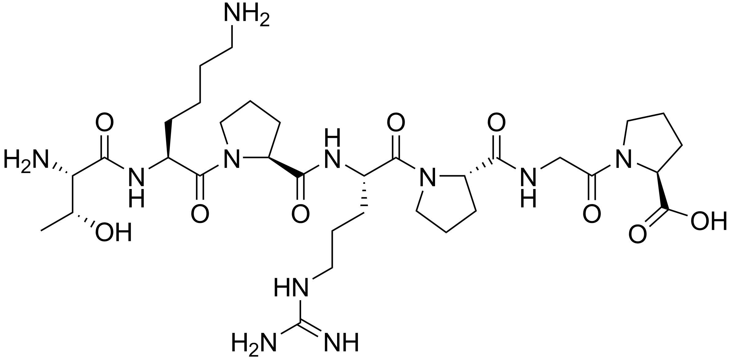
Every chiral residue is the L enantiomer, and the single glycine at position 6 is achiral, so PubChem reports seven defined atom stereocentres and none undefined for CID 11765600. Threonine contributes two of those centres, at the alpha carbon and at the beta carbon carrying the hydroxyl. The FDA substance register and an independent review that prints the systematic name both give the all-L composition. There is no cysteine, so no disulfide and no free thiol, and no sulfur anywhere in the molecule. The chain is linear and uncyclised, with no lipidation, no PEG, no glycosylation and no amidation. Three of the seven residues are proline, at positions 3, 5 and 7, which removes three backbone amide hydrogens; lysine at position 2 and arginine at position 4 give the molecule two basic side chains, and the computed partition coefficient of -6.1 reflects how polar the result is.[1,25,3]
What studies on Selank show
Each card names the experimental system and what was measured. A result in one system is not evidence about another.
radioligand binding on isolated membranes · plasma membranes isolated from brain cells, with tritiated GABA as the tracer
The peptide altered tritiated GABA binding in the pattern the authors read as positive allosteric modulation, and the combination with benzodiazepines was not additive, which they interpret as partly overlapping but non-identical sites. This is the single most cited molecular claim about the compound.[4]
- Material
- Selank, checked by high-performance liquid chromatography; benzodiazepines, diazepam and olanzapine as co-applied comparators
- Measured
- binding of tritiated GABA to the membrane preparation, with and without the peptide
- Compared with
- diazepam and olanzapine, alone and together with the peptide
- Limitation
- Read as an abstract only. A membrane preparation is not a defined receptor: no subunit composition, no dissociation constant, no half-maximal concentration and no functional recording at a named receptor are reported, so the allosteric reading is an inference from a binding shift rather than a characterisation of a site.
Vyunova TV et al., 2018, Protein and Peptide Letters
two-electrode voltage clamp screen · Xenopus oocytes expressing homomeric ASIC1a, ASIC1b, ASIC3, ASIC4 or BASIC
A laboratory with no connection to the compound's originators tested the sequence against defined recombinant channels. No peptide directly activated ASIC4 or BASIC, and the modulators resolved out of the active mixes were dynorphin A fragments, YFMRFamide and endomorphin-1. This sequence was not among them.[29]
- Material
- 109 rat neuropeptides under 20 residues, TKPRPGP among them, pooled into 13 mixes at 20 micromolar each as assay conditions
- Measured
- direct activation of the two orphan channels, and current amplitude and desensitisation time constant for the three proton-gated ones
- Compared with
- the other 108 peptides in the same screen, and the modulators the screen did resolve
- Limitation
- The peptide was tested inside a mix and single-peptide follow-up was run only for mixes that moved, so a small effect could have been missed; the paper's own power analysis was sized for large effects. A negative result at one channel family says nothing about any other target.
Vyvers A et al., 2018, Scientific Reports
enzyme inhibition in a biological fluid · human serum enkephalin-degrading enzyme activity
Half-maximal inhibition is reported at 20 micromolar for this heptapeptide and 10 micromolar for Semax as assay conditions, with both more inhibitory in this assay than puromycin or bacitracin. Pentapeptide fragments of each were inhibitory while tri-, tetra- and hexapeptide fragments were not, which is a structure-activity result rather than a potency claim. A companion plasma assay from the same laboratory gives 15 micromolar, also as an assay condition.[21]
- Material
- Selank and Semax, plus their tri-, tetra-, penta- and hexapeptide fragments
- Measured
- hydrolysis of enkephalin by serum enzymes as a function of peptide concentration
- Compared with
- Semax, puromycin and bacitracin
- Limitation
- Abstract only, and the preparation is a serum enzyme mixture: no purified peptidase is named, no inhibition mechanism is established, and micromolar figures in a mixture cannot be compared with receptor affinities.
Kost NV et al., 2001, Bioorganicheskaia Khimiia
tracer degradation chemistry · blood plasma in vitro, and rat brain tissue in vivo
The major plasma products of the heptapeptide are the pentapeptide TKPRP, the tripeptide TKP and the dipeptides Arg-Pro and Gly-Pro. Labelling every residue rather than one is what makes all products visible at once; Semax in the same study gave HFPGP and PGP.[5]
- Material
- TKPRPGP and the tripeptide PGP labelled uniformly with tritium at every residue, 50 to 150 Ci per mmol, by solid-phase catalytic isotope exchange, with chromatographic separation of products
- Measured
- identity and concentration of the labelled peptide and of each degradation product
- Compared with
- Semax, degraded in the presence of nerve cells in the same study
- Limitation
- Abstract only; the abstract gives no rate constants or half-lives, and products are assigned by chromatographic behaviour against unlabelled standards rather than by mass spectrometry.
Zolotarev IuA et al., 2006, Bioorganicheskaia Khimiia
whole-cell patch clamp in brain slices · rat hippocampal slices, CA1 pyramidal neurons
Both amplitude and discharge rate of spontaneous inhibitory currents rose, preceded in some cells by a transient fall. An earlier report from the same laboratory found the increase confined to the spike-dependent component, which points at inhibitory interneurons upstream rather than at the recorded cell.[32]
- Material
- Selank bath-applied across 1 to 8 micromolar as assay conditions
- Measured
- amplitude and frequency of spontaneous inhibitory postsynaptic currents
- Compared with
- the same cells before application
- Limitation
- Abstract only. The authors report no significant concentration dependence across the range tested, which is hard to reconcile with occupancy of a single site; no antagonist was used to identify the receptor carrying the effect, and the interneuron recording that would test the upstream interpretation was reported for a different compound.
Povarov IS et al., 2017, Bulletin of Experimental Biology and Medicine
radioreceptor assay with a behavioural arm · rat brain membranes with tritiated spiperone; mouse verticalisation test
The peptide did not displace tritiated spiperone, so no interaction with dopamine D2 sites was detected in this assay. The behavioural change it did produce was abolished by naloxone, which the authors attribute to the endogenous opioid system rather than to a dopamine receptor.[19]
- Material
- Selank; naloxone as an opioid antagonist; olanzapine as a comparator
- Measured
- displacement of tritiated spiperone from brain membrane binding sites, and apomorphine-induced behaviour
- Compared with
- olanzapine, and naloxone pretreatment
- Limitation
- Abstract only. A negative displacement result at one tracer concentration does not exclude a different site or a lower-affinity interaction, and naloxone sensitivity locates a pathway, not a molecule. The informative part is the absence of D2 binding.
Meshavkin VK et al., 2006, Bulletin of Experimental Biology and Medicine
quantitative PCR array · rat frontal cortex, 84 neurotransmission genes, 1 hour and 3 hours after a single exposure
Of 77 analysable genes, 45 changed at 1 hour and 22 at 3 hours. The largest moves were shared with GABA rather than specific to the peptide: Gabre, Gabrq and Hcrt fell about 20 to 25 fold at 1 hour and rose again by 3 hours. Four genes moved only under the peptide at 1 hour. Changes correlated positively with GABA at 1 hour and negatively at 3 hours.[33]
- Material
- Selank supplied as the diacetate salt; GABA as the comparator at the same amount per body mass
- Measured
- mRNA level of each gene relative to reference genes
- Compared with
- GABA, and vehicle
- Limitation
- Full text read. Tissue was pooled per group before RNA extraction, so between-animal variation is not represented, and the readout is mRNA with no protein or receptor-function confirmation. In human IMR-32 neuroblastoma cells the same laboratory found no transcript change from the peptide alone, so the cortical result does not establish a direct transcriptional action.
Volkova A et al., 2016, Frontiers in Pharmacology
cell differentiation assay · mouse embryonic stem cells and their neuronal derivatives
At the highest concentration tested the proportion of GABA-positive neurons fell by 61 percent, against 58 percent for thyroliberin and 87 percent for nerve growth factor in the same experiment, while proliferation and survival were little changed by this peptide. It is the only cell-level readout found that touches GABAergic phenotype directly rather than through transcripts.[34]
- Material
- Selank beside Semax, thyroliberin, HLDF-6, PGP, RPGP and PGLP, across 0.1 to 100 micromolar as assay conditions
- Measured
- proliferation, survival under serum deprivation, formation of neuronal precursors, and the proportion of GABA-positive neurons
- Compared with
- the other six peptides in the panel, and nerve growth factor
- Limitation
- Abstract only. One cell model, concentrations far above those implied by the whole-animal work, and no mechanism; the authors' own summary is a judgement that the panel is non-toxic rather than a measurement of anything about this peptide.
Kobylyanskii AG et al., 2017, Bulletin of Experimental Biology and Medicine
comparative clinical study, indexed as a randomised controlled trial · 62 adults with generalised anxiety disorder or neurasthenia, 30 given the peptide and 32 given medazepam, a benzodiazepine comparator
The authors report the anxiolytic scale changes of the two arms as similar, and additionally report antiasthenic and psychostimulant effects for the peptide. On the biochemical arm, patients in this population had a reduced leu-enkephalin half-life that correlated with illness duration, with symptom severity and with autonomic symptoms, and that figure rose during exposure to the peptide, most clearly in the generalised anxiety group. This is the serum-peptidase strand of the mechanism section measured in people rather than in a tube.[14]
- Material
- Selank, assessed against an active comparator rather than against placebo
- Measured
- Hamilton, Zung and Clinical Global Impression scale scores, and enkephalin-degrading activity in blood serum expressed as the half-life of leu-enkephalin
- Compared with
- medazepam
- Limitation
- Read as an abstract, published in Russian, from the originating network, with an active comparator and no placebo arm; the abstract reports no effect sizes, no confidence intervals and no blinding detail, and 62 participants split two ways cannot separate small differences. The scale findings are the authors' reading of their own scores and are reported here as such.
Zozulia AA et al., 2008, Zhurnal nevrologii i psikhiatrii imeni S.S. Korsakova
placebo-controlled resting-state functional MRI · 52 healthy adult participants, scanned three times: at baseline and twice after a single exposure
Between-group and between-condition differences appeared in connectivity between the right amygdala and a right-hemisphere region spanning fusiform, inferior and middle temporal and parahippocampal gyri. The authors separate general from compound-specific effects for the two peptides on that connection, and describe it as the first such analysis.[16]
- Material
- Selank, Semax or placebo in parallel groups, with the amygdala and the dorsolateral prefrontal cortex in each hemisphere as predefined regions of interest
- Measured
- whole-brain resting-state functional connectivity of each region of interest
- Compared with
- placebo, and the companion heptapeptide Semax
- Limitation
- Read as an abstract. Healthy volunteers, a single exposure and a short observation window; resting-state connectivity is a correlational measure, the regions of interest were predefined rather than the whole brain surveyed, and post hoc analysis carries the separation between the two peptides. It identifies no target and measures no concentration.
Panikratova YR et al., 2020, Doklady Biological Sciences
Selank compared with Semax and GABA
Four things about the 3 compounds most often read beside it, on axes that can be checked: what kind of substance it is, its formula and mass, what public records settle about which substance it is, and the strongest three-dimensional evidence that exists for it. Every figure is the one stated on that compound's own page, which is where it is cited, so the two can never disagree. The table compares no effect, no potency and nothing a compound has been reported to do, and these compounds are not alternatives to one another.
| Compound | Kind | Formula and mass | Identity on the public record | Strongest structure evidence |
|---|---|---|---|---|
| Selank | Peptide | 7 residues, C33H57N11O9, 751.9 g/mol | One species, pinned by public records | No three-dimensional model |
| Semax | Peptide | 7 residues, C37H51N9O10S, 813.9 g/mol | One species, pinned by public records | No three-dimensional model |
| GABA (gamma-aminobutyric acid) | Small molecule | C4H9NO2, 103.12 g/mol | One species, pinned by public records | Experimental structure, resolved inside a complex: 4MS3, X-ray diffraction at 2.5 Å |
| Selank + Semax (two-component vial) | Defined mixture | No single formula: a mixture of 2 components | One species, pinned by public records | No three-dimensional model |
What is actually measured when Selank is described as GABAergic
The GABAergic description rests on three kinds of measurement, and none of them is a receptor characterisation. First, a shift in tritiated GABA binding to isolated brain cell membranes, reported as positive allosteric modulation and as non-additive with benzodiazepines. Second, transcript-level work: in rat frontal cortex the peptide moved 29 of 77 neurotransmission genes at 1 hour, but the largest moves were shared with GABA itself; a microarray of rat hippocampus found 36 transcripts changed more than twofold after a single exposure, most of them encoding membrane or transmembrane proteins; and in human IMR-32 cells the peptide alone changed nothing while suppressing the changes GABA produced. Third, electrophysiology in hippocampal slices, where spontaneous inhibitory currents rose without a clear concentration dependence and with the effect confined to the spike-dependent component. Together these are consistent with something that alters GABAergic signalling; none of them names a subunit, a binding site or an affinity.[4,33,35,36,32,37]
A second strand runs through peptidases rather than receptors. The peptide inhibits enkephalin hydrolysis in serum and plasma at micromolar assay concentrations, with the activity apportioned preferentially to carboxypeptidase and dicarboxypeptidase rather than aminopeptidase routes, and it changes the activity of two basic-residue-removing carboxypeptidases in rat nervous tissue for up to 24 hours. Behavioural work from the same network reports that effects track plasma enkephalin half-life by mouse strain and are altered by the opioid antagonist naloxone. The same measurement was made in people: in the comparative clinical study the serum leu-enkephalin half-life was below the control value in this patient population and rose during exposure, which is the one place the peptidase strand and a human endpoint meet. This is a plausible route by which a polar heptapeptide could change signalling without binding a receptor at all, and it remains untested against any purified enzyme: no gene product has been named, and the concentrations involved are far above those a receptor mechanism would require.[18,21,38,39,40,41,14]
Whatever the route, attribution to the intact molecule is the open problem. The peptide is cut in plasma to a pentapeptide, a tripeptide and two dipeptides, and in mouse spleen the dipeptide Gly-Pro reproduced most of the transcript changes attributed to the parent, while pentapeptide fragments carried the enzyme inhibition and shorter ones did not. The same holds for the wider transcript work: in mouse spleen the parent and two of its fragments each moved the transcription factor Bcl6 together with its target and corepressor genes, out of 34 inflammation-related transcripts that changed. An effect measured in a whole animal may therefore belong to the heptapeptide, to a fragment, or to both, and the published work has not separated them.[5,6,42,43,21]
What is still unknown about Selank
No molecular target is established. There is no dissociation constant, no half-maximal concentration and no functional potency at any named receptor anywhere in this literature, and searches of ChEBI and ChEMBL return no record for the molecule at all, so there is not even a curated bioactivity table to consult. The strongest positive molecular observations are a binding shift in a membrane preparation and a change in radioligand binding-site density after repeated exposure over a five-day study period, which is an adaptive readout rather than evidence of occupancy. The one explicit negative at a defined receptor is the failure to displace tritiated spiperone from dopamine D2 sites, and the one independent test against defined recombinant channels, a screen of 109 neuropeptides on acid-sensing ion channels, did not find this sequence among its modulators.[4,44,19,29]
Most of the record also sits well above the molecular level. Typical endpoints are a protein content measured in dissected rat brain regions, where the peptide prevented an ethanol-associated rise in brain-derived neurotrophic factor in hippocampus and frontal cortex, or a transcript measured in infected animals, where interferon-alpha gene expression rose in influenza-infected mice without changes in the interleukin-4, interleukin-10 or tumour necrosis factor-alpha transcripts measured. Findings of that kind describe what changed in a named species; they do not identify what the molecule binds. The evidence base is also narrow in provenance. Almost every experiment read for this entry comes from one Moscow network, and several of the papers draw on overlapping datasets, so results that look like independent replication are often the same programme reported again. Much of it is published in Russian-language journals as abstracts with no accessible full text: of the 45 works counted here, 36 were read as abstracts, five as full texts and four are database records, and three further records relevant to the fragment and neurotrophin questions carry no abstract at all, so they could not be used. One abstract names a different peptide in its results paragraph than in its title and conclusion, a discrepancy that cannot be resolved from the public record. A review from outside that network describes the compound as poorly studied, which matches what the primary record shows.[45,46,17,47,3]
Human evidence exists and is thin. Three studies used human participants: a comparative study in 62 patients with generalised anxiety disorder or neurasthenia, indexed as a randomised controlled trial, against an active benzodiazepine comparator with no placebo arm; a cytokine study in the same patient population, reporting a shift in the serum Th1 to Th2 balance over a 14-day period and, in cell culture, complete suppression of interleukin-6 gene expression in peripheral blood from patients with depression at a stated assay concentration but not in healthy controls; and a placebo-controlled resting-state functional MRI study in 52 healthy volunteers. All three come from the originating network, two are Russian-language abstracts with no reachable full text, and none was registered on ClinicalTrials.gov. No independent controlled human study was found.[14,15,16,10]
The only independent analytical identification found is in two preparations examined by a national control laboratory, which confirmed the peptide was present. Which chemical form is being discussed also needs care: the studies that state it used the diacetate salt, PubChem carries a separate monoacetate record, and the formula and masses on this page are those of the free peptide.[28,33,48,1]
Structurally, the molecule is undescribed. No deposited structure, no computed conformer, and no solution-conformation study by nuclear magnetic resonance or circular dichroism was found. The nearest deposited coordinates contain the four-residue parent peptide bound to neuropilin-1, and the contact it makes is at the C-terminal carboxylate that this molecule's Pro-Gly-Pro extension replaces, so that structure is a reason to expect the two to differ rather than a model for this one.[20,31,30]
Key papers on Selank
- Peptide-based Anxiolytics: The Molecular Aspects of Heptapeptide Selank Biological Activity.
Vyunova TV, Andreeva L, Shevchenko K, Myasoedov N (2018), Protein and Peptide Letters · radioligand binding on isolated membranes
The paper the GABAergic description rests on, and the place to see exactly how much and how little the binding experiment establishes.
- [Evenly tritium-labeled peptides and their in vivo and in vitro biodegradation].
Zolotarev IuA, Dadaian AK, Dolotov OV, Kozik VS, Kost NV, Sokolov OIu, Dorokhova EM, Meshavkin VK, Inozemtseva LS, Gabaeva MV, Andreeva LA, and others (2006), Bioorganicheskaia Khimiia · tracer degradation chemistry
Names the degradation products by labelling every residue, which is the fact that constrains how any whole-animal result can be attributed.
- [Semax and selank inhibit the enkephalin-degrading enzymes from human serum]].
Kost NV, Sokolov OIu, Gabaeva MV, Grivennikov IA, Andreeva LA, Miasoedov NF, Zozulia AA (2001), Bioorganicheskaia Khimiia · enzyme inhibition in a biological fluid
The peptidase strand in its clearest form, with the fragment series that shows which lengths carry the inhibition.
- Selank Administration Affects the Expression of Some Genes Involved in GABAergic Neurotransmission.
Volkova A, Shadrina M, Kolomin T, Andreeva L, Limborska S, Myasoedov N, Slominsky P (2016), Frontiers in Pharmacology · quantitative PCR array
Open access, full methods, and the clearest look at a transcript-level dataset together with the comparator that makes it interpretable.
- [Efficacy and possible mechanisms of action of a new peptide anxiolytic selank in the therapy of generalized anxiety disorders and neurasthenia].
Zozulia AA, Neznamov GG, Siuniakov TS, Kost NV, Gabaeva MV, Sokolov OIu, et al. (2008), Zhurnal nevrologii i psikhiatrii imeni S.S. Korsakova · comparative clinical study
The clearest of the three human studies, and the only one with an active comparator; it is also where the serum peptidase measurement is made in patients.
- Screening of 109 neuropeptides on ASICs reveals no direct agonists and dynorphin A, YFMRFamide and endomorphin-1 as modulators.
Vyvers A, Schmidt A, Wiemuth D, Grunder S (2018), Scientific Reports · ion channel screen
The sequence tested against defined recombinant channels by a laboratory with no stake in it, which is rare in this literature.
- Tuftsin binds neuropilin-1 through a sequence similar to that encoded by exon 8 of vascular endothelial growth factor.
von Wronski MA, Raju N, Pillai R, Bogdan NJ, Marinelli ER, Nanjappan P, Ramalingam K, Arunachalam T, Eaton S, Linder KE, Yan F, Pochon S, Tweedle MF, Nunn AD (2006), Journal of Biological Chemistry · receptor binding and cell signalling
The best-characterised molecular interaction of the parent tetrapeptide, and the reason that interaction cannot be carried over to this molecule.
- Sedative-Hypnotic Agents That Impact Gamma-Aminobutyric Acid Receptors: Focus on Flunitrazepam, Gamma-Hydroxybutyric Acid, Phenibut, and Selank.
Doyno CR, White CM (2021), Journal of Clinical Pharmacology · review
An assessment of the evidence base from outside the originating network, useful as a check on how much of the above is settled.
References
- 1.National Center for Biotechnology Information. PubChem Compound Summary for CID 11765600, Selank. PubChem. source · database record
- 2.Ashmarin IP, Samonina GE, Lyapina LA, Kamenskii AA, Levitskaya NG, Grivennikov IA, Dolotov OV, Andreeva LA, Myasoedov NF. (2005). Natural and hybrid ("chimeric") stable regulatory glyproline peptides. Pathophysiology. 11(4):179-185. doi:10.1016/j.pathophys.2004.10.001 · abstract only
- 3.Deigin VI, Poluektova EA, Beniashvili AG, Kozin SA, Poluektov YM. (2022). Development of Peptide Biopharmaceuticals in Russia. Pharmaceutics. 14(4):716. doi:10.3390/pharmaceutics14040716 · full text read
- 4.Vyunova TV, Andreeva L, Shevchenko K, Myasoedov N. (2018). Peptide-based Anxiolytics: The Molecular Aspects of Heptapeptide Selank Biological Activity. Protein and Peptide Letters. 25(10):914-923. doi:10.2174/0929866525666180925144642 · abstract only
- 5.Zolotarev IuA, Dadaian AK, Dolotov OV, Kozik VS, Kost NV, Sokolov OIu, Dorokhova EM, Meshavkin VK, Inozemtseva LS, Gabaeva MV, Andreeva LA, and others. (2006). [Evenly tritium-labeled peptides and their in vivo and in vitro biodegradation]. Bioorganicheskaia Khimiia. 32(2):183-191. PMID 16637290 · abstract only
- 6.Kolomin T, Morozova M, Volkova A, Shadrina M, Andreeva L, Slominsky P, Limborska S, Myasoedov N. (2014). The temporary dynamics of inflammation-related genes expression under tuftsin analog Selank action. Molecular Immunology. 58(1):50-55. doi:10.1016/j.molimm.2013.11.002 · abstract only
- 7.U.S. Food and Drug Administration. (2026). Drugs@FDA: FDA-Approved Drugs. Drugs@FDA database. source · database record
- 8.European Medicines Agency. (2026). Medicines: search for human medicines (centrally authorised medicines). European Medicines Agency medicines database. source · database record
- 9.Health Canada. (2026). Drug Product Database online query. Health Canada Drug Product Database. source · database record
- 10.U.S. National Library of Medicine. (2026). ClinicalTrials.gov search by intervention: Selank. ClinicalTrials.gov. source · database record
- 11.World Anti-Doping Agency. (2025). World Anti-Doping Code International Standard: Prohibited List 2026. World Anti-Doping Agency. source · full text read
- 12.U.S. Food and Drug Administration. (2026). Certain Bulk Drug Substances for Use in Compounding that May Present Significant Safety Risks (content current as of 22 April 2026). U.S. Food and Drug Administration, Human Drug Compounding. source · full text read
- 13.Wardowska A, Dzierzbicka K, Mysliwski A. (2007). [Tuftsin--new analogues and properties]. Postepy Biochemii. 53(1):60-65. PMID 17718389 · abstract only
- 14.Zozulia AA, Neznamov GG, Siuniakov TS, Kost NV, Gabaeva MV, Sokolov OIu, et al. (2008). [Efficacy and possible mechanisms of action of a new peptide anxiolytic selank in the therapy of generalized anxiety disorders and neurasthenia]. Zhurnal nevrologii i psikhiatrii imeni S.S. Korsakova. 108:38-48. PMID 18454096 · abstract only
- 15.Uchakina ON, Uchakin PN, Miasoedov NF, Andreeva LA, Shcherbenko VE, Mezentseva MV, et al. (2008). [Immunomodulatory effects of selank in patients with anxiety-asthenic disorders]. Zhurnal nevrologii i psikhiatrii imeni S.S. Korsakova. 108:71-5. PMID 18577961 · abstract only
- 16.Panikratova YR, Lebedeva IS, Sokolov OY, Rumshiskaya AD, Kupriyanov DA, Kost NV, Myasoedov NF. (2020). Functional Connectomic Approach to Studying Selank and Semax Effects. Doklady Biological Sciences. 490(1):9-11. doi:10.1134/S001249662001007X · abstract only
- 17.Doyno CR, White CM. (2021). Sedative-Hypnotic Agents That Impact Gamma-Aminobutyric Acid Receptors: Focus on Flunitrazepam, Gamma-Hydroxybutyric Acid, Phenibut, and Selank. Journal of Clinical Pharmacology. 61 Suppl 2:S114-S128. doi:10.1002/jcph.1922 · abstract only
- 18.Zozulya AA, Kost NV, Sokolov OY, Gabaeva MV, Grivennikov IA, Andreeva LN, Zolotarev YA, Ivanov SV, Andryushchenko AV, Myasoedov NF, Smulevich AB. (2001). The inhibitory effect of Selank on enkephalin-degrading enzymes as a possible mechanism of its anxiolytic activity. Bulletin of Experimental Biology and Medicine. 131(4):315-317. doi:10.1023/a:1017979514274 · abstract only
- 19.Meshavkin VK, Kost NV, Sokolov OY, Zolotarev YA, Myasoedov NF, Zozulya AA. (2006). Naloxone-blocked depriming effect of anxiolytic selank on apomorphine-induced behavioral manifestations of hyperfunction of dopamine system. Bulletin of Experimental Biology and Medicine. 142(5):598-600. doi:10.1007/s10517-006-0428-1 · abstract only
- 20.Vander Kooi CW, Jusino MA, Perman B, Neau DB, Bellamy HD, Leahy DJ. (2007). RCSB PDB entry 2ORZ: Structural Basis for Ligand Binding and Heparin Mediated Activation of Neuropilin. RCSB Protein Data Bank. doi:10.2210/pdb2ORZ/pdb · database record
- 21.Kost NV, Sokolov OIu, Gabaeva MV, Grivennikov IA, Andreeva LA, Miasoedov NF, Zozulia AA. (2001). [Semax and selank inhibit the enkephalin-degrading enzymes from human serum]]. Bioorganicheskaia Khimiia. 27(3):180-183. doi:10.1023/a:1011373002885 · abstract only
- 22.Kozlovskaya MM, Kozlovskii II, Valdman EA, Seredenin SB. (2003). Selank and short peptides of the tuftsin family in the regulation of adaptive behavior in stress. Neuroscience and Behavioral Physiology. 33(9):853-860. doi:10.1023/a:1025988519919 · abstract only
- 23.Seredenin SB, Semenova TP, Kozlovskaia MM, Medvinskaia NI, Nezovibatko VN. (1995). [The characteristics of the anxiolytic action of taftsin and its analog TP-7 on behavior and serotonin metabolism in the brain of rats with chronic deprivation of serotoninergic system activity]. Eksperimentalnaia i Klinicheskaia Farmakologiia. 58(6):3-6. PMID 8704608 · abstract only
- 24.Czabak-Garbacz R, Cygan B, Wolanski L, Kozlovsky I. (2006). Influence of long-term treatment with tuftsin analogue TP-7 on the anxiety-phobic states and body weight. Pharmacological Reports. 58(4):562-567. PMID 16963804 · abstract only
- 25.United States Food and Drug Administration, Global Substance Registration System. Substance record SELANK, UNII TS9JR8EP1G. FDA Global Substance Registration System. source · database record
- 26.Ashmarin IP, Baglikova KE, Edeeva SE, Zolotarev IuA, Kozik VS, Dadaian AK, Dorokhova EM, Alfeeva LIu, Andreeva LA, Kopylova GN, and others. (2008). [A comparative analysis of distribution of glyprolines administered by various routes]. Bioorganicheskaia Khimiia. 34(4):464-470. doi:10.1134/s1068162008040043 · abstract only
- 27.Rogozinskaya EY, Lyapina MG. (2017). Anticoagulant Effects of Arginine-Containing Peptides of the Glyproline Family (His-Phe-Arg-Trp-Pro-Gly-Pro and Thr-Lys-Pro-Arg-Pro-Gly-Pro) Revealed by Thromboelastography. Bulletin of Experimental Biology and Medicine. 164(2):170-172. doi:10.1007/s10517-017-3950-4 · abstract only
- 28.Vanhee C, Francotte A, Janvier S, Deconinck E. (2020). The occurrence of putative cognitive enhancing research peptides in seized pharmaceutical preparations: An incentive for controlling agencies to prepare for future encounters of the kind. Drug Testing and Analysis. 12(3):371-381. doi:10.1002/dta.2717 · abstract only
- 29.Vyvers A, Schmidt A, Wiemuth D, Grunder S. (2018). Screening of 109 neuropeptides on ASICs reveals no direct agonists and dynorphin A, YFMRFamide and endomorphin-1 as modulators. Scientific Reports. 8(1):18000. doi:10.1038/s41598-018-36125-5 · full text read
- 30.von Wronski MA, Raju N, Pillai R, Bogdan NJ, Marinelli ER, Nanjappan P, Ramalingam K, Arunachalam T, Eaton S, Linder KE, Yan F, Pochon S, Tweedle MF, Nunn AD. (2006). Tuftsin binds neuropilin-1 through a sequence similar to that encoded by exon 8 of vascular endothelial growth factor. Journal of Biological Chemistry. 281(9):5702-5710. doi:10.1074/jbc.M511941200 · abstract only
- 31.Vander Kooi CW, Jusino MA, Perman B, Neau DB, Bellamy HD, Leahy DJ. (2007). Structural basis for ligand and heparin binding to neuropilin B domains. Proceedings of the National Academy of Sciences of the United States of America. 104(15):6152-6157. doi:10.1073/pnas.0700043104 · abstract only
- 32.Povarov IS, Kondratenko RV, Derevyagin VI, Myasoedov NF, Skrebitsky VG. (2017). Effect of Selank on Spontaneous Synaptic Activity of Rat Hippocampal CA1 Neurons. Bulletin of Experimental Biology and Medicine. 162(5):640-642. doi:10.1007/s10517-017-3676-3 · abstract only
- 33.Volkova A, Shadrina M, Kolomin T, Andreeva L, Limborska S, Myasoedov N, Slominsky P. (2016). Selank Administration Affects the Expression of Some Genes Involved in GABAergic Neurotransmission. Frontiers in Pharmacology. 7:31. doi:10.3389/fphar.2016.00031 · full text read
- 34.Kobylyanskii AG, Zolotarev YA, Andreeva LA, Grivennikov IA, Myasoedov NF. (2017). Studying the Toxic Effects of Some Biologically Active Peptides on the Model of Mouse Embryonic Stem Cells. Bulletin of Experimental Biology and Medicine. 163(6):731-736. doi:10.1007/s10517-017-3891-y · abstract only
- 35.Filatova E, Kasian A, Kolomin T, Rybalkina E, Alieva A, Andreeva L, Limborska S, Myasoedov N, Pavlova G, Slominsky P, Shadrina M. (2017). GABA, Selank, and Olanzapine Affect the Expression of Genes Involved in GABAergic Neurotransmission in IMR-32 Cells. Frontiers in Pharmacology. 8:89. doi:10.3389/fphar.2017.00089 · full text read
- 36.Kolomin TA, Agapova TIu, Agniullin IaV, Shram SI, Shadrina MI, Slominskii PA, Limborskaia SA, Miasoedov IF. (2013). [Transcriptome alteration in hippocampus under the treatment of tuftsin analog Selank]. Zhurnal Vysshei Nervnoi Deiatelnosti Imeni I P Pavlova. 63(3):365-374. doi:10.7868/s0044467713030052 · abstract only
- 37.Skrebitskii VG, Kondratenko RV, Povarov IS, Dereviagin VI. (2011). [Peptidergic modulation of the hippocampus synaptic activity]. Rossiiskii Fiziologicheskii Zhurnal Imeni I M Sechenova. 97(11):1169-1178. PMID 22390072 · abstract only
- 38.Zolotarev IuA, Sokolov OIu, Kost NV, Vaskovskii BV, Miasoedov NF, Zozulia AA. (2004). [Leu-enkephalin homogeneously labeled with tritium in studying the Selank inhibiting effect on the enkephalin-degrading enzymes of human plasma]. Bioorganicheskaia Khimiia. 30(3):234-240. doi:10.1023/b:rubi.0000030126.09208.c3 · abstract only
- 39.Soloviev VB, Gengin MT, Sollertinskaia TN, Latynova IV, Zhivaeva LV. (2012). [Effect of selank on the main carboxypeptidases in the rat nervous tissue]. Zhurnal Evoliutsionnoi Biokhimii i Fiziologii. 48(3):254-257. PMID 22827026 · abstract only
- 40.Sokolov OY, Meshavkin VK, Kost NV, Zozulya AA. (2002). Effects of Selank on behavioral reactions and activities of plasma enkephalin-degrading enzymes in mice with different phenotypes of emotional and stress reactions. Bulletin of Experimental Biology and Medicine. 133(2):133-135. doi:10.1023/a:1015582302311 · abstract only
- 41.Kozlovskii II, Andreeva LA, Kozlovskaia MM, Nadorova AV, Kolik LG. (2012). [The role of opioid system in peculiarities of anti-anxiety effect of peptide anxiolytic selank]. Eksperimentalnaia i Klinicheskaia Farmakologiia. 75(2):10-13. PMID 22550852 · abstract only
- 42.Kolomin TA, Shadrina MI, Slominskii PA, Limborskaia S, Miasoedov NF. (2011). [Changes in expression of the genes for chemokines, cytokines, and their receptors in response to selank and its fragments]. Genetika. 47(5):711-714. PMID 21786679 · abstract only
- 43.Kolomin T, Shadrina M, Andreeva L, Slominsky P, Limborska S, Myasoedov N. (2011). Expression of inflammation-related genes in mouse spleen under tuftsin analog Selank. Regulatory Peptides. 170(1-3):18-23. doi:10.1016/j.regpep.2011.05.001 · abstract only
- 44.Vasileva EV, Kondrakhin EA, Salimov RM, Kovalev GI. (2016). [COMPARISON OF PHARMACOLOGICAL EFFECTS OF HEPTAPEPTIDE SELANK AFTER INTRANASAL AND INTRAPERITONEAL ADMINISTRATION TO BALB/c AND C57BL/6 MICE.]. Eksperimentalnaia i Klinicheskaia Farmakologiia. 79(9):3-11. PMID 29787664 · abstract only
- 45.Kolik LG, Nadorova AV, Antipova TA, Kruglov SV, Kudrin VS, Durnev AD. (2019). Selank, Peptide Analogue of Tuftsin, Protects Against Ethanol-Induced Memory Impairment by Regulating of BDNF Content in the Hippocampus and Prefrontal Cortex in Rats. Bulletin of Experimental Biology and Medicine. 167(5):641-644. doi:10.1007/s10517-019-04588-9 · abstract only
- 46.Ershov FI, Uchakin PN, Uchakina ON, Mezentseva MV, Alekseeva LA, Miasoedov NF. (2009). [Antiviral activity of immunomodulator Selank in experimental influenza infection]. Voprosy Virusologii. 54(5):19-24. PMID 19882898 · abstract only
- 47.Leonidovna YA, Aleksandrovna SM, Aleksandrovna TA, Aleksandrovna BO, Fedorovich MN, Aleksandrovna AL. (2021). The Influence of Selank on the Level of Cytokines Under the Conditions of "Social" Stress. Current Reviews in Clinical and Experimental Pharmacology. 16(2):162-167. doi:10.2174/1574884715666200704152810 · abstract only
- 48.Kasian A, Kolomin T, Andreeva L, Bondarenko E, Myasoedov N, Slominsky P, Shadrina M. (2017). Peptide Selank Enhances the Effect of Diazepam in Reducing Anxiety in Unpredictable Chronic Mild Stress Conditions in Rats. Behavioural Neurology. 2017:5091027. doi:10.1155/2017/5091027 · full text read
- 49.Varentsov VE, Rumyanceva TA, Verzilina AD, Pshenisnov KK, Rudenko EE, Nikolenko VN, Shevchuk IV, Sinelnikov MY. (2021). Effect of a neurostimulator on postnatal neurogenesis in rodent olfactory bulbs. Neuropeptides. 89:102181. doi:10.1016/j.npep.2021.102181 · abstract only
- 50.Semenova TP, Kozlovskii II, Zakharova NM, Kozlovskaia MM. (2009). [Comparison of the effects of selank and tuftsin on the metabolism of serotonin in the brain of rats pretreated with PCPA]. Eksperimentalnaia i Klinicheskaia Farmakologiia. 72(4):6-8. PMID 19803361 · abstract only
- 51.Royal Society of Chemistry. (2026). ChemSpider record 9940290. ChemSpider. source · database record
