Thymosin alpha-1 in brief
Thymosin alpha-1 (INN thymalfasin) is a 28-residue acidic peptide, acetylated on its first serine and identical to residues 2 to 29 of the human protein prothymosin alpha (C129H215N33O55, 3108.3 g/mol). It was purified from a calf thymus extract in the 1970s, and a 2003 study showed that the protease legumain releases it from the precursor. In water it has no ordered structure, folding into a partial helix only on anionic surfaces or in helix-promoting solvent. No receptor is established: cellular work places its effects downstream of Toll-like receptor pathways, and the only receptor affinity located is a single micromolar figure.[1,2,3,4,5,6]
Thymosin alpha-1 regulatory status
No FDA, EMA (centralised procedure) or Health Canada marketing authorisation was found for thymalfasin in Drugs@FDA (queried through openFDA), the EMA medicines database or the Health Canada Drug Product Database (searched November 2026). It is authorised elsewhere: a 2023 review states that the synthetic peptide is approved in more than 35 countries for chronic hepatitis B and C, and no individual national register was read for this entry. ClinicalTrials.gov holds 66 registered studies naming it as an intervention, the highest registered phase being phase 3, for example a completed multicentre, quadruple-masked, randomised controlled trial in sepsis in China with a registered enrolment of 1,106 (NCT02867267). FDA lists thymosin-alpha 1 among bulk drug substances previously in category 2 whose nominations for compounding were withdrawn by the nominators, stating that compounded drugs containing it may pose a significant risk of immunogenicity and complexities with peptide-related impurities and characterisation, and that the safety-related information is inadequate for the agency to understand the extent of any safety issues raised (page current as of 22 April 2026). The peptide is not named on the World Anti-Doping Agency 2026 Prohibited List, whose only thymic entry is thymosin-beta4 and its derivatives under the growth-factor class S2.3.[7,8,9,10,11,12,13,14]
Thymosin alpha-1 at a glance
- A 28-residue peptide, acetylated at the N-terminal serine, with a markedly acidic side-chain composition: nine glutamates and aspartates against four lysines, and an isoelectric point reported as 4.2. No cysteine, therefore no disulfide.[2,15]
- The sequence is exactly residues 2 to 29 of human prothymosin alpha, and the acetyl group matches the N-acetylserine the processed precursor carries at that position.[16,15]
- It has no ordered structure in water. A partial helix appears in trifluoroethanol, in anionic detergent micelles, in vesicles carrying a negative head group, and in the presence of zinc ions; two solution NMR ensembles of the free peptide are deposited, both obtained under such conditions.[17,4,18,19]
- No receptor has been established for it. Pathway experiments in dendritic cells place its cellular readouts downstream of MyD88, TLR9 and the type I interferon receptor, and the only numeric receptor affinity located anywhere is a micromolar surface plasmon resonance constant against Toll-like receptor 2, measured as a comparator in a study of a fusion peptide.[5,20,6,10]
- Its international nonproprietary name is thymalfasin, and it is the one thymic peptide that became a medicine: a 2023 review states it is approved in more than 35 countries for chronic hepatitis B and C, although no authorisation appears in Drugs@FDA, the EMA centralised list or the Health Canada Drug Product Database. Clinical-grade material is made by solid-phase synthesis, not extracted from thymus.[10,7,8,9,21]
What is Thymosin alpha-1?
Thymosin alpha-1 was purified from calf thymus in the mid-1970s, out of a standardised tissue extract called thymosin fraction 5 prepared in the search for a thymic hormone. The 1977 report named it and established that it was 28 residues long, heat stable and highly acidic; two companion papers gave the residue-by-residue sequence, fixed the N terminus as acetylserine and measured an isoelectric point of 4.2.[1,2,22]
It is a fragment of a larger protein rather than a gene product in its own right. The human precursor, prothymosin alpha, is 111 residues long, is annotated as disordered over its entire length, and is found in the nucleus and cytosol in association with chromatin. Residues 2 to 29 of that protein are this peptide, and the processed precursor carries N-acetylserine at position 2, which is where the acetyl cap comes from. A 2003 study showed that a purified lysosomal asparaginyl endopeptidase, identified as mammalian legumain, cuts the precursor at Asn28-Gly29 to release this peptide and at Asn35-Gly36 to release the seven-residue-longer thymosin alpha-11, and reported the 28-residue product free in cytosol in several mammalian tissues at concentrations comparable to the precursor itself. The precursor is the product of a single human gene, PTMA, at 2q37.1, five exons on chromosome 2, whose record lists DNA-binding transcription factor binding, calcium ion binding and histone binding among its activities and places the protein in the cytosol and nucleoplasm.[16,3,23]
That paper exists because the molecule's status had been disputed. The objection, stated plainly in a 2003 review of the family, was that none of the peptides isolated from fraction 5 is really a thymic hormone, and that this one had never been found in extracts where proteolysis was blocked, which would make it an artifact of the preparation. The legumain work answers that charge directly. How a cytosolic peptide released by a lysosomal protease would reach a cell surface is not settled by it.[24,3]
Clinical-grade material is synthesised rather than extracted, which is the sharpest practical difference between this molecule and the thymus extracts it is confused with. Of the three routes described in the literature, tissue extraction, chemical solid-phase synthesis and recombinant expression, solid-phase synthesis is the only one a 2023 review reports as approved for clinical production. The sequence is a hard one to assemble: consecutive beta-branched residues, a tendency to form beta sheets and up to twenty side-chain protecting groups meant it was historically made by convergent solution chemistry, and a 2009 paper reports a stepwise solid-phase route on a polyethylene glycol support with the peptide anchored through a side chain. What the synthesis leaves behind has been measured: liquid chromatography with high-resolution mass spectrometry found 23 structurally related impurities above 0.1 mg per gram in a China Pharmacopoeia standard and in three commercial materials, including deamidation and amination products, succinimides, amino-acid insertions and deletions, dimers and isomers, and more than half of them arose directly or indirectly from the labile C-terminal asparagine.[10,21,25]
The thymosin name covers two unrelated families, which is the commonest confusion about this molecule, and the contrast is worth setting out. This peptide is 28 residues, acetylated on a serine, strongly acidic, cut from the 111-residue nuclear and cytosolic protein prothymosin alpha, and it has no established receptor. Thymosin beta-4 is a separate 43-residue protein from a separate gene, disordered along its whole length, whose established molecular partner is actin; the fragment sold under the name TB-500 is its residues 17 to 23. The two share no sequence, and they share a name only because both came out of the same calf thymus extract in the 1970s. Within the alpha family, thymosin alpha-11 is this peptide extended by seven residues, released from the same precursor by cleavage at a second asparagine-glycine bond.[26,24,3,16]
Two properties readers look for are measurable and worth stating plainly. The peptide is short-lived in plasma: a randomised three-way crossover study in nine healthy volunteers reported a peak serum concentration at one to two hours, peak concentrations of 30 to 80 micrograms per litre, an elimination half-life of under three hours, and an apparent volume of distribution of 30 to 40 litres, which the authors read as distribution within the extracellular volume. Endogenous concentrations were below that assay's limit of quantification of 0.10 micrograms per litre in most of the volunteers, which is worth noting on a molecule whose status as a circulating hormone has been disputed.[27,24]
The clinical literature is large enough to describe by its shape rather than by its conclusions. ClinicalTrials.gov holds 66 registered studies naming the peptide as an intervention: 7 registered as phase 1, 33 as phase 2, 8 as phase 3, 10 as phase 4, one as early phase 1 and 12 recording no applicable phase. Only 19 of the 66 record any masking, so most are open-label. The conditions studied are chronic hepatitis B and C, sepsis, a range of solid and haematological cancers, liver cirrhosis and liver failure, COVID-19, acute necrotising pancreatitis, aortic dissection, cardiopulmonary bypass, end-stage renal disease and HIV infection, and the sites are concentrated in China, the United States and Italy. This page does not summarise what those studies reported.[11]
Not to be confused with
- Thymosin beta-4, and the fragment listed elsewhere as TB-500
- A different protein family that happens to share the thymosin name because both were purified from the same calf thymus extract in the 1970s. The beta thymosins are actin-binding proteins of about 43 residues with their own sequence and their own structural literature; the alpha and beta thymosins are not variants of one molecule and share no sequence. The naming is historical, not chemical.[26,24,22]
- Thymosin alpha-11
- The same N-terminal stretch of prothymosin alpha extended by seven residues: positions 2 to 36 rather than 2 to 29. Both are produced in vitro when a lysosomal asparaginyl endopeptidase cuts the precursor, at Asn28-Gly29 for this peptide and at Asn35-Gly36 for thymosin alpha-11, with similar efficiency in that experiment.[3]
- Prothymosin alpha
- The 111-residue human precursor protein, a nuclear and cytosolic protein associated with chromatin, annotated as disordered along its whole length. This peptide is a fragment of it, not a shortened version of its function.[16,3]
- Thymosin fraction 5, and thymus extracts such as thymalin
- Fraction 5 is the standardised calf thymus extract this peptide was purified out of, described by its originators as a family of polypeptides spanning roughly 1,000 to 15,000 in molecular weight. An extract has no single sequence or mass; this entry is one defined molecule from one of them.[22,24]
- Thymopentin
- A pentapeptide taken from thymopoietin, a different thymic protein. It is frequently studied beside this peptide and has been fused to it in engineered constructs, which is where several of the receptor-binding numbers in the literature come from, but it is a separate molecule.[6]
- Polypeptide beta1, which turned out to be ubiquitin
- Worth knowing because it shows how provisional the early thymosin naming was. The second component characterised alongside this peptide in 1979 was given a thymosin-style name and then found to have the sequence of ubiquitin.[2]
Thymosin alpha-1 structure and sequence
The chain is Ac-SDAAVDTSSEITTKDLKEKKEVVEEAEN, all L residues, linear, with a free carboxylate at the C-terminal asparagine. Nine side chains are carboxylates and four are lysine amines, which is what the measured isoelectric point of 4.2 reflects, and the charge is not evenly spread: acidic residues cluster in the first fifteen positions and again at the far C terminus, with a basic stretch, Leu-Lys-Glu-Lys-Lys at positions 16 to 20, in between. That LKEKK motif is the part of the molecule most of the interaction work points at. There is no cysteine and therefore no disulfide, no cyclisation, no lipid arm, no polyethylene glycol and no sugar; the only modification is the acetyl on serine 1.[15,2,28,29]
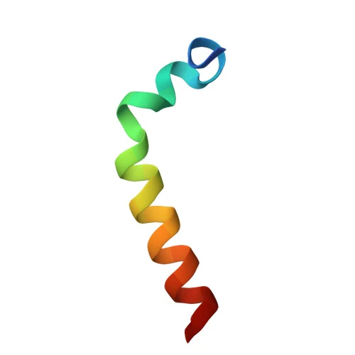
The important structural fact is a negative one: in water the peptide has no preferred conformation, which circular dichroism established in 1998 and which fits the precursor being annotated as disordered end to end. Structure can be induced, and which structure appears depends on what induces it. In water with trifluoroethanol an 800 MHz NMR study found a helix from residue 14 to residue 26 with two double beta-turns across the first twelve residues; in sodium dodecyl sulfate micelles the helix is broken near residues 9 and 14, and paramagnetic relaxation places residues 1 to 5 inside the hydrophobic interior of the micelle. An earlier study at a different cosolvent fraction put the helix at residues 17 to 24, and a later study of an engineered fusion reproduced a trifluoroethanol helix for the unfused peptide as a comparator. Zinc ions and anionic vesicles each produce partial structuring as well. A review of the whole family describes this as general behaviour rather than a peculiarity of this peptide: short, highly charged, unstructured natively, foldable by low pH, zinc, fluorinated alcohols, detergents or a binding partner. The 1998 study compared five environments directly: no preferred conformation in water, partial structuring in anionic vesicles, in sodium dodecyl sulfate and in the presence of zinc ions, and, in water with trifluoroethanol, a beta-turn between residues 5 and 8 with a helix between residues 17 and 24. It reported no zinc stoichiometry or binding constant, so the zinc effect there is spectroscopic only.[17,4,18,30,26,16]
Two solution NMR ensembles of the exact molecule are deposited, 2L9I in trifluoroethanol and 2MNQ in detergent micelles, and the viewer here shows the first. Both were obtained in environments chosen to make the peptide fold, and neither file contains the cosolvent, the micelle or any binding partner, so the picture shows one accessible conformation with nothing of the context that produced it. There is no deposited structure of this peptide bound to a receptor, and PubChem holds no computed conformer for it either. Synthetically the molecule is awkward for its size: consecutive beta-branched residues, a tendency to aggregate as beta sheet, and up to twenty side-chain protecting groups.[31,32,15,21]
N-terminus: Ac
- Ser1
- Asp2
- Ala3
- Ala4
- Val5
- Asp6
- Thr7
- Ser8
- Ser9
- Glu10
- Ile11
- Thr12
- Thr13
- Lys14
- Asp15
- Leu16
- Lys17
- Glu18
- Lys19
- Lys20
- Glu21
- Val22
- Val23
- Glu24
- Glu25
- Ala26
- Glu27
- Asn28
Modifications
- 1 N-terminal acetyl on the alpha-amino group of serine 1, giving N-acetylserine. This is not a synthetic cap added for stability: the precursor itself carries N-acetylserine at the equivalent position after its initiator methionine is removed, so the acetyl is part of the molecule's natural identity.[2,16,15]
Every stereocentre is L, as the systematic name in the PubChem record spells out residue by residue. There is no cysteine anywhere in the sequence, so there is no disulfide and no free thiol; the chain is linear, with no cyclisation, no branch point and no lipid, polyethylene glycol or sugar attached. No metal is part of the molecule. Zinc ions do change its circular dichroism spectrum in the same direction an anionic vesicle does, but no stoichiometry, binding constant or structure of a zinc complex has been published, so zinc is a condition that affects its folding rather than a component of it.[15,17,26]
What studies on Thymosin alpha-1 show
Each card names the experimental system and what was measured. A result in one system is not evidence about another.
Solution NMR with restrained molecular dynamics · 40% trifluoroethanol and 60% water, pH 4.9, 25 C, 800 MHz; 2D NOESY and TOCSY
A helix spanning residues 14 to 26 and two double beta-turns across the N-terminal twelve residues; the deposited ensemble is 20 lowest-energy conformers plus a minimised average.[4]
- Material
- Synthetic acetylated 28-residue peptide, acetyl group included in the model
- Measured
- NOE crosspeak pattern, converted into 549 restraints, and the resulting conformer ensemble
- Compared with
- Unrestrained simulations from a linear starting peptide
- Limitation
- Trifluoroethanol at 40% is a helix-promoting cosolvent, so the experiment shows that a helix is accessible, not that the peptide holds one in water. The coordinates come from restrained dynamics rather than direct structure calculation, and no binding partner was present.
Elizondo-Riojas MA et al., 2011, Biochemical and Biophysical Research Communications
Solution NMR and circular dichroism with paramagnetic relaxation · Sodium dodecyl sulfate micelles in aqueous buffer
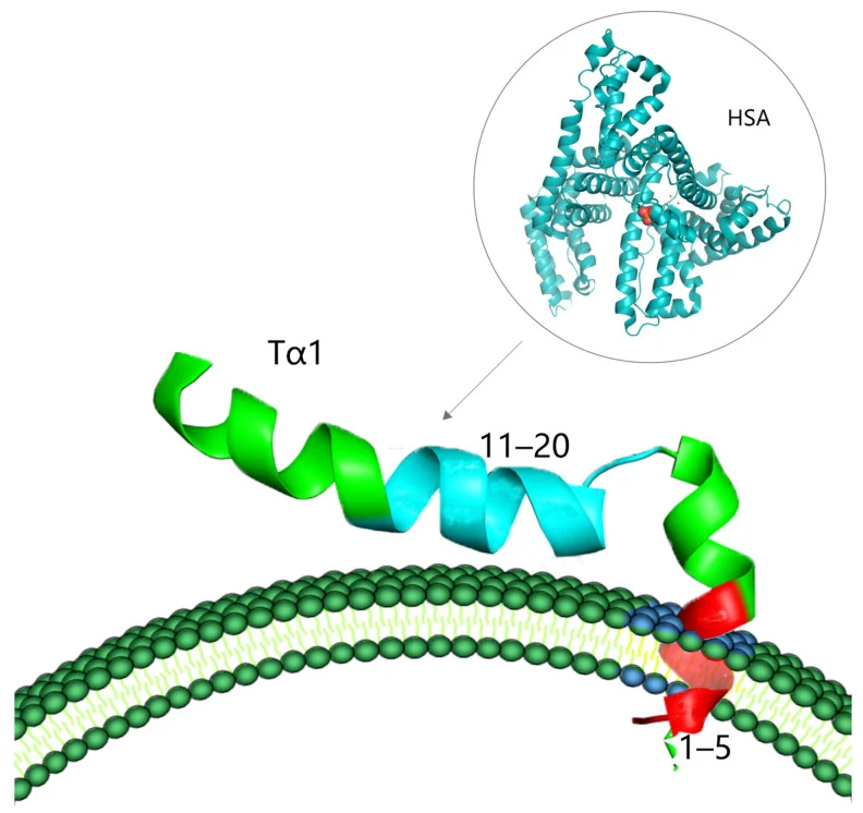
A helical conformation with a structural break near residues 9 and 14, with residues 1 to 5 positioned inside the hydrophobic region of the micelle. The peptide was confirmed unstructured in water in the same study.[18]
- Material
- Synthetic peptide
- Measured
- Secondary structure content and the depth of each segment relative to the micelle interior
- Compared with
- The same peptide in plain water, and the earlier trifluoroethanol work
- Limitation
- A detergent micelle is not a bilayer and its surface charge density is far higher than a membrane's. Only the first five residues insert, and the paper's closing suggestion that insertion initiates signalling is a hypothesis rather than a measurement. The result belongs to a series from one laboratory group, and no independent replication was located.
Nepravishta R et al., 2015, Expert Opinion on Biological Therapy
1H and natural-abundance 15N NMR, with saturation transfer, transferred NOE and diffusion-ordered spectroscopy · Mixed phosphatidylcholine and phosphatidylserine vesicles; K562 human leukaemia cells after resveratrol-induced apoptosis; human serum albumin in solution
Interaction with the vesicle required a negatively charged surface exposing phosphatidylserine. Separately, the C-terminal region of the peptide is the part engaged by human serum albumin.[33]
- Material
- Synthetic peptide
- Measured
- Whether an interaction occurs, and which part of the peptide is engaged
- Compared with
- Vesicles without the anionic phosphatidylserine component
- Limitation
- No dissociation constant for either interaction. The cellular part uses one leukaemia line with apoptosis chemically induced, and the inference that phosphatidylserine exposure is the physiological trigger is the authors' proposal, which the companion review states as an open question in its own title. It is part of the same single-laboratory series, with no independent replication located.
Mandaliti W et al., 2016, Biochemistry
Radioligand competition binding, with an enzyme activity readout · Human blood T lymphocytes; 125I-labelled cholera toxin B subunit, itself binding with Kd 3.3 nM; soluble and membrane-bound guanylate cyclase assays
The intact peptide competed with Ki 1.2 nM and the LKEKK pentapeptide with Ki 1.6 nM, while the reversed sequence was above 1 micromolar. LKEKK raised soluble guanylate cyclase activity and left the membrane-bound enzyme unchanged. The same LKEKK motif occurs at positions 16 to 20 of this peptide and at positions 131 to 135 of interferon-alpha2.[29]
- Material
- Unlabelled intact peptide, interferon-alpha2, the synthetic pentapeptide LKEKK, and the reversed pentapeptide KKEKL as a specificity control
- Measured
- Inhibition constant against the labelled toxin subunit, and guanylate cyclase activity
- Compared with
- The reversed pentapeptide KKEKL
- Limitation
- The binding site is defined only operationally, as whatever the labelled toxin subunit occupies on these cells, and was not identified molecularly. The figures are competition constants rather than a direct affinity measurement for this peptide, and the work is from a single laboratory.
Navolotskaya EV et al., 2017, Biochemistry (Moscow)
Surface plasmon resonance · Toll-like receptor 2 immobilised for binding measurement
A dissociation constant of 35.4 micromolar was reported for the unfused peptide, against 6.84 micromolar for the fusion. This is the only numeric receptor affinity located for this molecule.[6]
- Material
- A thymosin alpha-1 and thymopentin fusion peptide, with the unfused peptide and thymopentin measured alongside
- Measured
- Dissociation constant
- Compared with
- Thymopentin, and the fusion construct
- Limitation
- Read at abstract level, from a single laboratory, in a paper whose subject is the fusion rather than this peptide. A micromolar constant is weak for a proposed receptor interaction, no functional concentration-response at the receptor accompanies it, and no replication was found. The study's rodent tumour endpoints are outside what this page reports.
Li J et al., 2013, Cancer Letters
Enzyme kinetics, cell-free radical scavenging assays, and molecular docking · Purified angiotensin-converting enzyme; DPPH, ABTS, hydroxyl and superoxide radical assays; reactive oxygen species in 1321N1 human astrocytoma cells
Mixed-type inhibition of angiotensin-converting enzyme with IC50 0.8 micromolar and Ki 3.33 micromolar, and radical scavenging in the tens of micromolar; docking favoured the enzyme's N domain. This is the only measured enzyme inhibition constant found for the molecule.[34]
- Material
- Synthetic peptide
- Measured
- IC50, Ki and inhibition pattern against the enzyme; radical scavenging; cellular reactive oxygen species
- Compared with
- Assay controls; the enzyme's N and C domains compared in docking
- Limitation
- Abstract-level reading, one laboratory, no replication located. Non-specific electrostatic inhibition is a real possibility for a peptide this acidic, and docking a flexible 28-residue peptide is weak structural evidence. Scavenging figures are assay concentrations and describe the assay, not a property of a preparation.
Kharazmi-Khorassani J et al., 2019, Bioorganic Chemistry
In vitro proteolysis with a purified protease, plus tissue detection · Purified lysosomal asparaginyl endopeptidase identified as mammalian legumain, acting on prothymosin alpha; several mammalian tissues
Cleavage at Asn28-Gly29 released this 28-residue peptide and cleavage at Asn35-Gly36 released thymosin alpha-11 with similar in vitro efficiency, while the 28-residue product was the one mainly detected in vivo, free in the cytosol at concentrations similar to the precursor.[3]
- Material
- Prothymosin alpha as substrate
- Measured
- Cleavage site selectivity and efficiency; which product is present in tissue
- Compared with
- The Asn35-Gly36 site, which yields thymosin alpha-11
- Limitation
- Abstract-level reading. Cleavage by a purified enzyme in vitro does not establish that this is the route in an intact cell, and a cytosolic location is difficult to reconcile with a peptide proposed to act at a cell surface.
Sarandeses CS et al., 2003, The Journal of Biological Chemistry
Dendritic cell signalling and pathway genetics, with an animal infection model · Mouse dendritic cells pulsed with Aspergillus fumigatus; mice receiving haematopoietic transplants
The peptide drove maturation and interleukin-12 production in fungus-pulsed dendritic cells through a p38 mitogen-activated kinase and NF-kappaB pathway, and the effect required MyD88 and distinct Toll-like receptors. In the animals it shifted the antifungal response toward T-helper type 1 and improved survival in a model of aspergillosis after transplant.[5]
- Material
- The synthetic peptide added to fungus-pulsed dendritic cells; cells from mice lacking individual pathway components
- Measured
- Dendritic cell maturation markers and interleukin-12 production; which pathway components are required; antifungal T-helper responses and survival in the animals
- Compared with
- Fungus-pulsed cells with no peptide; knockout cells for MyD88 and individual Toll-like receptors
- Limitation
- Read as abstract only. The pathway requirement is genetic rather than a binding measurement, so it places the readout downstream of those components without identifying what the peptide binds. One laboratory, mouse cells, and the fungal stimulus is part of the signal being measured.
Romani L et al., 2004, Blood
Human primary cell assay across several receptor agonists · Human monocyte-derived dendritic cells in vitro
The direction of the effect depended on which receptor was stimulated: markers and cytokines rose with the TLR3 and TLR7/8 agonists and fell with the TLR2 and TLR4 agonists and with BCG. The authors describe this as a dual effect rather than a single immunological direction.[35]
- Material
- The peptide added alongside viral and bacterial Toll-like receptor agonists
- Measured
- HLA class I and class II expression and release of IL-6, TNF-alpha and IL-8
- Compared with
- Each agonist without the peptide
- Limitation
- Read as abstract only, one laboratory, human cells from donors in vitro. An effect that reverses with the stimulus is hard to attribute to a single molecular interaction, and no receptor binding was measured here.
Giacomini E et al., 2015, Expert Opinion on Biological Therapy
Thymosin alpha-1 compared with TB-500 and LL-37
Four things about the 3 compounds most often read beside it, on axes that can be checked: what kind of substance it is, its formula and mass, what public records settle about which substance it is, and the strongest three-dimensional evidence that exists for it. Every figure is the one stated on that compound's own page, which is where it is cited, so the two can never disagree. The table compares no effect, no potency and nothing a compound has been reported to do, and these compounds are not alternatives to one another.
| Compound | Kind | Formula and mass | Identity on the public record | Strongest structure evidence |
|---|---|---|---|---|
| Thymosin alpha-1 | Peptide | 28 residues, C129H215N33O55, 3108.3 g/mol | One species, pinned by public records | Experimental structure of the molecule itself: 2L9I, Solution NMR at 800 MHz (2D 1H-1H NOESY and TOCSY, pH 4.9, 298 K) with 549 distance and torsion restraints, refined by restrained molecular dynamics in an explicit 40% trifluoroethanol and 60% water box; 21 models deposited, being the 20 lowest-energy conformers and a minimised average |
| TB-500 | Peptide | 7 residues, C38H68N10O14, 889.0 g/mol | Which substance it is remains open | No three-dimensional model |
| LL-37 | Peptide | 37 residues, C205H340N60O53, 4493 g/mol | One species, pinned by public records | Experimental structure of the molecule itself: 5NMN, X-ray diffraction at 0.95 A |
| Thymalin | Heterogeneous preparation | No single formula: a mixture | Which substance it is remains open | No three-dimensional model |
What has been measured about thymosin alpha-1's molecular interactions, and why no receptor is settled
The interactions actually measured on this molecule are surface interactions rather than receptor interactions. Across detergent micelles and mixed phosphatidylcholine and phosphatidylserine vesicles, folding and insertion occur when the surface carries negative charge and do not occur, or occur without localisation, when the head groups are zwitterionic. Three partners engage three different parts of the chain: the inserting element is the N-terminal five residues, the stretch engaged by hyaluronan in a separate NMR experiment the authors call preliminary is the basic LKEKK motif, and the segment engaged by human serum albumin is the C-terminal region. All five of those membrane and carrier experiments (R06, R09, R10, R11, R26) come from one laboratory group, and no independent replication of the series was located, so the pattern rests on a single provenance. That pattern is consistent with a disordered peptide binding by charge complementarity, and it frames the physiological reading, recognition of phosphatidylserine exposed on a cell surface, as a question rather than a conclusion. Designers building hybrids from this molecule have treated the helical segment as its active stretch, which is a design choice and not a mapping result.[18,19,33,28,36,37]
The cellular literature points instead at Toll-like receptor pathways, and does so by pathway dependence rather than by binding. In mouse dendritic cells pulsed with Aspergillus fumigatus the peptide changed maturation markers and interleukin-12 production through p38 MAPK and NF-kappaB in a MyD88-dependent manner; in a follow-up, induction of indoleamine 2,3-dioxygenase in murine dendritic cells required both TLR9 and the type I interferon receptor, and a separate report on cultured bone marrow mesenchymal stem cells from children with aplastic anaemia described the same transcript direction for TLR9 and the same enzyme. Genetic requirement for a receptor is not evidence of binding to it, and TLR9 is an endosomal sensor of nucleic acids, so a peptide whose effect depends on it is a result in need of a mechanism rather than a mechanism.[5,20,38]
Human cells show that even the direction of the cellular readout depends on context. In human monocyte-derived dendritic cells, class I and II HLA surface expression and interleukin-6, tumour necrosis factor alpha and interleukin-8 secretion rose when the cells were given agonists of TLR3 or TLR7 and 8, and fell with agonists of TLR2 or TLR4 or with BCG infection. A 2023 review collects proposed involvement of TLR2, TLR3, TLR4, TLR7 and TLR9 with IRF3 and NF-kappaB downstream, which is best read as a map of how many receptors have been proposed rather than as five established ones. Against all of that, the only published affinity figure is a 35.4 micromolar surface plasmon resonance constant for TLR2, obtained as a comparator in a study of a fusion peptide.[35,10,6]
What is still unknown about Thymosin alpha-1
No receptor is established. Searches on receptor binding, affinity and surface plasmon resonance returned no direct binding study of the intact peptide against a named, purified receptor beyond the single micromolar figure above, and no structure of the peptide bound to anything is deposited. The radioligand work that does report nanomolar competition does so against a site defined only by what cholera toxin B subunit occupies on T lymphocytes, which is an operational definition rather than a molecular identification. Every statement that this peptide acts through a particular receptor should be read as a hypothesis with pathway evidence behind it.[6,29,10,31]
Both experimental structures required an environment that makes the peptide fold, and they do not fully agree about where the helix starts and stops. For an intrinsically disordered peptide these are measurements of different conditions rather than contradictions, but they mean no single conformation can be called the structure of the molecule, and a rendered model will look more definite than the evidence behind it.[4,32,18,17,26]
Whether this peptide is a hormone remains contested, and the dispute is about its existence as a distinct species rather than about its activity. A review of the family holds that none of the fraction 5 peptides is really a thymic hormone and that this one was absent from extracts prepared with proteolysis blocked; the 2003 legumain study answers that directly. That the peptide was found free in the cytosol, released by a lysosomal enzyme, leaves unexplained how it would reach the extracellular surfaces the interaction work studies.[24,3,16]
Finally, the identity of a given material is less determinate than the identity of the molecule. Registry records cover a free peptide, an acetate and a trifluoroacetate, and nothing read states which form a particular preparation is, so the formula and masses above describe the neutral peptide. The C-terminal asparagine is also chemically labile: liquid chromatography with high-resolution mass spectrometry identified and quantified 23 related impurities above 0.1 mg per gram in a pharmacopoeia reference standard, with the same method then applied to three commercial materials, and reported more than half of them as arising directly or indirectly at that residue. None of the masses here has been checked against a measured spectrum. The impurity work gives that limit a measured shape rather than leaving it abstract: 23 related peptides above 0.1 mg per gram were found in reference and commercial material, over half of them traceable to the C-terminal asparagine, so the question of what a given vial contains is a real one even for a fully defined molecule.[15,25,21]
Key papers on Thymosin alpha-1
- NMR structure of human thymosin alpha-1.
Elizondo-Riojas MA, Chamow SM, Tuthill CW, Gorenstein DG, Volk DE (2011), Biochemical and Biophysical Research Communications · Solution NMR with restrained molecular dynamics
The dedicated structure determination, and the paper behind the model shown here. Read the methods as well as the result: the 40% trifluoroethanol box is the reason the helix exists.
- Thymosin α1 inserts N terminus into model membranes assuming a helical conformation.
Nepravishta R, Mandaliti W, Eliseo T, Sinibaldi Vallebona P, Pica F, Garaci E, Paci M (2015), Expert Opinion on Biological Therapy · Solution NMR with paramagnetic relaxation
The complementary structure, in detergent micelles rather than cosolvent, and the one that locates which five residues insert into a hydrophobic interior.
- Prothymosin alpha is processed to thymosin alpha 1 and thymosin alpha 11 by a lysosomal asparaginyl endopeptidase.
Sarandeses CS, Covelo G, Díaz-Jullien C, Freire M (2003), The Journal of Biological Chemistry · In vitro proteolysis with a purified enzyme
The paper that answers the standing objection that this peptide was an extraction artifact, by naming the protease and the bond and reporting the free peptide in tissue.
- The thymosins. Prothymosin alpha, parathymosin, and beta-thymosins: structure and function.
Hannappel E, Huff T (2003), Vitamins and Hormones · Review of the thymosin family
The objection itself, stated by authors who do not accept the thymic hormone framing. Worth reading before the discovery papers rather than after them.
- The chemistry and biology of thymosin. II. Amino acid sequence analysis of thymosin alpha1 and polypeptide beta1.
Low TL, Goldstein AL (1979), The Journal of Biological Chemistry · Protein sequencing
The residue-by-residue sequence, the acetylserine N terminus and the isoelectric point, together with the ubiquitin episode that shows how provisional the family's naming was.
- Dual effect of Thymosin α 1 on human monocyte-derived dendritic cell in vitro stimulated with viral and bacterial toll-like receptor agonists.
Giacomini E, Severa M, Cruciani M, Etna MP, Rizzo F, Pardini M, Scagnolari C, Garaci E, Coccia EM (2015), Expert Opinion on Biological Therapy · In vitro cell stimulation
Human primary cells, and the clearest demonstration that the cellular readout changes direction with which Toll-like receptor is engaged, which is what makes a single mechanism hard to state.
- Identification and determination of structurally related peptide impurities in thymalfasin by liquid chromatography-high-resolution mass spectrometry.
Cheng Y, Wu P, Kan Y, Li M, Li H (2022), Analytical and Bioanalytical Chemistry · Liquid chromatography with high-resolution mass spectrometry
For anyone concerned with what is actually in a preparation: the impurity profile, and why the C-terminal asparagine is the residue that governs it.
- Structures of Thymosin Proteins.
Hoch K, Volk DE (2016), Vitamins and Hormones · Structural biology review
Sets this peptide in its family and makes plain that the alpha and beta thymosins are structurally unrelated, which is the commonest confusion about the name.
- Thymosin alpha 1 activates dendritic cells for antifungal Th1 resistance through toll-like receptor signaling.
Romani L, Bistoni F, Gaziano R, Bozza S, Montagnoli C, Perruccio K, Pitzurra L, Bellocchio S, Velardi A, Rasi G, Di Francesco P, Garaci E (2004), Blood · Cell signalling with pathway knockouts
The cellular experiment the mechanism section rests on: dendritic cell maturation placed downstream of MyD88 and Toll-like receptor signalling by pathway genetics.
- Pharmacokinetics of thymosin alpha1 after subcutaneous injection of three different formulations in healthy volunteers.
Rost KL, Wierich W, Masayuki F, Tuthill CW, Horwitz DL, Herrmann WM (1999), International journal of clinical pharmacology and therapeutics · Pharmacokinetics in healthy volunteers
The only pharmacokinetic study located, and the source of the half-life and distribution figures.
References
- 1.Goldstein AL, Low TL, McAdoo M, McClure J, Thurman GB, Rossio J, Lai CY, Chang D, Wang SS, Harvey C, Ramel AH, Meienhofer J. (1977). Thymosin alpha1: isolation and sequence analysis of an immunologically active thymic polypeptide. Proceedings of the National Academy of Sciences of the United States of America. 74:725-729. doi:10.1073/pnas.74.2.725 · abstract only
- 2.Low TL, Goldstein AL. (1979). The chemistry and biology of thymosin. II. Amino acid sequence analysis of thymosin alpha1 and polypeptide beta1. The Journal of Biological Chemistry. 254:987-995. PMID 762108 · abstract only
- 3.Sarandeses CS, Covelo G, Díaz-Jullien C, Freire M. (2003). Prothymosin alpha is processed to thymosin alpha 1 and thymosin alpha 11 by a lysosomal asparaginyl endopeptidase. The Journal of Biological Chemistry. 278:13286-13293. doi:10.1074/jbc.M213005200 · abstract only
- 4.Elizondo-Riojas MA, Chamow SM, Tuthill CW, Gorenstein DG, Volk DE. (2011). NMR structure of human thymosin alpha-1. Biochemical and Biophysical Research Communications. 416:356-361. doi:10.1016/j.bbrc.2011.11.041 · full text read
- 5.Romani L, Bistoni F, Gaziano R, Bozza S, Montagnoli C, Perruccio K, Pitzurra L, Bellocchio S, Velardi A, Rasi G, Di Francesco P, Garaci E. (2004). Thymosin alpha 1 activates dendritic cells for antifungal Th1 resistance through toll-like receptor signaling. Blood. 103:4232-4239. doi:10.1182/blood-2003-11-4036 · abstract only
- 6.Li J, Cheng Y, Zhang X, Zheng L, Han Z, Li P, Xiao Y, Zhang Q, Wang F. (2013). The in vivo immunomodulatory and synergistic anti-tumor activity of thymosin α1-thymopentin fusion peptide and its binding to TLR2. Cancer Letters. 337:237-247. doi:10.1016/j.canlet.2013.05.006 · abstract only
- 7.U.S. Food and Drug Administration. (2026). Drugs@FDA data, openFDA drugsfda endpoint, active-ingredient search for THYM* (thymalfasin, thymosin) (searched November 2026). openFDA. source · database record
- 8.European Medicines Agency. (2026). Medicines data: all centrally processed human and veterinary medicines (JSON report), retrieved November 2026. European Medicines Agency. source · database record
- 9.Health Canada. (2026). Drug Product Database, active ingredient search for thym (thymalfasin, thymosin) (searched November 2026). Health Canada Drug Product Database. source · database record
- 10.Tao N, Xu X, Ying Y, Hu S, Sun Q, Lv G, Gao J. (2023). Thymosin α1 and Its Role in Viral Infectious Diseases: The Mechanism and Clinical Application. Molecules. 28:3539. doi:10.3390/molecules28083539 · full text read
- 11.U.S. National Library of Medicine. (2026). ClinicalTrials.gov registered studies with thymalfasin or thymosin alpha 1 as an intervention. ClinicalTrials.gov. source · database record
- 12.ClinicalTrials.gov. (2016). NCT02867267: The efficacy and safety of thymosin alpha 1 for sepsis: a multicenter, double-blinded, randomized and controlled clinical trial. ClinicalTrials.gov. source · database record
- 13.U.S. Food and Drug Administration. (2026). Certain Bulk Drug Substances for Use in Compounding that May Present Significant Safety Risks (Category 2 of the bulk substances nominated under sections 503A or 503B), content current as of 04/22/2026. U.S. Food and Drug Administration. source · full text read
- 14.World Anti-Doping Agency. (2025). World Anti-Doping Code International Standard: Prohibited List 2026. World Anti-Doping Agency. source · full text read
- 15.PubChem, National Center for Biotechnology Information. Compound Summary: Thymalfasin, CID 16130571. PubChem. source · database record
- 16.UniProt Consortium. UniProtKB P06454 (PTMA_HUMAN): Prothymosin alpha. UniProtKB. source · database record
- 17.Grottesi A, Sette M, Palamara T, Rotilio G, Garaci E, Paci M. (1998). The conformation of peptide thymosin alpha 1 in solution and in a membrane-like environment by circular dichroism and NMR spectroscopy. A possible model for its interaction with the lymphocyte membrane. Peptides. 19:1731-1738. doi:10.1016/s0196-9781(98)00132-6 · abstract only
- 18.Nepravishta R, Mandaliti W, Eliseo T, Sinibaldi Vallebona P, Pica F, Garaci E, Paci M. (2015). Thymosin α1 inserts N terminus into model membranes assuming a helical conformation. Expert Opinion on Biological Therapy. 15 Suppl 1:S71-S81. doi:10.1517/14712598.2015.1009034 · abstract only
- 19.Mandaliti W, Nepravishta R, Sinibaldi Vallebona P, Pica F, Garaci E, Paci M. (2016). New studies about the insertion mechanism of Thymosin α1 in negative regions of model membranes as starting point of the bioactivity. Amino Acids. 48:1231-1239. doi:10.1007/s00726-016-2169-4 · abstract only
- 20.Romani L, Bistoni F, Perruccio K, Montagnoli C, Gaziano R, Bozza S, Bonifazi P, Bistoni G, Rasi G, Velardi A, Fallarino F, Garaci E, Puccetti P. (2006). Thymosin alpha1 activates dendritic cell tryptophan catabolism and establishes a regulatory environment for balance of inflammation and tolerance. Blood. 108:2265-2274. doi:10.1182/blood-2006-02-004762 · abstract only
- 21.García-Ramos Y, Giraud M, Tulla-Puche J, Albericio F. (2009). Optimized Fmoc solid-phase synthesis of Thymosin alpha1 by side-chain anchoring onto a PEG resin. Biopolymers. 92:565-572. doi:10.1002/bip.21317 · abstract only
- 22.Low TL, Thurman GB, McAdoo M, McClure J, Rossio JL, Naylor PH, Goldstein AL. (1979). The chemistry and biology of thymosin. I. Isolation, characterization, and biological activities of thymosin alpha1 and polypeptide beta1 from calf thymus. The Journal of Biological Chemistry. 254:981-986. PMID 216684 · abstract only
- 23.National Center for Biotechnology Information. (2026). Gene record 5757, PTMA, prothymosin alpha (Homo sapiens). NCBI Gene. source · database record
- 24.Hannappel E, Huff T. (2003). The thymosins. Prothymosin alpha, parathymosin, and beta-thymosins: structure and function. Vitamins and Hormones. 66:257-296. doi:10.1016/s0083-6729(03)01007-0 · abstract only
- 25.Cheng Y, Wu P, Kan Y, Li M, Li H. (2022). Identification and determination of structurally related peptide impurities in thymalfasin by liquid chromatography-high-resolution mass spectrometry. Analytical and Bioanalytical Chemistry. 414:8035-8045. doi:10.1007/s00216-022-04336-5 · abstract only
- 26.Hoch K, Volk DE. (2016). Structures of Thymosin Proteins. Vitamins and Hormones. 102:1-24. doi:10.1016/bs.vh.2016.04.009 · abstract only
- 27.Rost KL, Wierich W, Masayuki F, Tuthill CW, Horwitz DL, Herrmann WM. (1999). Pharmacokinetics of thymosin alpha1 after subcutaneous injection of three different formulations in healthy volunteers. International journal of clinical pharmacology and therapeutics. 37:51-7. PMID 10027483 · abstract only
- 28.Mandaliti W, Nepravishta R, Pica F, Sinibaldi Vallebona P, Garaci E, Paci M. (2017). Thymosin α1 Interacts with Hyaluronic Acid Electrostatically by Its Terminal Sequence LKEKK. Molecules. 22:1843. doi:10.3390/molecules22111843 · abstract only
- 29.Navolotskaya EV, Sadovnikov VB, Zinchenko DV, Zolotarev YA, Lipkin VM, Zav'yalov VP. (2017). Interaction of Cholera Toxin B-subunit with Human T-lymphocytes. Biochemistry (Moscow). 82:1036-1041. doi:10.1134/S0006297917090061 · abstract only
- 30.Wang F, Li B, Fu P, Li Q, Zheng H, Lao X. (2018). Immunomodulatory and enhanced antitumor activity of a modified thymosin α1 in melanoma and lung cancer. International Journal of Pharmaceutics. 547:611-620. doi:10.1016/j.ijpharm.2018.06.041 · abstract only
- 31.Elizondo-Riojas MA, Volk DE, Gorenstein DG. (2011). NMR structure of thymosin alpha-1. RCSB Protein Data Bank, entry 2L9I. doi:10.2210/pdb2L9I/pdb · database record
- 32.Nepravishta R, Mandaliti W, Eliseo T, Paci M. (2015). 1H, 13C, and 15N Chemical Shift Assignments for Thymosin alpha 1. RCSB Protein Data Bank, entry 2MNQ. doi:10.2210/pdb2MNQ/pdb · database record
- 33.Mandaliti W, Nepravishta R, Sinibaldi Vallebona P, Pica F, Garaci E, Paci M. (2016). Thymosin α1 Interacts with Exposed Phosphatidylserine in Membrane Models and in Cells and Uses Serum Albumin as a Carrier. Biochemistry. 55:1462-1472. doi:10.1021/acs.biochem.5b01345 · abstract only
- 34.Kharazmi-Khorassani J, Asoodeh A, Tanzadehpanah H. (2019). Antioxidant and angiotensin-converting enzyme (ACE) inhibitory activity of thymosin alpha-1 (Thα1) peptide. Bioorganic Chemistry. 87:743-752. doi:10.1016/j.bioorg.2019.04.003 · abstract only
- 35.Giacomini E, Severa M, Cruciani M, Etna MP, Rizzo F, Pardini M, Scagnolari C, Garaci E, Coccia EM. (2015). Dual effect of Thymosin α 1 on human monocyte-derived dendritic cell in vitro stimulated with viral and bacterial toll-like receptor agonists. Expert Opinion on Biological Therapy. 15 Suppl 1:S59-S70. doi:10.1517/14712598.2015.1019460 · abstract only
- 36.Nepravishta R, Mandaliti W, Sinibaldi Vallebona P, Pica F, Garaci E, Paci M. (2016). Mechanism of Action of Thymosinα1: Does It Interact with Membrane by Recognition of Exposed Phosphatidylserine on Cell Surface? A Structural Approach. Vitamins and Hormones. 102:101-119. doi:10.1016/bs.vh.2016.04.002 · abstract only
- 37.Zhang L, Wei X, Zhang R, Petitte JN, Si D, Li Z, Cheng J, Du M. (2019). Design and Development of a Novel Peptide for Treating Intestinal Inflammation. Frontiers in Immunology. 10:1841. doi:10.3389/fimmu.2019.01841 · abstract only
- 38.Hou F, Huang JM, Li G. (2010). [Regulatory effect of thymosin α1 on expression of tlr9/ido mRNA in bone marrow mesenchymal stem cells from children with aplastic anemia]. Zhongguo Shi Yan Xue Ye Xue Za Zhi. 18:1564-1567. PMID 21176371 · abstract only
- 39.ChEBI, European Bioinformatics Institute. thymalfasin (CHEBI:135915). ChEBI. source · database record
- 40.U.S. Food and Drug Administration. (2026). Global Substance Registration System record for thymalfasin (UNII W0B22ISQ1C), registry codes. FDA Global Substance Registration System. source · database record
