TB-500 in brief
TB-500 is the name used for Ac-LKKTETQ, a seven-residue N-acetylated peptide matching residues 17 to 23 of the 43-residue protein thymosin beta-4 (C38H68N10O14, 889.0 g/mol); it is not the protein itself. Its identity was settled in 2012 by two doping control laboratories that analysed circulating material by mass spectrometry, and it is prohibited in sport at all times under WADA class S2.3. Its claimed activity is inherited from work on the full-length protein, where this stretch forms part of the actin-binding site. Work on the free peptide is sparse and none of it is a published human study: in rats and human serum it is quickly cut into shorter acetylated fragments, and most cell and animal studies used the unacetylated form. No study compares the two forms at a shared endpoint, so the acetyl group that defines the compound has no characterised consequence.[1,2,3,4,5,6,7]
TB-500 regulatory status
No FDA, EMA or Health Canada marketing authorisation was found for TB-500 or for thymosin beta-4 in Drugs@FDA (queried through openFDA), the EMA medicines database or the Health Canada Drug Product Database (searched November 2026); the highest-phase registered human study found is a Phase 1/2 trial in adults with stable atherosclerotic cardiovascular disease, listed as recruiting (NCT07487363). FDA lists 'thymosin beta-4, fragment (LKKTETQ), also known as TB-500' among bulk drug substances previously in category 2 whose nominations for compounding were withdrawn by the nominators, stating that compounded drugs containing it may pose a risk of immunogenicity because of the potential for aggregation and peptide-related impurities, that it has not identified any human exposure data, and that it lacks important information on whether it would cause harm in humans (page current as of 22 April 2026). The 2026 WADA Prohibited List names 'thymosin-β4 and its derivatives e.g. TB-500' under S2.3, growth factors and growth factor modulators, prohibited at all times.[8,9,10,11,12,6]
TB-500 at a glance
- TB-500 is a name for a molecule, not a protein: material sold under it was characterised by high-resolution mass spectrometry as the acetylated 17-23 fragment of human thymosin beta-4, Ac-LKKTETQ, and a reference standard of that peptide was synthesised to confirm it. A second laboratory, working on equine samples, independently named N-acetylated LKKTETQ as the key ingredient.[1,2]
- Seven residues, C38H68N10O14, average mass 889.0 g/mol, monoisotopic 888.4916 Da, all-L, linear, with one acetyl group and nothing else attached. The 43-residue protein it is cut from is about five and a half times heavier.[5,13]
- The shape of the evidence matters more than any single finding in it. No completed or published human study of this peptide was located; one phase 1/2 placebo-controlled trial of TB-500, described in its registration as the thymosin beta 4 17-23 fragment, is registered as recruiting in adults with stable atherosclerotic cardiovascular disease. The only in vivo work located on the acetylated molecule itself is a rat metabolism study and a rat Achilles tendon study with eight animals per group. Almost everything else was done on the unacetylated heptapeptide or on the full-length protein, and a 2026 review states the fragment and the protein are routinely conflated with no validated biologic equivalence.[4,14,7,11]
- The sequence was named the central actin-binding site of thymosin beta-4 on the strength of mutational and cross-linking work on the whole protein, where Lys18 inside this stretch forms an electrostatic contact with actin's N-terminal acidic residues. That work tested substituted full-length protein, not a free heptapeptide.[15,3,16]
- The best-characterised property of the peptide itself is how quickly it is cut down. In rats and in human serum it is degraded to shorter acetylated fragments, with Ac-LK most abundant early and Ac-LKK still detectable at 72 hours, and in a fibroblast scratch assay in the same study it was the metabolite Ac-LKKTE rather than the parent that changed the measured readout.[4]
What is TB-500?
Thymosin beta-4 is a 43-residue, highly polar protein of the beta-thymosin family, present in most cell types and body fluids, which binds monomeric actin and keeps a reservoir of it out of filaments. In the 1990s its actin-binding site was mapped to a short internal stretch, and by the 2000s a seven-residue peptide covering that stretch, LKKTETQ, was being tested in its own right beside the parent protein.[13,3,16,17]
TB-500 is the trade-style name that grew up around the acetylated version of that stretch. Its identity was settled by analytical chemistry rather than by a discovery paper: in 2012 a doping control laboratory in Ghent analysed a TB-500 formulation by HPLC with Orbitrap mass spectrometry, identified the N-terminally acetylated 17-23 fragment, and synthesised Ac-LKKTETQ to confirm the assignment. A Hong Kong racing laboratory reached the same conclusion from a veterinary preparation in the same year. Both papers exist because the material was circulating in sport, which is also why much of what is known about its fate in the body comes from antidoping work.[1,2,18]
The name is the main source of confusion and the reason this page exists. Thymosin B4 Acetate, a name that also circulates for this material, describes the protein, while the material the name TB-500 has been shown to contain is a seven-residue fragment of it; a 2026 narrative review puts it plainly, calling TB-500 a distinct seven amino-acid fragment frequently conflated with full-length thymosin beta-4 in the absence of validated biologic equivalence. The word acetate adds a second ambiguity, since it could name a counter-ion or the acetyl group that defines the peptide, and no record for a defined acetate salt of it was found.[7,1,5]
Two regulators have taken a position on it, and both are worth stating plainly because they are the reason much of the analytical literature exists. The 2026 World Anti-Doping Agency Prohibited List names thymosin-beta4 and its derivatives, giving TB-500 as the example, under class S2.3, growth factors and growth factor modulators, prohibited at all times in and out of competition. The FDA lists thymosin beta-4, fragment (LKKTETQ), also known as TB-500, among bulk drug substances previously in category 2 whose nominations for compounding were withdrawn by the nominators; its stated concern is a risk of immunogenicity from aggregation and peptide-related impurities, that it has identified no human exposure data for drug products containing the fragment, and that it therefore lacks important information on whether the substance would cause harm in humans. No marketing authorisation for the fragment or the protein was found in the FDA, EMA or Health Canada databases. The regulatory section below carries the citations and the search dates.[6,12,8,9,10]
Identity on this page is therefore recorded as partially verified. What is verified is the molecule the name refers to in the published record, confirmed by two independent laboratories and consistent with PubChem CID 62707662. What is open is the name itself, which circulates for both the fragment and the full-length protein.[1,2,5,7]
Not to be confused with
- Thymosin beta-4 (the 43-residue protein, UniProt P62328, CAS 77591-33-4)
- The protein is six times the mass of this peptide and carries interactions the fragment does not. Its mature chain is SDKPDMAEIEKFDKSKLKKTETQEKNPLPSKETIEQEKQAGES, annotated as disordered along its whole length, and the acetyl group it carries naturally sits on its first residue (an N-acetylserine), not on the leucine that begins this peptide. A 2026 narrative review states directly that TB-500 is a distinct seven-residue fragment frequently conflated with the full-length protein with no validated biologic equivalence. Figures published for the protein, including actin-binding geometry and mass, do not transfer.[13,7,24]
- LKKTETQ, the unacetylated heptapeptide (PubChem CID 10169788)
- Identical sequence, 42 Da lighter: average mass 847.0 against 889.0, formula C36H66N10O13 against C38H68N10O14. The single acetyl group is the whole chemical difference, and it matters for reading the literature, because most of the experimental work on the seven-residue sequence, including the dermal and hepatic stellate cell studies, used the unacetylated form.[20,5,17,23]
- Thymosin alpha-1
- A 28-residue peptide from a separate precursor (prothymosin alpha) and a separate lineage, sharing only the historical word thymosin. It has no sequence relationship to the beta-thymosin family and nothing in this page applies to it.[13]
- Ac-SDKP and the N-terminal 15-residue fragment of thymosin beta-4
- Two other short sequences cut from the same protein, with different attributed activities. In human hepatic stellate cells the 1-15 fragment and the 17-23 fragment gave different molecular readouts in the same experiment, so the fragments are not interchangeable with each other any more than with the parent.[16,23]
TB-500 structure and sequence
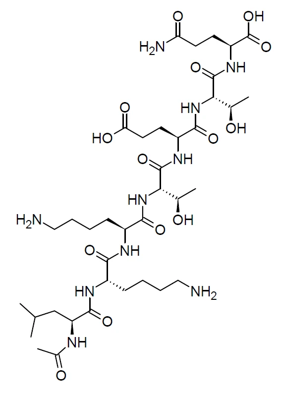
The chain is leucine, two lysines, threonine, glutamate, threonine, glutamine, read N to C, with an acetyl cap on the leucine nitrogen and a free acid at the glutamine. Counting the mature thymosin beta-4 sequence in UniProt P62328, which is stored as 44 residues and loses its initiator methionine, places that stretch exactly at positions 17 to 23. Thr22 of the mature protein, the threonine immediately before the terminal glutamine here, is an annotated phosphosite in the parent, recorded by UniProt at position 23 of its stored 44-residue sequence; the mature and stored numberings differ by one throughout. The terminal glutamine is chemically distinctive in its own right: transglutaminase labels Gln23 and Gln36 of the mature protein while leaving Gln39 alone.[13,19,5]
Two numbers do most of the work in telling the molecules apart. The acetylated peptide is C38H68N10O14 at 889.0 g/mol average and 888.4916 Da monoisotopic; the unacetylated peptide is C36H66N10O13 at 847.0 and 846.4811. The 42 Da gap is one acetyl group, and it is the only chemical difference between the compound sold as TB-500 and the peptide used in most of the published experiments on this sequence. The parent protein, at 43 residues, is in a different mass regime altogether, which is how a mass spectrometer settles the question in a single measurement.[5,20,13]
There is nothing three-dimensional to display, and the reason is interesting rather than merely inconvenient. Free thymosin beta-4 is unstructured in solution and folds on binding monomeric actin, taking up a central extended region flanked by two helices; its helix I runs residues 5 to 16 and extending that helix over the LKK segment causes a loss of activity, so the behaviour of this sequence in the protein depends on where a neighbouring helix stops. None of that geometry is a property the excerpt can carry on its own, and seven polar residues in water have no single preferred conformation to deposit.[21,22,15]
Modifications
- 1 (Leu, equivalent to Leu17 of thymosin beta-4) N-terminal acetyl on the alpha-amino group, added synthetically. It accounts for the whole 42 Da difference from the unacetylated heptapeptide and is a different site from the protein's own N-acetylserine, which lies sixteen residues upstream and is absent from this molecule.[1,5,20,13]
The chain is all-L with no D-residues: the PubChem IUPAC name for CID 62707662 assigns (2S) at every alpha carbon and (2S,3R) at both threonines. There is no cysteine, so no disulfide; the peptide is linear and uncyclised, with no lipidation, no PEG, no glycosylation and no metal coordination. The only non-backbone chemistry is the single N-terminal acetyl. The two lysine side chains and the glutamate carboxylate make it strongly polar and charged at neutral pH, which is a family trait of the beta-thymosins.[5,15]
What studies on TB-500 show
Each card names the experimental system and what was measured. A result in one system is not evidence about another.
Analytical identification with synthesis of a reference standard · A commercial TB-500 formulation analysed by HPLC with high-resolution Orbitrap mass spectrometry; the reference peptide made by solid-phase synthesis
The peptide in the formulation is the N-terminally acetylated 17-23 fragment of human thymosin beta-4. The authors also proposed a triple-quadrupole strategy for detecting it in plasma and urine.[1]
- Material
- Molecule tested: Ac-LKKTETQ, the acetylated heptapeptide. The formulation as sold, against synthetic Ac-LKKTETQ
- Measured
- Accurate mass and fragmentation of the peptide present in the formulation
- Compared with
- The synthesised reference peptide
- Limitation
- One formulation examined in one laboratory in 2012. It establishes what the name has been shown to mean, not the contents of any other vial, and the abstract reports no purity figure.
Esposito S et al., 2012, Drug Testing and Analysis
Liquid chromatography-mass spectrometry in a biological matrix, with in vitro metabolite generation · Equine urine and plasma, ion-exchange solid-phase extraction, plus post-administration samples from horses
Independent confirmation of the identity, with confirmation at 0.02 ng/mL in equine plasma and 0.01 ng/mL in urine, and the first identification of the peptide and its metabolites in post-administration horse samples.[2]
- Material
- Molecule tested: Ac-LKKTETQ, the acetylated heptapeptide. N-acetylated LKKTETQ, named as the key ingredient of a veterinary TB-500 preparation
- Measured
- Retention time and product-ion abundances for the parent peptide and its metabolites; confirmation limits
- Compared with
- In vitro generated metabolites and blank matrix
- Limitation
- An analytical study. The activity claims its introduction repeats are attributed to the product's own marketing, and nothing about molecular interaction was measured here.
Ho EN et al., 2012, Journal of Chromatography A
Metabolite identification and quantification by UHPLC-Orbitrap MS/MS, with cell assays · Human serum, several in vitro enzyme systems, urine from treated rats; cytotoxicity and scratch (closure) assays in fibroblasts
Ac-LK was the most abundant metabolite in rats over the first six hours and Ac-LKK was still detectable at 72 hours. Neither parent nor metabolites were cytotoxic. Of everything tested, only Ac-LKKTE changed the scratch-assay readout significantly against control, and the authors note that the biological effects of TB-500 itself had not previously been documented.[4]
- Material
- Molecule tested: Ac-LKKTETQ, the acetylated heptapeptide, against its own synthesised metabolites. Ac-LKKTETQ with synthesised authentic standards of each metabolite
- Measured
- Concentrations of parent and metabolites over time, cytotoxicity, and closure in the fibroblast assay
- Compared with
- Parent peptide against its own metabolites, and untreated controls
- Limitation
- The abstract gives no effect size for the assay, which is cell-layer closure in culture and not tissue repair. The suggestion that activity reported for the parent belongs to a metabolite is the authors' interpretation, not an established mechanism.
Rahaman KA et al., 2024, Journal of Chromatography B
Cell signalling study with fragment comparison · Early-passage human hepatic stellate cell cultures (passages 4 to 6, isolated from human liver biopsies) stimulated with platelet-derived growth factor BB at 50 ng/mL, with each peptide at 10 ng/mL (assay conditions)
The 17-23 fragment, but not 1-15, blocked the growth-factor-driven rise in PDGF-beta receptor, alpha-SMA and alpha-1(I) collagen mRNA, cutting the induced levels by 65, 53 and 55 percent respectively (p below 0.05 for each) against the growth-factor-only group, and reduced Akt phosphorylation at both T308 and S473, with PRAS40 phosphorylation following. It also prevented the reappearance of the PDGF-beta receptor protein at 72 hours after the growth factor had degraded it, and cut induced proliferation by 35 percent at 24 hours. The 1-15 fragment touched only S473, through mTOR, and changed neither proliferation nor migration.[23]
- Material
- Molecule tested: LKKTETQ, the UNACETYLATED heptapeptide, beside an unrelated fragment of the same protein. Thymosin beta-4 fragments 17-23 (LKKTETQ, unacetylated) and 1-15 (Ac-SDKPDMAEIEKFDKS)
- Measured
- PDGF-beta receptor, alpha-SMA and collagen 1 expression; Akt phosphorylation at T308 and S473; PRAS40 phosphorylation; proliferation and migration
- Compared with
- The two fragments against each other and against PDGF-BB alone
- Limitation
- One cell type from donors undergoing bariatric surgery, blot- and PCR-based readouts, no binding measurement, and the peptide is the unacetylated heptapeptide rather than the acetylated molecule named TB-500. Read in full text.
Shah R et al., 2018, Expert Opinion on Biological Therapy
Mutational mapping with cross-linking and sequestering assays · Chemically synthesised full-length thymosin beta-4 variants with monomeric actin; native gels, chemical cross-linking, circular dichroism
Residues 1-16 and the hexapeptide 17-22 behave as separate structural entities, and the contacts that matter are a hydrophobic patch on one face of the N-terminal helix plus electrostatic contributions from Lys18 in the motif and Lys14 in the helix. The N-terminal part must adopt a helix for binding.[3]
- Material
- Molecule tested: full-length 43-residue thymosin beta-4, not a peptide. Point-substituted full-length protein
- Measured
- Complex formation, cross-linking efficiency and actin-sequestering activity per variant
- Compared with
- Wild-type protein and variants against each other
- Limitation
- Every variant is full-length. The motif was not tested as a free peptide, so the study locates a site within a protein rather than showing what the isolated sequence does.
Van Troys M et al., 1996, The EMBO Journal
Zero-length cross-linking with peptide sequencing, plus circular dichroism and NMR · Purified thymosin beta-4 and monomeric actin in 1:1 complex
Lys3 of the protein cross-linked to actin Glu167, Lys18 (inside this page's sequence) to one of actin Asp1-Glu4, and Lys38 to actin Gln41, placing contacts on both the barbed and pointed faces and requiring the C-terminal half to be extended. Free protein was predominantly unstructured with at most six helical residues.[15]
- Material
- Molecule tested: full-length 43-residue thymosin beta-4, not a peptide. Full-length thymosin beta-4
- Measured
- Which residue pairs cross-link, and helical content free and bound
- Compared with
- Two cross-linking chemistries, carbodiimide and transglutaminase
- Limitation
- Cross-linking reports proximity rather than atomic geometry, and the distances constrain the whole 43-residue chain. Nothing here measures a seven-residue peptide.
Safer D et al., 1997, Biochemistry
X-ray crystallography with biochemical assays and molecular dynamics · Crystals of actin-thymosin beta-4 fusion constructs, deposited as 4PL7 and 4PL8
The protein engages actin through two helices contacting the barbed and pointed faces, which sterically prevents the bound monomer joining a filament; a second form shows an opened nucleotide cleft with the C-terminal helix contact disrupted, and exchange with profilin has both steric and allosteric components.[24]
- Material
- Molecule tested: a covalent actin and full-length thymosin beta-4 fusion chain, not a peptide. A covalent actin-thymosin beta-4 hybrid chain, not the free protein and not a peptide
- Measured
- Atomic structure at 2.3 A for 4PL7, nucleotide-cleft geometry, and exchange behaviour between the two sequestering proteins
- Compared with
- The two structures against each other, and against published profilin:actin structures
- Limitation
- A designed fusion of yeast actin with the 43-residue protein. Its thymosin chain is modelled over mature residues 5 to 40, so the 17 to 23 stretch is present, but only inside the whole chain and alongside the flanking helices that make the contacts. It is the closest structural work to this sequence and still contains no free LKKTETQ peptide, acetylated or otherwise, which is why this page shows no 3D view.
Xue B et al., 2014, Proceedings of the National Academy of Sciences of the United States of America
Animal model with biomechanical, histopathological and immunohistochemical endpoints · Thirty-two male Sprague-Dawley rats, standardised Achilles tendon transection and repair, four groups of eight, four-week endpoint
Maximum load to failure was higher in the TB-500 group than in controls (p<0.05), total Bonar score was lower (p=0.016) and total Movin score was lower (p=0.017). Collagen I expression did not differ significantly between groups while collagen III did. The combination with BPC-157 conferred nothing beyond either agent alone.[14]
- Material
- Molecule tested: material described by its authors as synthetic thymosin beta-4 (TB-500), with no analytical confirmation reported that it was the acetylated heptapeptide. Material described as synthetic thymosin beta-4 (TB-500), alone and combined with BPC-157
- Measured
- Maximum load to failure; Bonar and Movin histological scores; Sirius red birefringence; collagen I and III expression by H-score
- Compared with
- Untreated controls, BPC-157 alone, and the combination
- Limitation
- Eight animals per group at a single four-week endpoint, described by its own authors as exploratory, in rats. The abstract does not state that the material was verified as the acetylated heptapeptide, and no molecular endpoint was measured. The absence of an additive effect is a negative result in one model, not a statement about any mixture.
Biçer O et al., 2026, Joint Diseases and Related Surgery
What is known about the actin-binding motif, and what the free heptapeptide has been shown to do
The mechanistic case for this sequence is inherited rather than measured. Thymosin beta-4 is the major actin-sequestering protein of higher eukaryotes: it forms a 1:1 complex with monomeric actin and blocks its incorporation into filaments, and mutational and cross-linking work on the whole protein identified residues 17 to 22 as one of two separate binding entities, with Lys18 contributing an electrostatic contact to actin's N-terminal acidic residues. A review of the protein's short active sites named LKKTETQ its central actin-binding domain, and that phrase is what most writing about TB-500 is ultimately quoting.[15,3,16]
Later structural work complicates the inheritance. Crystallography and NMR across the beta-thymosin and WH2 family put the strongest actin contact on an amphipathic N-terminal helix that recognises the ATP-bound conformation of actin, a helix lying outside residues 17 to 23 and absent from this peptide; whether a family domain sequesters actin or feeds filament growth is set by the dynamics of its C-terminal half, with a single salt bridge beside the central LKKT/V motif deciding sequestration at physiological ionic strength. On that account the motif is necessary company rather than a self-sufficient binding module, and no dissociation constant for the free heptapeptide against actin was found in any source read here.[25,26,24]
What has been measured on seven residues is narrower and mostly used the unacetylated form. In human hepatic stellate cells the 17-23 peptide reduced PDGF-BB-driven Akt phosphorylation and matrix gene expression where the 1-15 peptide did not; in full-thickness dermal wounds in aged mice the unacetylated peptide promoted repair comparably to the parent protein, though the itemised endpoints in that study, wound contracture, keratinocyte migration and collagen deposition, were reported for the protein and not measured separately for the peptide; and a corneal study used the sequence built into a self-assembling hydrogel, where the construct rather than a free peptide was the thing applied. For the acetylated molecule specifically, the clearest finding is metabolic: it is cut to shorter acetylated fragments quickly, and in the one study to test parent against metabolites in the same assay it was Ac-LKKTE, not Ac-LKKTETQ, that moved the readout.[23,17,27,4]
What is still unknown about TB-500
The central gap is a missing comparison. No study was located that measures Ac-LKKTETQ against unacetylated LKKTETQ at the same endpoint, and no study measures either against the full-length protein at a molecular endpoint such as actin binding. So the acetyl group, the one feature that defines the compound, has no characterised consequence: it is reasonable to expect it to change the molecule's susceptibility to aminopeptidases and its charge at the N-terminus, and neither expectation was found tested. Until that work exists, inferences from the protein to the fragment and from the unacetylated peptide to the acetylated one both remain assumptions.[4,1,7]
The identity of any particular material is a separate question from the identity of the name. Two independent laboratories established what TB-500 contained in the samples they analysed in 2012. The name Thymosin B4 Acetate names the protein and not the fragment, and the word acetate is itself ambiguous between a counter-ion and the acetyl group; no record of a defined acetate salt of this peptide was found. A mass measurement settles which of the two a material is, since the fragment and the protein differ by roughly 4,000 Da.[1,2,7,5]
There is no structure of this peptide and there is unlikely to be one soon; the molecular structure section above gives the reason in full. The one point worth repeating here is for a reader who goes looking: the deposited thymosin beta-4 complexes do contain this sequence, and they are structures of the 43-residue protein inside a fusion construct rather than of the compound.[21,28,24]
Finally, the parent protein's biology does not reduce to actin, which bears on how much a single-function reading of the fragment can carry, and part of that record is unwelcome rather than merely complicated. The protein binds Fe(II) and Fe(III) at four regions spread along its chain, which is consistent with its composition (it is acidic, pI 5.1, with eleven carboxylate side chains and nine lysines) and happens without any change in its secondary structure: eighteen residues lost intensity in the presence of Fe(II) and fifteen with Fe(III) in carbon-proton HSQC NMR, and its presence in the growth medium of the J774 macrophage line reduced erastin- and glutamate-induced ferroptosis. Its nuclear distribution was found to depend neither on actin binding nor on its basic lysine cluster. Separately, the protein has been reported in tumour tissue and in tumour-cell experiments: immunostaining of 76 human colorectal adenocarcinomas found it in 59 of them, strongest in invading cells at the deep margin where E-cadherin was falling, and overexpressing it in two oral squamous carcinoma cell lines increased proliferation, colony formation, invasion and matrix metalloproteinase 2 activity with a shift in epithelial and mesenchymal markers. Those are measurements on the 43-residue protein, in tissue and in cell lines, and not on this seven-residue peptide, for which no tumour-biology data were located at all. Properties distributed over 43 residues give no grounds either for attributing them to any seven of them or for assuming the seven are free of them.[29,30,31,32]
Key papers on TB-500
- Synthesis and characterization of the N-terminal acetylated 17-23 fragment of thymosin beta 4 identified in TB-500, a product suspected to possess doping potential.
Esposito S, Deventer K, Goeman J, Van der Eycken J, Van Eenoo P (2012), Drug Testing and Analysis · Analytical identification with reference synthesis
The identity paper. It is where the name TB-500 was tied to a specific molecule, by mass spectrometry on a formulation and by synthesising the peptide to check.
- Simultaneous quantification of TB-500 and its metabolites in in-vitro experiments and rats by UHPLC-Q-Exactive orbitrap MS/MS and their screening by wound healing activities in-vitro.
Rahaman KA, Muresan AR, Min H, Son J, Han HS, Kang MJ, Kwon OS (2024), Journal of Chromatography B · Metabolite quantification with cell assays
The most informative study of the acetylated peptide itself: metabolite identification with authentic standards, and the finding that a metabolite rather than the parent moved the cell assay.
- Investigational Peptides in Orthopaedic Practice: Evidence, Safety, and Patient Counseling for BPC-157, Thymosin Beta-4, and TB-500.
Tham A, Hodgkins CW, Kennedy JG (2026), The Journal of the American Academy of Orthopaedic Surgeons · Narrative review
States the fragment-versus-protein confusion explicitly and surveys what direct evidence exists, which is the shortest route to seeing how thin the ground under the name is.
- The actin binding site of thymosin beta 4 mapped by mutational analysis.
Van Troys M, Dewitte D, Goethals M, Carlier MF, Vandekerckhove J, Ampe C (1996), The EMBO Journal · Mutational mapping of a binding site
Where residues 17 to 22 were first mapped as an actin-binding entity, and worth reading for how carefully it keeps the claim inside the full-length protein.
- Structural basis of thymosin-β4/profilin exchange leading to actin filament polymerization.
Xue B, Leyrat C, Grimes JM, Robinson RC (2014), Proceedings of the National Academy of Sciences of the United States of America · X-ray crystallography of fusion constructs
The structural account of how the parent protein holds monomeric actin, and the clearest demonstration of why no structure here is a structure of the compound.
- The beta-thymosin/WH2 domain; structural basis for the switch from inhibition to promotion of actin assembly.
Hertzog M, van Heijenoort C, Didry D, Gaudier M, Coutant J, Gigant B, Didelot G, Préat T, Knossow M, Guittet E, Carlier MF (2004), Cell · Crystallography and NMR with mutagenesis
The reason to be cautious about the fragment: it places the family's strongest actin contact on an N-terminal helix that this peptide does not contain.
- Thymosin β4 inhibits PDGF-BB induced activation, proliferation, and migration of human hepatic stellate cells via its actin-binding domain.
Shah R, Reyes-Gordillo K, Rojkind M (2018), Expert Opinion on Biological Therapy · Cell signalling study
A rare clean comparison of two fragments of the same protein at molecular endpoints in one experiment, showing they are not interchangeable.
References
- 1.Esposito S, Deventer K, Goeman J, Van der Eycken J, Van Eenoo P. (2012). Synthesis and characterization of the N-terminal acetylated 17-23 fragment of thymosin beta 4 identified in TB-500, a product suspected to possess doping potential. Drug Testing and Analysis. 4:733-8. doi:10.1002/dta.1402 · abstract only
- 2.Ho EN, Kwok WH, Lau MY, Wong AS, Wan TS, Lam KK, Schiff PJ, Stewart BD. (2012). Doping control analysis of TB-500, a synthetic version of an active region of thymosin β₄, in equine urine and plasma by liquid chromatography-mass spectrometry. Journal of Chromatography A. 1265:57-69. doi:10.1016/j.chroma.2012.09.043 · abstract only
- 3.Van Troys M, Dewitte D, Goethals M, Carlier MF, Vandekerckhove J, Ampe C. (1996). The actin binding site of thymosin beta 4 mapped by mutational analysis. The EMBO Journal. 15:201-10. PMID 8617195 · abstract only
- 4.Rahaman KA, Muresan AR, Min H, Son J, Han HS, Kang MJ, Kwon OS. (2024). Simultaneous quantification of TB-500 and its metabolites in in-vitro experiments and rats by UHPLC-Q-Exactive orbitrap MS/MS and their screening by wound healing activities in-vitro. Journal of Chromatography B. 1235:124033. doi:10.1016/j.jchromb.2024.124033 · abstract only
- 5.National Center for Biotechnology Information. PubChem Compound Summary for CID 62707662, TB-500 (Ac-Leu-Lys-Lys-Thr-Glu-Thr-Gln-OH). PubChem. source · database record
- 6.World Anti-Doping Agency. (2025). World Anti-Doping Code International Standard: Prohibited List 2026. World Anti-Doping Agency. source · full text read
- 7.Tham A, Hodgkins CW, Kennedy JG. (2026). Investigational Peptides in Orthopaedic Practice: Evidence, Safety, and Patient Counseling for BPC-157, Thymosin Beta-4, and TB-500. The Journal of the American Academy of Orthopaedic Surgeons. doi:10.5435/JAAOS-D-26-00049 · abstract only
- 8.U.S. Food and Drug Administration. (2026). Drugs@FDA data, openFDA drugsfda endpoint, active-ingredient search for THYM* (thymosin) (searched November 2026). openFDA. source · database record
- 9.European Medicines Agency. (2026). Medicines data: all centrally processed human and veterinary medicines (JSON report), retrieved November 2026. European Medicines Agency. source · database record
- 10.Health Canada. (2026). Drug Product Database, active ingredient search for thym (thymosin) (searched November 2026). Health Canada Drug Product Database. source · database record
- 11.ClinicalTrials.gov. (2026). NCT07487363: A Phase 1/2, randomized, double-blind, placebo-controlled, sequential dose-escalation study of TB-500 (thymosin beta 4 17-23 fragment) in adults with stable atherosclerotic cardiovascular disease. ClinicalTrials.gov. source · database record
- 12.U.S. Food and Drug Administration. (2026). Certain Bulk Drug Substances for Use in Compounding that May Present Significant Safety Risks (Category 2 of the bulk substances nominated under sections 503A or 503B), content current as of 04/22/2026. U.S. Food and Drug Administration. source · full text read
- 13.UniProt Consortium. UniProtKB entry P62328 (TYB4_HUMAN), Thymosin beta-4, Homo sapiens. UniProtKB. source · database record
- 14.Biçer O, Adanir O, Güleryaz Y, Balci EC, Dinçel YM, Yenigün MY, Aydin C, Bayrak BY. (2026). Effects of BPC-157 and TB-500 on Achilles tendon healing in rats: A histopathological and biomechanical study. Joint Diseases and Related Surgery. 37:822-837. doi:10.52312/jdrs.2026.2951 · abstract only
- 15.Safer D, Sosnick TR, Elzinga M. (1997). Thymosin beta 4 binds actin in an extended conformation and contacts both the barbed and pointed ends. Biochemistry. 36:5806-16. doi:10.1021/bi970185v · abstract only
- 16.Sosne G, Qiu P, Goldstein AL, Wheater M. (2010). Biological activities of thymosin beta4 defined by active sites in short peptide sequences. The FASEB Journal. 24:2144-51. doi:10.1096/fj.09-142307 · abstract only
- 17.Philp D, Badamchian M, Scheremeta B, Nguyen M, Goldstein AL, Kleinman HK. (2003). Thymosin beta 4 and a synthetic peptide containing its actin-binding domain promote dermal wound repair in db/db diabetic mice and in aged mice. Wound Repair and Regeneration. 11:19-24. doi:10.1046/j.1524-475x.2003.11105.x · abstract only
- 18.Kwok WH, Ho EN, Lau MY, Leung GN, Wong AS, Wan TS. (2013). Doping control analysis of seven bioactive peptides in horse plasma by liquid chromatography-mass spectrometry. Analytical and Bioanalytical Chemistry. 405:2595-606. doi:10.1007/s00216-012-6697-9 · abstract only
- 19.App C, Knop J, Huff T, Seebahn A, Becker CM, Iavarone F, Castagnola M, Hannappel E. (2014). Peptide labeling with photoactivatable trifunctional cadaverine derivative and identification of interacting partners by biotin transfer. Analytical Biochemistry. 456:14-21. doi:10.1016/j.ab.2014.04.003 · abstract only
- 20.National Center for Biotechnology Information. PubChem Compound Summary for CID 10169788, the unacetylated heptapeptide LKKTETQ. PubChem. source · database record
- 21.Domanski M, Hertzog M, Coutant J, Gutsche-Perelroizen I, Bontems F, Carlier MF, Guittet E, van Heijenoort C. (2004). Coupling of folding and binding of thymosin beta4 upon interaction with monomeric actin monitored by nuclear magnetic resonance. The Journal of Biological Chemistry. 279:23637-45. doi:10.1074/jbc.M311413200 · abstract only
- 22.Simenel C, Van Troys M, Vandekerckhove J, Ampe C, Delepierre M. (2000). Structural requirements for thymosin beta4 in its contact with actin. An NMR-analysis of thymosin beta4 mutants in solution and correlation with their biological activity. European Journal of Biochemistry. 267:3530-8. doi:10.1046/j.1432-1327.2000.01380.x · abstract only
- 23.Shah R, Reyes-Gordillo K, Rojkind M. (2018). Thymosin β4 inhibits PDGF-BB induced activation, proliferation, and migration of human hepatic stellate cells via its actin-binding domain. Expert Opinion on Biological Therapy. 18:177-184. doi:10.1080/14712598.2018.1478961 · full text read
- 24.Xue B, Leyrat C, Grimes JM, Robinson RC. (2014). Structural basis of thymosin-β4/profilin exchange leading to actin filament polymerization. Proceedings of the National Academy of Sciences of the United States of America. 111:E4596-605. doi:10.1073/pnas.1412271111 · abstract only
- 25.Hertzog M, van Heijenoort C, Didry D, Gaudier M, Coutant J, Gigant B, Didelot G, Préat T, Knossow M, Guittet E, Carlier MF. (2004). The beta-thymosin/WH2 domain; structural basis for the switch from inhibition to promotion of actin assembly. Cell. 117:611-23. doi:10.1016/s0092-8674(04)00403-9 · abstract only
- 26.Didry D, Cantrelle FX, Husson C, Roblin P, Moorthy AM, Perez J, Le Clainche C, Hertzog M, Guittet E, Carlier MF, van Heijenoort C, Renault L. (2012). How a single residue in individual β-thymosin/WH2 domains controls their functions in actin assembly. The EMBO Journal. 31:1000-13. doi:10.1038/emboj.2011.461 · abstract only
- 27.Lu P, Shan M, Peng C, Ji W, Yang T, Yang Z, Zhang Z, Wang Y. (2025). Alkaline Phosphatase-Triggered Spatiotemporal Repair of Corneal Injury with TB500 Peptide Hydrogel. ACS Applied Materials and Interfaces. 17:67503-67518. doi:10.1021/acsami.5c14652 · abstract only
- 28.RCSB Protein Data Bank. PDB entry 4PL7: Structure of Komagataella pastoris actin-thymosin beta4 hybrid. Protein Data Bank. source · database record
- 29.Lachowicz JI, Pichiri G, Piludu M, Fais S, Orrù G, Congiu T, Piras M, Faa G, Fanni D, Dalla Torre G, Lopez X, Chandra K, Szczepski K, Jaremko L, Ghosh M, Emwas AH, Castagnola M, Jaremko M, Hannappel E, Coni P. (2022). Thymosin β4 Is an Endogenous Iron Chelator and Molecular Switcher of Ferroptosis. International Journal of Molecular Sciences. 23:551. doi:10.3390/ijms23010551 · full text read
- 30.Zoubek RE, Hannappel E. (2007). Subcellular distribution of thymosin beta4. Annals of the New York Academy of Sciences. 1112:442-50. doi:10.1196/annals.1415.031 · abstract only
- 31.Nemolato S, Restivo A, Cabras T, Coni P, Zorcolo L, Orru G, Fanari M, Cau F, Gerosa C, Fanni D, Messana I, Castagnola M, Casula G, Faa G. (2012). Thymosin beta 4 in colorectal cancer is localized predominantly at the invasion front in tumor cells undergoing epithelial mesenchymal transition. Cancer Biology and Therapy. 13:191-7. doi:10.4161/cbt.13.4.18691 · abstract only
- 32.Hong KO, Lee JI, Hong SP, Hong SD. (2016). Thymosin beta4 induces proliferation, invasion, and epithelial-to-mesenchymal transition of oral squamous cell carcinoma. Amino Acids. 48:117-27. doi:10.1007/s00726-015-2070-6 · abstract only
- 33.U.S. Food and Drug Administration. (2026). Global Substance Registration System record for TB-500 (UNII QHK6Z47GTG), registry codes. FDA Global Substance Registration System. source · database record
- 34.RCSB Protein Data Bank. PDB entry 3U9D: Crystal structure of a chimera containing the N-terminal domain (residues 8-24) of Drosophila Ciboulot and the C-terminal domain (residues 13-44) of bovine thymosin-beta4, bound to G-actin-ATP. Protein Data Bank. source · database record
- 35.RCSB Protein Data Bank. PDB entry 3M3N: Structure of a longitudinal actin dimer assembled by tandem W domains. Protein Data Bank. source · database record
- 36.RCSB Protein Data Bank. PDB entry 1HJ0: Thymosin beta9. Protein Data Bank. source · database record
Related entries
- BPC-157BPC-157 is a synthetic 15-residue peptide, named in 1993 after a gastric-juice protein whose own sequence has never been published. It has been studied almost entirely in rodent and cell systems, has no identified molecular target, and is named by the World Anti-Doping Agency in its 2026 Prohibited List.
- Thymosin alpha-1Thymosin alpha-1 is a 28-residue acidic peptide, acetylated on its first serine, cut out of the larger human protein prothymosin alpha. It was first isolated from a calf thymus extract in the mid-1970s.
