hCG in brief
Human chorionic gonadotropin is the placental glycoprotein hormone of pregnancy, built from two non-covalently associated chains: a 92-residue alpha chain shared with luteinizing hormone, follitropin and thyrotropin, and a 145-residue beta chain unique to it. Each chain folds round a cystine knot, and eight attached glycans make up roughly 25 to 30 percent of its mass, so it has no single formula or mass. It acts at the luteinizing hormone and choriogonadotropin receptor, seen in 2021 cryo-EM complexes. Its beta C-terminal peptide and the O-glycans on it account for the long circulating half-life that distinguishes it from luteinizing hormone, and no deposited structure models either that segment or the carbohydrate. A preparation specified only in International Units states a potency against a reference standard, not an amount of substance, although the unit convention does point to the pooled-source rather than the recombinant substance.[1,2,3,4,5,6,7,8]
hCG regulatory status
Urinary chorionic gonadotropin is a prescription medicine in the United States, licensed for prepubertal cryptorchidism not due to anatomical obstruction, selected cases of hypogonadotropic hypogonadism in males and induction of ovulation in anovulatory infertile women; its label states that it has not been shown to be effective in the treatment of obesity. In the EU a recombinant form, choriogonadotropin alfa, holds a central authorisation for triggering final follicular maturation in assisted reproduction, and Health Canada lists several marketed chorionic gonadotropin products (checked November 2026). The WADA 2026 Prohibited List prohibits chorionic gonadotrophin in males at all times, under S2.2.1. Chorionic gonadotropin appears on neither FDA compounding list: it is in no category of the list of bulk drug substances nominated under section 503A (updated 14 May 2026), and in neither the category 2 table nor the table of withdrawn nominations on the page of bulk substances that may present significant safety risks under the 503A and 503B interim policies (content current as of 22 April 2026). The two substances are also distinguished by how their strength is expressed: the recombinant one is authorised by protein mass in micrograms, while the pooled-source product is specified in International Units. No retraction or expression of concern was found for any study cited here; the 1990 erratum attached to R35 is recorded with that reference.[9,10,11,12,10,13,14]
hCG at a glance
- Two different chains, not one: a 92-residue alpha chain identical in human chorionic gonadotropin, luteinizing hormone, follitropin and thyrotropin, and a 145-residue beta chain unique to this hormone. They are held together without a covalent bond between them, and each is cross-linked internally, five disulfides in the alpha chain and six in the beta chain.[1,2,15]
- It is a hormone of pregnancy. Placental syncytiotrophoblast cells secrete it, pituitary gonadotrope cells make it too, and it acts on the same receptor as luteinising hormone; it is also the analyte that pregnancy immunoassays detect. A review of the molecule describes five commonly distinguished species in circulation: the hormone itself, a sulfated form, a hyperglycosylated form, the free beta subunit and a hyperglycosylated free beta subunit, with the first group made by syncytiotrophoblast and pituitary cells and the second by cytotrophoblast cells and by malignancies.[8]
- Each chain folds round a cystine knot, three disulfides threaded so that one passes through a ring formed by the other two, the same motif found in some protein growth factors. The two chains reach that fold from only about 10 percent sequence identity with each other, and a beta-chain segment wraps round the alpha chain tethered by the Cys26 to Cys110 disulfide, which the 1994 structure paper named the seat belt.[3,16]
- Eight attachment points for sugars: N-linked at alpha Asn52, alpha Asn78, beta Asn13 and beta Asn30, and O-linked at beta Ser121, Ser127, Ser132 and Ser138. Carbohydrate accounts for roughly 25 to 30 percent of the mass, and because those structures vary the molecule has no single formula or mass.[1,2,15,4]
- What the deposited structures do not show: the beta C-terminal peptide (beta 112 to 145) has no coordinates in either 1994 crystal form or in the 2021 receptor complexes, and essentially none of the carbohydrate is modelled anywhere.[17,18,19,16]
- A potency in International Units is not an amount of substance. An International Unit is assigned by comparison with a reference ampoule, and the current WHO standard carries two different figures for one ampoule, 159 IU by bioassay and 186 IU by immunoassay, with the immunoassay value corresponding to 0.41 nmol per ampoule (expanded uncertainty 0.40 to 0.43). The instructions state that the approximate mass content was not experimentally derived and must not be used as a formal mass assignment.[7]
What is hCG?
Human chorionic gonadotropin belongs to the glycoprotein hormone family, four human hormones that share one alpha chain and differ in a second, hormone-specific beta chain. The shared alpha chain is 92 residues once its signal peptide is removed; the beta chain of this hormone is 145 residues, and the same beta sequence is encoded by more than one near-identical gene, CGB3, CGB5 and CGB8. The chains associate without any covalent bond between them, so the intact hormone is a complex held together by surface contacts rather than a single polypeptide.[1,2]
Biologically it is a placental hormone. Syncytiotrophoblast cells of the placenta secrete it, pituitary gonadotrope cells make it as well, and it signals through the luteinising hormone and choriogonadotropin receptor, which is what makes it the analyte detected by pregnancy immunoassays. A review of the molecule separates five commonly reported species into two groups by origin: the hormone, a sulfated form and a hyperglycosylated form, together with the free beta subunit and a hyperglycosylated free beta subunit; the first group acts on the hormone's own receptor while the second is described as acting on a transforming growth factor beta receptor instead. Its regulatory status is set out in the Regulatory status section: the pooled-source and the recombinant substances are separately registered prescription medicines, and the 2026 WADA Prohibited List names chorionic gonadotrophin in males.[8,5,9,10,12]
Its protein architecture was settled in 1994 by two crystal structures published within weeks of each other. Both found that the two chains, which share only about 10 percent sequence identity, adopt similar folds, each with three disulfides threaded into a cystine knot, and that the interface between them is unusually large, involving two inter-chain beta sheets plus a tethered beta-chain arm wrapped round the alpha chain. Thirty years on the hCG beta chain is still the search model used to solve related structures: the 2019 crystal structure of the bovine luteinizing hormone beta subunit was phased by molecular replacement with it.[3,16,20]
The point that most needs stating is that this entry names a protein whose sequence is exact and whose carbohydrate is not. Eight glycans attach at defined positions, but the structures occupying those positions are a distribution. Glycomic analysis of hormone purified from urine at two points in the same pregnancies found mono-, bi-, tri- and tetra-antennary N-glycans throughout, in different proportions at 7 and at 20 weeks, including bisected glycans and LewisX antigens earlier surveys had not reported. Material described as hyperglycosylated carries the same two polypeptide chains with larger N-glycans and hexasaccharide in place of trisaccharide O-glycans, so it is not a sequence variant and no sequence check distinguishes it.[21,4]
Two consequences follow for identity. The regulatory registries hold the pooled-source substance and the recombinant substance separately, with different UNII codes, INN list numbers and DrugBank accessions, but CAS 9002-61-3 is listed on both, so a CAS number does not say which a label means. And a vial specified only in International Units states a potency relative to a reference ampoule rather than an amount of substance: the WHO instructions for the current standard assign one ampoule 159 IU for bioassay and 186 IU for immunoassay, and say the approximate mass printed on the leaflet was not experimentally derived and must not be used as a mass assignment, because preparations from different sources may differ in post-translational modification such as glycosylation. What a unit specification does narrow is the source: choriogonadotropin alfa is authorised and labelled by protein mass in micrograms, so material specified in International Units is the pooled-source substance rather than the recombinant one.[15,22,7,10]
Not to be confused with
- Choriogonadotropin alfa
- The recombinant substance, registered separately from pooled-source chorionic gonadotropin: UNII 6413W06WR3 against 20ED16GHEB, INN list number 7473 against 1972, DrugBank DB00097 against DB09126, and its own ChEBI entry. Both records carry the same two chains of 145 and 92 residues, so the polypeptide is the same molecule; what differs is the production route and therefore the carbohydrate. A mass spectrometric study of recombinant gonadotropins expressed in Chinese hamster ovary cells reported complex-type glycans matching the natural counterparts except that sulphated terminal glycosylation was absent. Note that CAS 9002-61-3 is listed on BOTH substance records, so a CAS number does not tell the two apart.[15,22,33,34]
- Human luteinizing hormone (lutropin, LH)
- A different hormone that activates the same receptor. It shares the identical alpha chain, and its beta chain is about 85 percent identical to the hCG beta chain over the first 114 residues but lacks the 31-residue C-terminal extension that hCG beta carries. The two are also assembled differently: in hCG the beta chain folds before the chains dock and the alpha chain threads into the preassembled beta chain, whereas the LH beta chain is incompletely folded when it meets the alpha chain, which then acts as a chaperone for it.[27,32,1]
- Human follitropin (FSH) and thyrotropin (TSH)
- Two more hormones sharing the same alpha chain with their own beta chains. Measured at the recombinant human TSH receptor in a cAMP assay, purified human LH was about ten times as potent as purified hCG, and a recombinant hCG lacking the beta C-terminal peptide was close to LH, so the beta chain differences are measurable at a receptor that is not the hormone's own.[1,27]
- hCG free beta subunit, and the beta-core fragment
- Separate molecular species, not forms of the heterodimer. The beta-core fragment is the terminal urinary degradation product and is two peptides, beta6 to beta40 and beta55 to beta92, still held together by disulfides, of which only the first retains truncated N-linked sugars. Mass spectrometry of material purified from pregnancy urine resolved at least ten species in it, so it is itself a glycoform population rather than one compound.[28,4]
- Hyperglycosylated hCG
- The same two polypeptide chains with a different carbohydrate population: larger triantennary N-glycans and hexasaccharide rather than trisaccharide O-glycans. Glycomic analysis of hCG purified from urine at two points in one pregnancy found the same N-glycan structures throughout but in different proportions, with bisected glycans and LewisX antigens abundant in pregnancy samples. Because the peptide sequence is identical, no sequence-based identity check can distinguish it.[21,4]
- Gonadorelin (GnRH)
- A ten-residue linear peptide acting at a different receptor family entirely. It is sometimes grouped with hCG because both belong to the literature of the reproductive axis, but it shares no sequence, no fold and no receptor with this molecule.
hCG structure and sequence
Read the sequence above as two separate chains. Numbering is the trap: the structural and glycosylation literature counts from the start of each mature chain, while UniProt features are listed on the precursors, so its alpha positions run 24 higher and its beta positions 20 higher than the numbers used here. The glycan the signalling literature calls alpha Asn52 is Asn76 in the UniProt record, and a site quoted without saying which numbering is meant is ambiguous by exactly that offset.[1,2,23]
Within each chain three disulfides form a cystine knot, one passing through a ring closed by the other two. That makes the fold hard to unpick, and it is why the hormone survives proteolytic cleavage in a recognisable form: when leukocyte elastase cuts the beta chain at Val44 to 45 or Gly47 to 48, the pieces stay associated because disulfides still span the cut. The one segment with no disulfide anchor is the beta C-terminal peptide, residues 93 to 145, which is why proteolysis at beta Leu92 releases it cleanly. That segment is the hormone's only large structural departure from LH, FSH and TSH, and it carries all four O-glycans.[3,4,2]
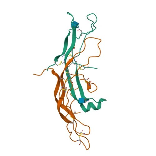
The 3D view shows the protein core and almost nothing else. It is PDB 1HCN at 2.6 angstroms, a recombinant selenomethionyl protein that had to be partially deglycosylated to crystallise, so a single N-acetylglucosamine residue stands in for eight glycans and beta residues 112 to 145 have no coordinates at all. The companion entry 1HRP, at 3.0 angstroms from independent crystals, models two GlcNAc residues and loses the same beta segment. The view is therefore a reliable picture of the fold, the knots and the seat belt, and silent about the part of the molecule that differs between preparations. A 1999 structure that caged the hormone between two antibody Fv fragments at 3.5 angstroms found the protein conformation close to that of the deglycosylated hormone, with neither antibody contacting the sugars, which is the evidence that removing the glycans does not greatly distort the scaffold being shown.[17,16,18,24]
Alpha chain (92 residues): APDVQDCPECTLQENPFFSQPGAPILQCMGCCFSRAYPTPLRSKKTMLVQKNVTSESTCCVAKSYNRVTVMGGFKVENHTACHCSTCYYHKS | Beta chain (145 residues): SKEPLRPRCRPINATLAVEKEGCPVCITVNTTICAGYCPTMTRVLQGVLPALPQVVCNYRDVRFESIRLPGCPRGVNPVVSYAVALSCQCALCRRSTTDCGGPKDHPLTCDDPRFQDSSSSKAPPPSLPSPSRLPGPSDTPILPQ
Modifications
- alpha chain, 5 disulfide bonds Cys7-Cys31, Cys10-Cys60, Cys28-Cys82, Cys32-Cys84 and Cys59-Cys87 in mature numbering (UniProt lists them as 31-55, 34-84, 52-106, 56-108 and 83-111 in precursor numbering). Three of them form the cystine knot at the core of the chain.[1,3]
- beta chain, 6 disulfide bonds Cys9-Cys57, Cys23-Cys72, Cys26-Cys110, Cys34-Cys88, Cys38-Cys90 and Cys93-Cys100 in mature numbering (UniProt precursor numbering: 29-77, 43-92, 46-130, 54-108, 58-110, 113-120). Three form the beta chain's own cystine knot. Cys26-Cys110 is the latch of the seat belt described below. The FDA substance record counts 11 disulfide links in the heterodimer, matching 5 plus 6.[2,3,15]
- alpha Asn52 and alpha Asn78 Two N-linked glycosylation sites on the alpha chain (precursor Asn76 and Asn102). On the free alpha chain the two sites are processed independently and differently: glycans at Asn52 were almost entirely concanavalin-A-unbound and trisialylated, while most Asn78 glycans were concanavalin-A-bound and disialylated.[1,35,15]
- beta Asn13 and beta Asn30 Two N-linked glycosylation sites on the beta chain (precursor Asn33 and Asn50). Hydrogen-deuterium exchange work found that the glycans at these two sites changed the affinity of one anti-hCG monoclonal antibody whose epitope maps to beta residues 65 to 83, while a second antibody was unaffected.[2,31,15]
- beta Ser121, Ser127, Ser132 and Ser138 Four O-linked glycosylation sites, all within the beta chain's C-terminal extension (precursor Ser141, 147, 152 and 158).[2,15]
- beta chain, residues 93 to 145 The beta C-terminal peptide, a 31-residue-plus proline-rich and serine-rich extension that the other three human glycoprotein hormone beta chains do not have, carrying all four O-glycans. It is not held by any disulfide to the rest of the molecule and is released by proteolysis at beta Leu92.[4,27,2]
Both chains are ordinary all-L polypeptides with no D-residues, no non-proteinogenic amino acids and no cyclisation of the backbone. The stereochemical complexity is in the eleven disulfide bonds and in the eight glycans: a glycosidic linkage carries its own anomeric configuration and branch point, and those are the part of the molecule that is not fixed by the gene. There is no metal coordination.[1,2,3]
What studies on hCG show
Each card names the experimental system and what was measured. A result in one system is not evidence about another.
X-ray crystallography · Crystals of deglycosylated human chorionic gonadotropin
Each subunit's three disulfides form a cystine knot, a motif shared with some protein growth factors, and a beta-chain segment wraps around the alpha chain tethered by the Cys26 to Cys110 disulfide, which the authors named the seat belt.[3]
- Material
- The intact heterodimer, deglycosylated
- Measured
- The three-dimensional fold of both chains and the inter-chain contacts
- Limitation
- Read here as an abstract only; resolution and refinement figures were taken from the PDB entry. Deglycosylated material, so the structure describes the protein core and not the glycoprotein.
Lapthorn AJ et al., 1994, Nature
X-ray crystallography with multiwavelength anomalous diffraction · Crystals of recombinant selenomethionyl hCG produced in mammalian cells and partially deglycosylated
The two chains have similar tertiary folds at about 10 percent sequence identity, the interface includes two inter-chain beta sheets and a disulfide-tethered beta-chain arm, and the O-glycan-rich beta C-terminal peptide is disordered. Segments of both chains previously implicated in receptor binding lie together on one face, with a glycosylation site implicated in signalling but not in binding nearby.[16]
- Material
- The intact heterodimer, selenomethionine substituted
- Measured
- Structure at 2.6 Å; subunit fold similarity; the extent and composition of the interface
- Compared with
- Other cystine-knot proteins, notably platelet-derived growth factor
- Limitation
- Abstract only. The protein was both selenomethionylated and partially deglycosylated, so neither the native methionines nor the native glycans are represented.
Wu H et al., 1994, Structure
Site-directed mutagenesis with receptor binding and cell-based readouts · Chinese hamster ovary cells expressing hCG glycosylation mutants; receptor binding, cAMP accumulation and steroidogenesis assays
Removing any or all N-linked glycans changed receptor affinity only slightly, and removing the beta glycans or alpha Asn78 alone did not change the cAMP or steroidogenesis readouts. Removing the alpha Asn52 glycan reduced both. Dimers combining a deglycosylated beta chain with an alpha chain lacking Asn52 still bound the receptor but raised neither readout, behaving as competitive antagonists in that assay.[23]
- Material
- hCG derivatives with individual or combined N-glycosylation sites removed
- Measured
- Receptor affinity, cAMP accumulation and steroidogenesis
- Compared with
- Fully glycosylated recombinant hCG
- Limitation
- Abstract only. This separates binding from the downstream readout in one recombinant cell system; it does not establish the mechanism of the separation, and the antagonism is a property of engineered derivatives in that assay.
Matzuk MM et al., 1989, Journal of Biological Chemistry
Subunit recombination with circular dichroism and NMR spectroscopy · Purified subunits recombined in solution
Recombination reached about 80 percent after 16 hours either way and the dissociation constant was below 1 x 10⁻⁵ per second in both cases, so the Asn52 glycan contributes little to holding the dimer together. The spectra showed a local conformational difference apparently involving beta-chain residues near that glycan.[25]
- Material
- Beta chain plus either the intact alpha chain or an alpha chain enzymically deglycosylated at Asn52 only
- Measured
- Recombination efficiency, dimer dissociation constant, CD and NMR spectra
- Compared with
- The glycosylated dimer
- Limitation
- Abstract only. The authors' proposal that the glycan is needed to stabilise a receptor-bound conformational change is an interpretation; no structure of either dimer bound to the receptor was determined.
Erbel PJ et al., 2002, Biochemical Journal
Single-particle cryo-electron microscopy · Purified full-length human luteinizing hormone and choriogonadotropin receptor in complex with heterotrimeric Gs
The authors describe a push-and-pull activation in which the hormone pushes the receptor's extracellular domain while an extended hinge loop pulls it, and identify a conserved ten-residue hinge fragment they call P10 that acts as a tethered agonist on the transmembrane domain. Org43553 occupies a transmembrane pocket and contacts P10.[5]
- Material
- Wild-type receptor in inactive and active states and a constitutively active mutant; active states bound to chorionic gonadotropin, one also with the small molecule Org43553
- Measured
- Four structures and the conformational changes between them
- Compared with
- The inactive-state receptor, and the other glycoprotein hormone receptors in the same study
- Limitation
- Abstract only; resolutions of 3.2 to 4.3 Å in the deposited entries, and the hormone's own beta C-terminal peptide is unmodelled in all of them. Detergent-solubilised, nanobody- and mutation-stabilised complexes.
Duan J et al., 2021, Nature
Antibody epitope accessibility mapping with molecular modelling · hCG bound to lutropin receptors, probed with monoclonal antibodies
The alpha chain's N-terminal region, the exposed face of its cystine knot and the outer parts of its first and third loops stayed accessible in the complex, so those surfaces are not buried in the interface. The authors combined this with beta-chain data and the deglycosylated crystal structure into a model of the interaction.[26]
- Material
- Intact hCG with defined alpha-chain epitopes
- Measured
- Which epitopes remained antibody-accessible while the hormone was receptor-bound
- Compared with
- The free hormone
- Limitation
- Abstract only. Epitope accessibility is indirect evidence about an interface, and the resulting model predates the cryo-EM structures that now show the complex directly.
Moyle WR et al., 1995, Journal of Biological Chemistry
Review of cAMP accumulation assays at a recombinant receptor · CHO cells expressing the recombinant human thyrotropin receptor
Purified LH was about ten times as potent as purified hCG at that receptor; a recombinant hCG lacking the beta C-terminal peptide was close to LH; nicked preparations gave roughly 1.5 to 2 fold more stimulation than intact hormone; and deglycosylation or desialylation raised the measured potency.[27]
- Material
- Purified hCG, purified human LH, a recombinant hCG lacking the beta C-terminal peptide, nicked hCG, and deglycosylated or desialylated hCG
- Measured
- cAMP accumulation at the thyrotropin receptor
- Compared with
- Intact purified hCG
- Limitation
- Abstract of a review, so the underlying assays were not read at source. Every figure is cAMP in a recombinant cell line at a receptor other than this hormone's own, and the comparisons are between purified laboratory preparations.
Yoshimura M et al., 1995, Thyroid
MALDI-TOF mass spectrometry with on-target reduction · Beta-core fragment purified from human pregnancy urine
The fragment is two disulfide-linked peptides, beta6 to beta40 and beta55 to beta92, only the first glycosylated. Intact spectra gave a broad triple peak at m/z 8700 to 11300; after reduction, a peak at m/z 4156.8 matched the bare beta55 to beta92 peptide and nine further peaks indicated glycoforms both smaller and larger than the trimannosyl core, with no completely unglycosylated beta6 to beta40 seen.[28]
- Material
- The terminal urinary degradation product of the hormone
- Measured
- Masses of the intact fragment and of its reduced constituent peptides
- Compared with
- The calculated mass of the unglycosylated beta55 to beta92 peptide
- Limitation
- Abstract only. Carbohydrate compositions were inferred by subtracting monosaccharide masses, not sequenced.
Jacoby ES et al., 2000, Clinical Chemistry
Site-directed mutagenesis and gene transfer with receptor binding, cell and animal assays · Chinese hamster ovary cells, including an O-glycosylation mutant line, with mouse LH and hCG receptor binding, in vitro steroidogenesis and a rat ovulation assay
Neither removing the beta O-glycans nor truncating the C-terminal extension made much difference to receptor binding or to signal transduction in vitro. In the rat assay the truncated derivative was about three-fold less active than wild type, which the authors read as the extension and its O-glycans being unimportant for binding and in vitro signalling but critical for the response in an animal.[29]
- Material
- Intact dimer lacking the beta-subunit O-linked oligosaccharides, and a dimer truncated at beta codon 115 so that beta residues 115 to 145 are absent
- Measured
- Receptor binding, cAMP accumulation and steroidogenesis in vitro, and ovulation in rats
- Compared with
- Wild-type recombinant hCG
- Limitation
- Read as an abstract only, and the record carries a 1990 erratum that was not read. Engineered derivatives in a recombinant cell line plus one rodent endpoint; the exposure conditions reported in the paper are deliberately not reproduced here.
Matzuk MM et al., 1990, Endocrinology
Chimeric gene construction with receptor binding, cell assays and circulating half-life measurement · Expressed chimeric gonadotropins; receptor binding and steroidogenesis assays, and circulating half-life measurement
Adding the C-terminal peptide did not significantly affect assembly with the alpha subunit or secretion of the dimer, and receptor binding and steroidogenic activity in vitro matched wild-type follitropin, while both the potency in an animal and the circulating half-life of dimers carrying one or two copies were raised. The authors state that this region of the chorionic gonadotropin beta subunit is important for maintaining the prolonged plasma half-life of the chorionic gonadotropin dimer.[6]
- Material
- Follitropin beta subunit carrying one or two copies of the chorionic gonadotropin beta C-terminal peptide, assembled with the common alpha subunit
- Measured
- Assembly and secretion, receptor binding, steroidogenic activity, potency in an animal and half-life in circulation
- Compared with
- Wild-type follitropin
- Limitation
- Read as an abstract only. The measurement is on a chimeric follitropin rather than on chorionic gonadotropin itself, so the inference to this hormone's own half-life is the authors' and rests on the transferred segment behaving the same way in both contexts.
Fares FA et al., 1992, Proceedings of the National Academy of Sciences of the United States of America
What the receptor structures show, and where the hormone sits in them
This hormone and luteinizing hormone act at the same receptor, the luteinizing hormone and choriogonadotropin receptor, a leucine-rich repeat G protein-coupled receptor with a large extracellular domain. Until 2021 the interaction was inferred rather than seen: the available evidence was which hormone surfaces stayed accessible to antibodies while the hormone was receptor-bound, combined with the deglycosylated crystal structure, which produced a model rather than coordinates.[26,3]
The cryo-EM study of full-length receptor complexes supplied the coordinates. In PDB 7FII the hormone is present as two chains, the alpha chain as label chain E and auth chain X and the beta chain as label chain F and auth chain Y, beside the receptor (auth chain R) and an engineered Gs heterotrimer. Reading the deposited model rather than the text: the alpha chain has no coordinates for its first four mature residues, the beta chain none for residues 132 to 165 in precursor numbering, and the receptor is missing residues 277 to 319 and 635 to 697. The authors describe activation as the extracellular domain being pushed by the bound hormone and pulled by an extended hinge loop, with a ten-residue hinge fragment acting as a tethered agonist on the transmembrane domain, and they place the small-molecule agonist Org43553 in a transmembrane pocket contacting that fragment. A separate study reported terphenyl derivatives as the first low molecular weight allosteric inhibitors of this receptor; its functional assay used LH, so it bears on the receptor rather than on this ligand.[19,5,30]
The beta C-terminal peptide is the reason this hormone outlasts luteinising hormone, and it does that without doing much at the receptor. Site-directed mutagenesis and gene transfer in Chinese hamster ovary cells produced a dimer with no beta O-glycans and a separate dimer truncated at beta codon 115, and neither change made much difference to receptor binding or to cAMP and steroidogenesis in vitro; in a rat ovulation assay the truncated derivative was about three-fold less active than the wild type, which the authors read as the extension and its O-glycans mattering for the response in an animal rather than at the receptor. The complementary experiment transferred the segment: fusing the chorionic gonadotropin beta C-terminal peptide onto the follitropin beta subunit left assembly, secretion, receptor binding and in vitro steroidogenic activity unchanged while raising both the circulating half-life and the in vivo potency of the resulting dimer, and the authors state the region is important for maintaining the prolonged plasma half-life of the chorionic gonadotropin dimer itself. So the segment that no deposited structure resolves is the one that accounts for the main functional difference from luteinising hormone.[29,6,27]
The glycan experiments complicate any simple mechanism statement. Removing N-linked glycans one at a time barely changed receptor affinity, yet removing the glycan at alpha Asn52 reduced the cAMP and steroidogenesis readouts, and a doubly deglycosylated derivative bound the receptor while raising neither, acting as a competitive antagonist in that assay. Later spectroscopic work found that same glycan contributes little to dimer stability but is associated with a local conformational difference near it. Binding and the downstream readout are separable in this system, and the carbohydrate is on the separating side of the line. No structure yet shows the glycans in a receptor complex, so the structural basis of that separation is unobserved.[23,25,19]
What is still unknown about hCG
The identity of a preparation specified only in International Units cannot be resolved from public records. The WHO instructions for the 6th International Standard put the limit of the unit plainly: the immunoassay value of 186 IU corresponds to 0.41 nmol per ampoule with an expanded uncertainty of 0.40 to 0.43 nmol, the approximate mass content was not experimentally derived and must not be used as a formal mass assignment, and calibrating in vitro cell-based assays with the material needs caution because sources may differ in post-translational modification. An older review notes that for the earlier 3rd and 4th standards 1 microgram of pure urinary hormone was taken as 9.3 IU; that equivalence belongs to those standards and those assays and cannot be carried across to arbitrary material.[7,4]
Glycoform is the live analytical question and sequence analysis cannot answer it. Hyperglycosylated material has the same two polypeptide chains, so distinguishing it requires glycan analysis or a glycan-sensitive antibody, and antibodies differ in whether they notice: hydrogen-deuterium exchange work found one monoclonal whose affinity shifted with the N-glycan population while a second was insensitive to it. That is also why an immunoassay figure and a bioassay figure for the same ampoule are not interchangeable.[21,31,7]
No deposited structure models the glycosylated molecule. Both 1994 crystal structures needed deglycosylated protein and between them model one and two N-acetylglucosamine residues against the eight glycans the molecule carries; the 2021 receptor complexes do not model the carbohydrate either, and the beta C-terminal peptide is absent from every one of those entries. So the segment that distinguishes this hormone's beta chain from the three related ones, and that carries all four O-glycans, has never been resolved in a deposited model.[17,18,19,24,16]
One smaller loose end survives. The deposited alpha-chain sequence of 1HRP reads Thr at position 4 where UniProt P01215 and the 1HCN deposition both read Val, and nothing in the entry metadata explains the difference.[18,17,1]
Key papers on hCG
- Crystal structure of human chorionic gonadotropin.
Lapthorn AJ, Harris DC, Littlejohn A, Lustbader JW, Canfield RE, Machin KJ, Morgan FJ, Isaacs NW (1994), Nature · X-ray crystallography
The paper that named the seat belt and the cystine knot. Two pages, and the vocabulary of the whole hormone family comes from it.
- Structure of human chorionic gonadotropin at 2.6 A resolution from MAD analysis of the selenomethionyl protein.
Wu H, Lustbader JW, Liu Y, Canfield RE, Hendrickson WA (1994), Structure · X-ray crystallography with MAD phasing
The companion 1994 structure, at 2.6 Å and the one displayed here. Read it for the interface description and for the explicit note that the beta C-terminal peptide is disordered.
- Structures of full-length glycoprotein hormone receptor signalling complexes.
Duan J, Xu P, Cheng X, Mao C, Croll T, He X, Shi J, Luan X, Yin W, You E, Liu Q, Zhang S, Jiang H, Zhang Y, Jiang Y, Xu HE (2021), Nature · Cryo-electron microscopy
The first structures of the full-length receptor with the hormone and Gs bound, which replaced three decades of inferred interaction models.
- Site specificity of the chorionic gonadotropin N-linked oligosaccharides in signal transduction.
Matzuk MM, Keene JL, Boime I (1989), Journal of Biological Chemistry · Site-directed mutagenesis in cell culture
The experiment that separated receptor binding from the downstream readout, and localised the effect to one glycan out of four.
- WHO International Standard, 6th International Standard for Chorionic Gonadotrophin, human, NIBSC code 18/244: Instructions for use (Version 4.0, 20/10/2025)
National Institute for Biological Standards and Control (2025), NIBSC, Medicines and Healthcare products Regulatory Agency · WHO reference standard documentation
Four pages that explain why an International Unit is not an amount of substance, including one ampoule carrying two different IU values.
- Insights into the hyperglycosylation of human chorionic gonadotropin revealed by glycomics analysis.
Ibeto L, Antonopoulos A, Grassi P, Pang PC, Panico M, Bobdiwala S, Al-Memar M, Davis P, Davis M, Norman Taylor J, Almeida P, Johnson MR, Harvey R, Bourne T, Seckl M, Clark G, Haslam SM, Dell A (2020), PLoS One · MALDI-TOF glycomics
A modern glycomic survey showing the glycan population and how its proportions move, which is the part of this molecule no sequence states.
- Human lutropin (hLH) and choriogonadotropin (CG) are assembled by different pathways: a model of hLH assembly.
Bernard MP, Lin W, Kholodovych V, Moyle WR (2014), Journal of Biological Chemistry · Mutational analysis of subunit assembly
Shows that this hormone and luteinizing hormone, which act at one receptor and share a chain, are assembled by different routes.
- hCG, the wonder of today's science
Cole LA (2012), Reproductive biology and endocrinology · Review
A short review of what hCG is biologically and of the five species usually distinguished in circulation, which is the orientation this page otherwise assumed.
References
- 1.UniProt Consortium. UniProtKB P01215 (GLHA_HUMAN): Glycoprotein hormones alpha chain, Homo sapiens. UniProt Knowledgebase. source · database record
- 2.UniProt Consortium. UniProtKB P0DN86 (CGB3_HUMAN): Choriogonadotropin subunit beta 3, Homo sapiens. UniProt Knowledgebase. source · database record
- 3.Lapthorn AJ, Harris DC, Littlejohn A, Lustbader JW, Canfield RE, Machin KJ, Morgan FJ, Isaacs NW. (1994). Crystal structure of human chorionic gonadotropin. Nature. 369:455-461. doi:10.1038/369455a0 · abstract only
- 4.Cole LA. (2009). New discoveries on the biology and detection of human chorionic gonadotropin. Reproductive Biology and Endocrinology. 7:8. doi:10.1186/1477-7827-7-8 · full text read
- 5.Duan J, Xu P, Cheng X, Mao C, Croll T, He X, Shi J, Luan X, Yin W, You E, Liu Q, Zhang S, Jiang H, Zhang Y, Jiang Y, Xu HE. (2021). Structures of full-length glycoprotein hormone receptor signalling complexes. Nature. 598:688-692. doi:10.1038/s41586-021-03924-2 · abstract only
- 6.Fares FA, Suganuma N, Nishimori K, LaPolt PS, Hsueh AJ, Boime I. (1992). Design of a long-acting follitropin agonist by fusing the C-terminal sequence of the chorionic gonadotropin beta subunit to the follitropin beta subunit. Proceedings of the National Academy of Sciences of the United States of America. 89:4304-8. doi:10.1073/pnas.89.10.4304 · abstract only
- 7.National Institute for Biological Standards and Control. (2025). WHO International Standard, 6th International Standard for Chorionic Gonadotrophin, human, NIBSC code 18/244: Instructions for use (Version 4.0, 20/10/2025). NIBSC, Medicines and Healthcare products Regulatory Agency. source · full text read
- 8.Cole LA. (2012). hCG, the wonder of today's science. Reproductive biology and endocrinology. 10:24. doi:10.1186/1477-7827-10-24 · abstract only
- 9.U.S. Food and Drug Administration. (2026). Chorionic gonadotropin for injection, prescribing information (BLA 017067). DailyMed / openFDA drug label. source · full text read
- 10.European Medicines Agency. (2026). Ovitrelle (choriogonadotropin alfa): EPAR. European Medicines Agency. source · full text read
- 11.Health Canada. (2026). Drug Product Database: products containing chorionic gonadotropin. Health Canada Drug Product Database. source · database record
- 12.World Anti-Doping Agency. (2025). World Anti-Doping Code International Standard: Prohibited List 2026. World Anti-Doping Agency. source · full text read
- 13.U.S. Food and Drug Administration. (2026). Bulk Drug Substances Nominated for Use in Compounding Under Section 503A of the Federal Food, Drug, and Cosmetic Act (updated May 14, 2026). U.S. Food and Drug Administration. source · full text read
- 14.U.S. Food and Drug Administration. (2026). Certain Bulk Drug Substances for Use in Compounding that May Present Significant Safety Risks (content current as of 22 April 2026). U.S. Food and Drug Administration, Human Drug Compounding. source · full text read
- 15.US Food and Drug Administration, Global Substance Registration System. Substance record: CHORIONIC GONADOTROPIN, UNII 20ED16GHEB. FDA GSRS. source · database record
- 16.Wu H, Lustbader JW, Liu Y, Canfield RE, Hendrickson WA. (1994). Structure of human chorionic gonadotropin at 2.6 A resolution from MAD analysis of the selenomethionyl protein. Structure. 2:545-558. doi:10.1016/s0969-2126(00)00054-x · abstract only
- 17.Wu H, Lustbader JW, Liu Y, Canfield RE, Hendrickson WA. (1994). RCSB PDB entry 1HCN: Structure of human chorionic gonadotropin at 2.6 angstroms resolution from MAD analysis of the selenomethionyl protein. RCSB Protein Data Bank. source · database record
- 18.Lapthorn AJ, Harris DC, Littlejohn A, Lustbader JW, Canfield RE, Machin KJ, Morgan FJ, Isaacs NW. (1994). RCSB PDB entry 1HRP: Crystal structure of human chorionic gonadotropin. RCSB Protein Data Bank. source · database record
- 19.Duan J, Xu P, Cheng X, Mao C, Croll T, He X, Shi J, Luan X, Yin W, You E, Liu Q, Zhang S, Jiang H, Zhang Y, Jiang Y, Xu HE. (2021). RCSB PDB entry 7FII: luteinizing hormone/choriogonadotropin receptor-chorionic gonadotropin-Gs complex. RCSB Protein Data Bank. source · database record
- 20.Larson SB, McPherson A. (2019). The crystal structure of the β subunit of luteinizing hormone and a model for the intact hormone. Current Research in Structural Biology. 1:1-5. doi:10.1016/j.crstbi.2019.07.001 · abstract only
- 21.Ibeto L, Antonopoulos A, Grassi P, Pang PC, Panico M, Bobdiwala S, Al-Memar M, Davis P, Davis M, Norman Taylor J, Almeida P, Johnson MR, Harvey R, Bourne T, Seckl M, Clark G, Haslam SM, Dell A. (2020). Insights into the hyperglycosylation of human chorionic gonadotropin revealed by glycomics analysis. PLoS One. 15:e0228507. doi:10.1371/journal.pone.0228507 · abstract only
- 22.US Food and Drug Administration, Global Substance Registration System. Substance record: CHORIOGONADOTROPIN ALFA, UNII 6413W06WR3. FDA GSRS. source · database record
- 23.Matzuk MM, Keene JL, Boime I. (1989). Site specificity of the chorionic gonadotropin N-linked oligosaccharides in signal transduction. Journal of Biological Chemistry. 264:2409-2414. PMID 2536708 · abstract only
- 24.Tegoni M, Spinelli S, Verhoeyen M, Davis P, Cambillau C. (1999). Crystal structure of a ternary complex between human chorionic gonadotropin (hCG) and two Fv fragments specific for the alpha and beta-subunits. Journal of Molecular Biology. 289:1375-1385. doi:10.1006/jmbi.1999.2845 · abstract only
- 25.Erbel PJ, Haseley SR, Kamerling JP, Vliegenthart JF. (2002). Studies on the relevance of the glycan at Asn-52 of the alpha-subunit of human chorionic gonadotropin in the alphabeta dimer. Biochemical Journal. 364:485-495. doi:10.1042/BJ20011482 · abstract only
- 26.Moyle WR, Campbell RK, Rao SN, Ayad NG, Bernard MP, Han Y, Wang Y. (1995). Model of human chorionic gonadotropin and lutropin receptor interaction that explains signal transduction of the glycoprotein hormones. Journal of Biological Chemistry. 270:20020-20031. doi:10.1074/jbc.270.34.20020 · abstract only
- 27.Yoshimura M, Hershman JM. (1995). Thyrotropic action of human chorionic gonadotropin. Thyroid. 5:425-434. doi:10.1089/thy.1995.5.425 · abstract only
- 28.Jacoby ES, Kicman AT, Laidler P, Iles RK. (2000). Determination of the glycoforms of human chorionic gonadotropin beta-core fragment by matrix-assisted laser desorption/ionization time-of-flight mass spectrometry. Clinical Chemistry. 46:1796-1803. PMID 11067815 · abstract only
- 29.Matzuk MM, Hsueh AJ, Lapolt P, Tsafriri A, Keene JL, Boime I. (1990). The biological role of the carboxyl-terminal extension of human chorionic gonadotropin [corrected] beta-subunit. Endocrinology. 126:376-83. doi:10.1210/endo-126-1-376 · abstract only · corrected: Erratum: Endocrinology 1990 Apr;126(4):2204. The erratum itself was not read.
- 30.Heitman LH, Narlawar R, de Vries H, Willemsen MN, Wolfram D, Brussee J, Ijzerman AP. (2009). Substituted terphenyl compounds as the first class of low molecular weight allosteric inhibitors of the luteinizing hormone receptor. Journal of Medicinal Chemistry. 52:2036-2042. doi:10.1021/jm801561h · abstract only · corrected: PubMed lists an erratum: J Med Chem. 2010 Jan 14;53(1):520. The erratum itself was not read, so what it corrects is unknown.
- 31.Li D, Zhang P, Li F, Chi L, Zhu D, Zhang Q, Chi L. (2015). Recognition of N-glycoforms in human chorionic gonadotropin by monoclonal antibodies and their interaction motifs. Journal of Biological Chemistry. 290:22715-22723. doi:10.1074/jbc.M115.657072 · abstract only
- 32.Bernard MP, Lin W, Kholodovych V, Moyle WR. (2014). Human lutropin (hLH) and choriogonadotropin (CG) are assembled by different pathways: a model of hLH assembly. Journal of Biological Chemistry. 289:14360-14369. doi:10.1074/jbc.M113.535609 · abstract only
- 33.ChEBI, European Bioinformatics Institute. ChEBI entries CHEBI:749511 (choriogonadotropin alfa) and CHEBI:749576 (gonadotropin, chorionic). Chemical Entities of Biological Interest. source · database record
- 34.Amoresano A, Siciliano R, Orrù S, Napoleoni R, Altarocca V, De Luca E, Sirna A, Pucci P. (1996). Structural characterisation of human recombinant glycohormones follitropin, lutropin and choriogonadotropin expressed in Chinese hamster ovary cells. European Journal of Biochemistry. 242:608-618. doi:10.1111/j.1432-1033.1996.0608r.x · abstract only
- 35.Bielinska M, Matzuk MM, Boime I. (1989). Site-specific processing of the N-linked oligosaccharides of the human chorionic gonadotropin alpha subunit. Journal of Biological Chemistry. 264:17113-17118. PMID 2477364 · abstract only
- 36.Duan J, Xu P, Cheng X, Mao C, Croll T, He X, Shi J, Luan X, Yin W, You E, Liu Q, Zhang S, Jiang H, Zhang Y, Jiang Y, Xu HE. (2021). RCSB PDB entry 7FIG: luteinizing hormone/choriogonadotropin receptor(S277I)-chorionic gonadotropin-Gs complex. RCSB Protein Data Bank. source · database record
- 37.Duan J, Xu P, Cheng X, Mao C, Croll T, He X, Shi J, Luan X, Yin W, You E, Liu Q, Zhang S, Jiang H, Zhang Y, Jiang Y, Xu HE. (2021). RCSB PDB entry 7FIH: luteinizing hormone/choriogonadotropin receptor(S277I)-chorionic gonadotropin-Gs-Org43553 complex. RCSB Protein Data Bank. source · database record
- 38.PubChem, National Center for Biotechnology Information. PUG-REST compound name lookup for "chorionic gonadotropin", and the property record of the compound it returns (CID 1108). PubChem. source · database record
Related entries
- GonadorelinGonadorelin is the synthetic form of GnRH, the hypothalamic decapeptide that releases luteinising hormone and follicle-stimulating hormone. It acts on gonadotroph cells of the anterior pituitary, is a ten-residue chain blocked at both ends, and is the natural agonist of the class A G protein-coupled GnRH receptor.
- SomatropinSomatropin is recombinant human growth hormone, originally produced in Escherichia coli: one chain of 191 amino acids matching the mature pituitary hormone. It is folded as a four-helix bundle and closed by two disulfide bonds.
