Gonadorelin in brief
Gonadorelin is the synthetic form of the mammalian hypothalamic decapeptide GnRH, pGlu-His-Trp-Ser-Tyr-Gly-Leu-Arg-Pro-Gly-NH2 (1182.3 g/mol), blocked by a pyroglutamate ring at one end and a glycinamide at the other, and the natural agonist of the GnRH receptor, which it binds at the cloned human receptor with a Ki of 4.8 nanomolar. Its sequence was established in 1971 and 1972 for the porcine and ovine peptides. Intermittent and continuous exposure have opposite consequences, an observation first made in lesioned monkeys and traced at the receptor to internalisation and down-regulation rather than to classical desensitisation. The molecule is short-lived, with a rapid-component plasma half-time of 7.8 minutes measured in healthy adults against 18.7 minutes for a position-6 D-tryptophan analogue, which is why the analogue family exists. Cryo-EM structures from 2025 show it bound in a U shape to pig and frog receptors, but no human receptor complex has been reported, and its free conformation in water is unresolved. The material is sold as the acetate, a salt both pharmacopoeias write with an unspecified number of equivalents and bound by an acetic-acid and water range instead.[1,2,3,4,5,6,7,8]
Gonadorelin regulatory status
In the United States, Drugs@FDA lists two gonadorelin applications, a hydrochloride approved in 1982 and an acetate approved in 1989, with every product under both now discontinued; a Phase 3 trial of gonadorelin acetate in primary amenorrhoea with hypogonadotropic hypogonadism was completed in 2018 (NCT01976728). No centrally authorised EU medicine was found in the EMA medicines dataset, and in the Health Canada Drug Product Database every human gonadorelin product is cancelled except one acetate product, authorised for induction of ovulation in primary hypothalamic amenorrhoea, that is listed as dormant, while several veterinary gonadorelin products are marketed (both searched November 2026). The WADA 2026 Prohibited List names gonadorelin under S2.2.1, peptide hormones and releasing factors prohibited at all times and applying to males. Gonadorelin appears on neither FDA compounding list: it is in no category of the list of bulk drug substances nominated under section 503A (updated 14 May 2026), and it is in neither the category 2 table nor the table of withdrawn nominations on the page of bulk substances that may present significant safety risks under the 503A and 503B interim policies (content current as of 22 April 2026). It carries pharmacopoeial monographs on both sides of the Atlantic, Gonadorelini acetas in the European Pharmacopoeia and Gonadorelin Acetate in the USP, whose labelling section directs that the material be marked for veterinary use only. No retraction or expression of concern was found for any study cited here; the 1990 erratum attached to R13 is recorded with that reference.[9,10,11,12,13,14,15,8,16,17,18]
Gonadorelin at a glance
- A ten-residue peptide with both termini chemically blocked: a pyroglutamate ring at position 1 in place of a free amine, and a glycinamide at position 10 in place of a free carboxylate.[19,20,21]
- What it does is release gonadotropins. It binds the GnRH receptor on gonadotroph cells of the anterior pituitary, a seven-transmembrane receptor that couples predominantly through Gq, and that signal releases luteinising hormone and follicle-stimulating hormone. Intermittent and continuous exposure give opposite outcomes, which is the single most consequential fact about the molecule.[22,23,24,6]
- Measured affinity at the cloned human type I receptor was a Ki of 4.8 nanomolar in competition binding, and 4.1 nanomolar at the cloned chicken receptor in the same study; at the cloned sea lamprey receptor it was 12.9 nanomolar, well behind that receptor's own preferred ligand.[3,25]
- Cryo-EM structures published in 2025 show the decapeptide bound to pig and frog receptors in a U shape, gripping the pocket by both ends while the middle points outward, unlike most peptide complexes of G protein-coupled receptors. No structure of the human receptor with the peptide bound has been reported.[4,26,27]
- Status: approved in the United States as a hydrochloride in 1982 and an acetate in 1989, with every product under both applications now discontinued; one Canadian human acetate product remains authorised but dormant while veterinary products are marketed; and the substance has its own monographs in the European Pharmacopoeia and the USP, the USP one directing that the label state veterinary use only. The 2026 WADA Prohibited List names it under S2.2.1, prohibited at all times and applying to males.[9,10,13,8,16,15]
- It does not last. Measured in eight healthy adults with a specific radioimmunoassay, plasma clearance was biphasic and the half-time of the rapid component was 7.8 plus or minus 1.1 minutes, against 18.7 plus or minus 1.8 minutes for the position-6 D-tryptophan analogue measured alongside it, with metabolic clearance rates of 1766.6 plus or minus 404.3 and 503.4 plus or minus 196.4 mL/min. That difference is the reason the analogue family exists.[7]
What is Gonadorelin?
Gonadorelin is the synthetic form of the hypothalamic decapeptide that controls gonadotropin release from the anterior pituitary. It was isolated from porcine hypothalamic material, found to be ten amino acids long and then synthesised. Its sequence was proposed for the porcine peptide in 1971 and independently established for the ovine peptide in 1972, by enzymatic digestion, Edman-dansylation sequencing and mass spectrometry of the derived phenylthiohydantoins, which found the two species' peptides identical.[28,1,2]
The molecule is not a free gene product. The human GNRH1 gene encodes a 92-residue precursor, progonadoliberin-1: a 23-residue signal sequence, the decapeptide at residues 24 to 33, a Gly-Lys-Arg triplet, and a second peptide, GnRH-associated peptide 1, at residues 37 to 92. That arrangement is the one expected for proteolytic release and C-terminal amidation, which is where the glycinamide comes from.[29,30]
Its receptor was identified by cDNA cloning in 1992, first from mouse and then from human tissue, as a seven-transmembrane G protein-coupled receptor that binds the decapeptide with high affinity when expressed and that lacks the cytoplasmic C-terminal tail most class A receptors carry. The human receptor is 328 residues long and ends immediately after transmembrane helix 7. It couples predominantly through Gq.[22,23,31,24]
Names are a recurring source of confusion here. The same molecule is GnRH, GnRH-I, LHRH, LRF, luliberin and gonadoliberin I, and it is listed by the supplier as the acetate. It is also the parent of a large analogue family filed under the same heading: leuprolide, triptorelin, buserelin, nafarelin and goserelin each substitute the glycine at position 6 with a D-residue and most alter the C terminus too, and the small-molecule antagonists at the same receptor share no chemistry with it.[21,20,4,32,33,34]
It is also a regulated drug substance with an unusual split between human and veterinary records. Drugs@FDA holds two applications, a hydrochloride approved in 1982 and an acetate approved in 1989, and every product under both is now discontinued; the Health Canada database carries one human acetate product that is authorised but dormant alongside marketed veterinary products; and both the European Pharmacopoeia and the USP carry monographs for the acetate, the USP monograph instructing that the label state veterinary use only. The European monograph adds two facts that bear on identity: the substance is obtained by chemical synthesis, and the label states the mass of peptide in the container rather than the gross mass of the salt. None of the analogues named above has an entry in this library.[9,10,13,8,16]
Not to be confused with
- Leuprolide (leuprorelin)
- A nine-residue analogue with an ethylamide cap. Its deposited sequence reads pGlu-His-Trp-Ser-Tyr-D-Leu-Leu-Arg-Pro-NHEt: D-leucine replaces the glycine at position 6 and the Pro9-Gly10-amide pair is replaced by a prolyl ethylamide, so it is two chemical changes away from gonadorelin rather than a formulation of it. The two sit side by side as separate depositions in one NMR series, 1YY1 for the decapeptide and 1YY2 for leuprolide.[5,32]
- Triptorelin
- The same decapeptide with D-tryptophan at position 6. That one substitution is the whole subject of the conformational argument in this family, and it is the molecule behind the only atomic-resolution crystal structure here (0.85 angstrom), which is often reproduced as though it were a structure of gonadorelin.[33,4]
- GnRH II
- A separate vertebrate decapeptide, [His5,Trp7,Tyr8]GnRH, conserved in higher vertebrates including humans. Three residues differ. At the cloned human type I receptor it is a much weaker ligand than gonadorelin, while at the chicken receptor it bound with a Ki of 0.6 nanomolar against 4.8 nanomolar for gonadorelin at the human receptor; the human gene for its own cognate receptor carries a frame shift and a stop codon.[3,24]
- Chicken GnRH and the lamprey GnRH forms
- Further natural variants of the same ten-position scaffold: chicken GnRH is [Gln8]GnRH, and lamprey GnRH-I and GnRH-III are more distant. At the cloned lamprey receptor, lamprey GnRH-III bound with a Ki of 0.708 nanomolar and the mammalian decapeptide with 12.9 nanomolar, so the scaffold is shared but the molecules and their receptor preferences are not interchangeable. Twenty-three structural variants of GnRH have been catalogued across protochordates and vertebrates.[3,25,24]
- Progonadoliberin-1 and GnRH-associated peptide
- The decapeptide is encoded inside a 92-residue human precursor. After a 23-residue signal sequence, residues 24 to 33 are the decapeptide and residues 37 to 92 are a second peptide, GnRH-associated peptide 1. A reference to the GNRH1 gene product is therefore not a reference to this molecule.[30,29]
- Small-molecule GnRH receptor antagonists
- Elagolix, relugolix, linzagolix and related compounds are not peptides at all and occupy a partly overlapping but differently placed pocket in the same receptor, closer to transmembrane helices 5 to 7, leaving room for the receptor's own N terminus to stay buried. They are frequently filed under the same receptor heading as gonadorelin and share no chemistry with it.[34,4]
Gonadorelin structure and sequence
The backbone is unremarkable: ten residues, all L apart from the two achiral glycines, with no cysteine, no disulfide, no cyclisation of the chain, no sugar, lipid or metal. What distinguishes it is the two ends. Position 1 is pyroglutamate, a glutamate whose side-chain carbonyl has closed onto its own alpha-amino group to give a 5-oxoproline ring, so there is no free N-terminal amine; position 10 is a glycine amide rather than a free acid. Reconstructing the formula residue by residue reproduces PubChem's C55H75N17O13 and 1182.3 exactly, while the plain ten-letter chain as a free acid would be C55H77N17O14: one reason database records here do not all agree.[35,21,20]
The blocked termini were inferred before the sequence was known. Chymotrypsin, subtilisin, papain and thermolysin abolished the activity of the porcine preparation while pepsin, trypsin and a panel of exopeptidases did not, and a purified pyrrolidone carboxylyl peptidase did: a profile fitting a peptide with both ends capped and aromatic residues in the middle. Chemical modification of the tyrosine, histidine, tryptophan or arginine also abolished activity. The ring is a conformational element as well as a cap: force-field calculations on position-2 and position-3 analogues found every low-energy conformer sharing a directional change at its cis peptide bond.[19,36,37,4]
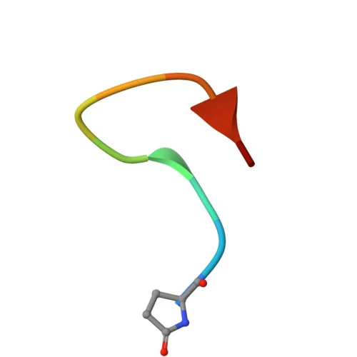
The three-dimensional view on this page is the deposited NMR ensemble of the peptide on its own, in DMSO: 21 conformers restrained by 138 NOE-derived distances. It shows the U or hairpin-like fold this family is known for, and should be read as a set of shapes consistent with a small restraint set rather than one determined structure. It is not aqueous, and a free-peptide shape is a different question from the bound one, which the 2025 receptor complexes answer instead.[5,38,4,33]
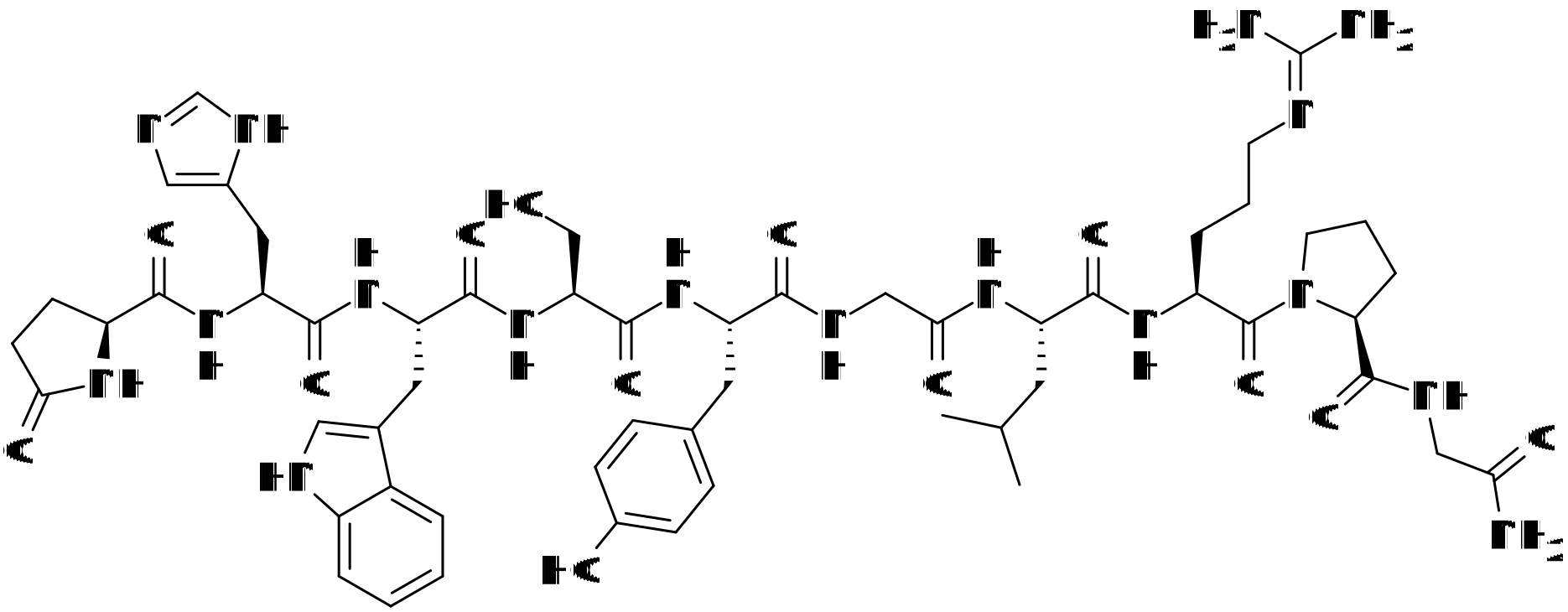
C-terminus: NH2
- pGlu1
- His2
- Trp3
- Ser4
- Tyr5
- Gly6
- Leu7
- Arg8
- Pro9
- Gly10
Modifications
- 1 N-terminal pyroglutamate: the glutamate side chain is cyclised onto its own alpha-amino group as a 5-oxoproline ring, so there is no free N-terminal amine. Chemical component PCA in the deposited structures, and recorded as pidolic acid on the FDA substance record.[5,20,21]
- 10 C-terminal glycinamide in place of a free carboxylate. In the precursor the decapeptide is followed by Gly-Lys-Arg, the arrangement expected for proteolytic release and amidation of the C terminus, so the amide is the product of precursor processing rather than a synthetic cap added later.[29,30,20]
Every chiral residue is the L enantiomer, including the pyroglutamate at position 1; the two glycines, at positions 6 and 10, are achiral. There is no cysteine and therefore no disulfide, no cyclisation of the backbone, no glycosylation, no lipid arm and no metal coordination: the only ring closure in the molecule is the pyroglutamate side chain onto its own amino group. The absence of a D-residue is the chemical line between gonadorelin and the analogue family built on it, which substitutes position 6 with a D-amino acid.[21,20,5,4]
What studies on Gonadorelin show
Each card names the experimental system and what was measured. A result in one system is not evidence about another.
Single-particle cryo-EM with site-directed mutagenesis and inositol monophosphate accumulation assays · Purified complexes of Sus scrofa and Xenopus laevis GnRH receptors with an engineered mini-Gq chimera, G beta 1, G gamma 2 and the scFv16 fiducial, expressed in High Five insect cells; separately, human receptor point mutants assayed in cells for IP1 accumulation
The peptide binds as a U, contacting the receptor through both its N- and C-terminal ends while the middle of the chain points into the extracellular space, which the authors contrast with the usual peptide-GPCR arrangement. pGlu1 contacts the residues at positions 1.35, 3.32, 6.51 and 7.39; His2 contacts 3.36, 5.42, 6.51 and 6.52; Trp3 contacts 6.51, 6.59 and 7.39. The transmembrane domains of the two complexes superimpose to 0.89 angstrom. Against the inactive human receptor, the receptor's own N terminus swings out of the pocket and lies over the ligand as a lid, transmembrane helices 1 and 7 move inward by about 4.0 angstrom, helix 2 outward by 3.3 angstrom and helix 5 by 9.1 angstrom. His2 does not touch the conserved toggle residue at 6.48 directly; it shifts the tyrosines at 6.51 and 6.52, which rotates the toggle. Alanine substitution at 6.51, 6.52 and 7.39 reduced the peptide's potency in the IP1 assay.[4]
- Material
- The unmodified decapeptide, stated to be identical in human, pig and frog, against receptors carrying an N-terminal prolactin-precursor and FLAG extension
- Measured
- Structures at 3.18 angstrom (pig) and 2.67 angstrom (frog); the residue-by-residue contact set in the orthosteric pocket; helix displacements between active and inactive states; and the change in potency (delta pEC50) for IP1 accumulation at human receptor mutants relative to wild type
- Compared with
- The antagonist-bound inactive human receptor (PDB 7BR3); wild-type receptors for the mutagenesis; the triptorelin crystal structure (PDB 4D5M) for the turn comparison
- Limitation
- No structure of the human receptor with the peptide bound; the two receptors are pig and frog, both as fusion constructs, and intracellular loops 1 and 3 of the pig receptor and loop 3 of the frog receptor were unresolved. Every functional reading is IP1 accumulation in a recombinant cell system, so the work establishes nothing about arrestin recruitment, receptor trafficking or pathway bias. The analogue binding poses in the same paper are modelling and docking rather than structures. One laboratory, one deposition set, so the contacts are unreplicated.
Shen S et al., 2025, Proceedings of the National Academy of Sciences of the United States of America
X-ray crystallography with functional assays and docking · Crystallised human GnRH1R
The receptor's own N terminus co-occupies an enlarged orthosteric site alongside the antagonist, and the receptor lacks the cytoplasmic C-terminal helix carried by most class A receptors, giving it different microswitch features. This is the inactive-state reference against which the peptide-bound structures were later compared, and it is where the lid behaviour of the N terminus comes from.[34]
- Material
- Human receptor with the small-molecule antagonist elagolix; the peptide is not present
- Measured
- Structure at 2.8 angstrom resolution and the occupancy of the orthosteric site
- Compared with
- Other class A G protein-coupled receptors, for the microswitch comparison
- Limitation
- Read as an abstract only. The compound is absent from this structure: it constrains the pocket the peptide has to enter rather than showing the peptide in it, and it is an inactive-state crystal rather than an agonist complex.
Yan W et al., 2020, Nature Communications
Site-directed mutagenesis with radioligand binding and phosphatidylinositol hydrolysis assays · Expressed receptor, with receptor density varied by irreversible antagonism to separate affinity from receptor reserve
Arginine at position 121 preserved high-affinity agonist binding, glutamine dropped binding below detection and raised the EC50 to 282 plus or minus 52 nanomolar against the 2.4 plus or minus 0.2 nanomolar maximum obtained by thinning out wild-type receptor, which the authors read as a loss of agonist affinity greater than 100-fold; leucine and aspartate abolished both binding and signal. Antagonist affinity was comparable at wild type and at both the Arg121 and Gln121 mutants. The conclusion, that a charge-strengthened hydrogen bond donor is needed at this position for agonist but not antagonist binding, anticipated by thirty years the 3.32 contact with pGlu1 that the cryo-EM maps show.[39]
- Material
- Wild-type receptor and Lys121 replaced by Arg, Gln, Leu or Asp
- Measured
- Agonist and antagonist binding, and EC50 for phosphatidylinositol hydrolysis
- Compared with
- Wild type at reduced receptor density; an antagonist at each mutant
- Limitation
- Read as an abstract only. Mutagenesis infers a contact rather than showing one, and a single substitution can act through folding or expression as well as through binding; the structural confirmation came separately.
Zhou W et al., 1995, The Journal of Biological Chemistry
Competition radioligand binding and functional assays on cloned receptors, with a receptor chimera · Cloned chicken pituitary GnRH receptor and the cloned human receptor
The decapeptide bound both receptors comparably, Ki 4.8 plus or minus 1.2 nanomolar at the human receptor and 4.1 plus or minus 1.2 nanomolar at the chicken receptor, while the chicken receptor additionally bound chicken GnRH (5.3 plus or minus 0.5 nanomolar) and GnRH II (0.6 plus or minus 0.01 nanomolar) with high affinity and the human receptor did not. Three antagonists carrying D-Lys6 or D-isopropyl-Lys6 were pure antagonists at the human receptor but agonists at the chicken one, and transplanting extracellular loop two into the human receptor transferred that behaviour. This is the clearest published figure for the compound's affinity at the human receptor.[3]
- Material
- The decapeptide, chicken GnRH ([Gln8]GnRH), GnRH II ([His5,Trp7,Tyr8]GnRH) and six antagonists, against wild-type receptors and a human receptor carrying the chicken extracellular loop two
- Measured
- Ki in competition binding, and whether each ligand behaved as agonist or antagonist
- Compared with
- The two receptors against each other, and the loop-swap chimera against the parent human receptor
- Limitation
- Read as an abstract only. Affinity was measured by displacement of a radiolabelled analogue rather than of the compound itself, and the agonist-versus-antagonist switch concerns the analogues, not gonadorelin.
Sun YM et al., 2001, The Journal of Biological Chemistry
Saturation and intact-cell competition binding, cAMP accumulation and internalisation assays on a cloned receptor and three C-terminal truncations · Cloned sea lamprey (Petromyzon marinus) type II GnRH receptor, which carries an unusually long 120-residue cytoplasmic tail, plus 80, 40 and 0 residue truncations of it
The receptor preferred lamprey GnRH-III (Ki 0.708 plus or minus 0.245 nanomolar) and chicken GnRH-II (0.765 plus or minus 0.160 nanomolar) over the mammalian decapeptide (12.9 plus or minus 1.96 nanomolar) and lamprey GnRH-I (118 plus or minus 23.6 nanomolar), so the compound is a comparatively weak ligand at this ancestral receptor. The membrane-proximal 40 residues of the tail were required for cAMP accumulation, and the tail-less receptor internalised much less.[25]
- Material
- Lamprey GnRH-I, lamprey GnRH-III, chicken GnRH-II and the mammalian decapeptide
- Measured
- Ki in intact-cell competition binding, cAMP accumulation, and ligand-dependent internalisation
- Compared with
- The four peptides against one another; the truncations against the full-length receptor
- Limitation
- Read as an abstract only. A non-mammalian receptor; it bounds how receptor-specific the compound's affinity is and says nothing about the human receptor. The cAMP readout is this receptor's, not a claim about mammalian coupling.
Silver MR et al., 2006, Journal of Molecular Endocrinology
Endocrine experiment in lesioned animals · Rhesus monkeys carrying hypothalamic lesions that had abolished gonadotropic hormone release by the pituitary
Continuous exposure failed to restore sustained gonadotropin secretion; intermittent exposure matching the animals' physiological rhythm re-established it. The authors state the effect is attributable to the pattern of exposure rather than to the quantity the pituitary cells were exposed to, and that beginning continuous exposure in an animal already restored by intermittent exposure can produce desensitisation or down-regulation of the machinery responsible for gonadotropin release.[6]
- Material
- The synthetic decapeptide, presented either continuously or intermittently at the frequency of the animals' own gonadotropin rhythm
- Measured
- Pituitary gonadotropin secretion
- Compared with
- Continuous against intermittent exposure in the same lesioned animals, and the switch from one to the other
- Limitation
- Read as an abstract only. Lesioned rhesus monkeys, a deliberately artificial preparation chosen to remove the animals' own hypothalamic input, and the readout is a circulating hormone concentration rather than anything measured at the receptor. The exposure conditions reported in the paper are deliberately not reproduced here.
Belchetz PE et al., 1978, Science (New York, N.Y.)
Plasma clearance measurement with specific radioimmunoassay · Eight healthy adults
Clearance of both peptides was biphasic, with a rapid and a slow component. The half-time of the rapid component was 7.8 plus or minus 1.1 minutes for the decapeptide and 18.7 plus or minus 1.8 minutes for the D-tryptophan analogue, a difference significant at p less than 0.001, and the metabolic clearance rates were 1766.6 plus or minus 404.3 and 503.4 plus or minus 196.4 mL/min. Chromatography of plasma from one subject given the analogue alone gave a single peak coeluting with the intact analogue. The authors attribute the analogue's longer half-time to a slower rate of degradation.[7]
- Material
- The decapeptide and the position-6 D-tryptophan analogue, each quantified by its own radioimmunoassay that did not cross-react with the other peptide or with degradation fragments
- Measured
- Plasma half-disappearance time and metabolic clearance rate
- Compared with
- The two peptides against one another in the same subjects
- Limitation
- Read as an abstract only, and a 1982 radioimmunoassay study. It measures a property of the molecule in people and says nothing about the receptor; the exposure conditions reported in the paper are deliberately not reproduced here. A separately compiled database record gives a wider range, an initial half-life of 2 to 10 minutes and a terminal one of 10 to 40 minutes.
Barron JL et al., 1982, The Journal of clinical endocrinology and metabolism
Automated imaging of receptor expression and trafficking · HeLa cells expressing haemagglutinin-tagged human, mouse and Xenopus GnRH receptors and chimeras, by adenoviral transduction
Agonist exposure drove down-regulation or internalisation of mouse and Xenopus receptors. Human receptors were mostly intracellular, and the agonist drove their internalisation or down-regulation only once surface expression was raised by adding the Xenopus C-terminal tail or by a chaperone. The effects were not reproduced by a peptide antagonist and were abolished by a mutation that prevents receptor signalling, so they depend on signalling as well as on occupancy.[40]
- Material
- Tagged receptors, including human receptors carrying an added Xenopus C-terminal tail
- Measured
- Receptor expression and agonist-driven internalisation and down-regulation
- Compared with
- Agonist against a peptide antagonist; a signalling-dead receptor mutant; receptors with and without a pharmacological chaperone
- Limitation
- Read as an abstract only. HeLa cells rather than gonadotrophs, with receptors introduced by transduction and carrying an epitope tag, and the human result required an engineered tail or a chaperone to be visible at all.
Finch AR et al., 2009, American journal of physiology. Cell physiology
Enzyme-inhibition experiment with plasma gonadotropin measurement in rats · Rat, in vivo
The study names the Tyr5-Gly6 bond as the bond this enzyme cleaves in the decapeptide, which is a chemical fact about the molecule: the scissile bond sits in the middle of the chain, at the position the analogue family substitutes. With the enzyme inhibited, the measured plasma concentrations at two and four hours were about an order of magnitude above control and comparable to those obtained with the enzyme-resistant analogues, and the authors attribute the analogues' greater activity in that system largely to that resistance.[41]
- Material
- The native decapeptide, with and without two inhibitors of endopeptidase 24.15
- Measured
- Plasma luteinizing hormone and follicle-stimulating hormone concentrations at two and four hours
- Compared with
- Two D-amino-acid analogues at position 6 that resist the enzyme, and untreated controls
- Limitation
- Read as an abstract only, and the record carries an erratum that was not read. A measured endpoint in rats, which says nothing about any other species, and the readout is a gonadotropin concentration rather than anything about the peptide's structure. The exposure conditions reported in the paper are deliberately not reproduced here.
Lasdun A et al., 1990, The Journal of Pharmacology and Experimental Therapeutics
Enzyme purification with substrate and inhibition profiling · Pyroglutamyl aminopeptidase type II purified about 3000-fold from the membrane fraction of bovine brain
The enzyme removes N-terminal pyroglutamate only from tri- and tetrapeptides carrying histidine in the penultimate position. The decapeptide and its 1-5 fragment were not substrates but were competitive inhibitors, with Ki values of 8 and 11 micromolar. So sharing the pGlu-His opening with thyrotropin-releasing hormone does not make the decapeptide's pyroglutamate removable by this activity: chain length, not the motif, decides.[42]
- Material
- The decapeptide and its 1-5 fragment, tested against the enzyme alongside tripeptide and tetrapeptide substrates
- Measured
- Whether each peptide was hydrolysed, and Ki for those that inhibited
- Compared with
- Thyrotropin-releasing hormone and related short pGlu-His peptides, which are substrates
- Limitation
- Read as an abstract only. One enzyme from one tissue of one species; it says nothing about other pyroglutamyl peptidases or about stability in any other setting.
Gallagher SP et al., 1998, The International Journal of Biochemistry and Cell Biology
Two-dimensional NMR in two solvents with molecular dynamics, plus a cell assay on the two linear analogues · DMSO-d6 and D2O solutions; MCF-7 cells for the two analogues
NOE data indicated a type I beta-turn over residues 2 to 5 in DMSO-d6 and a type II beta-turn over residues 3 to 6 in D2O for the same molecules. Within this chemical family, therefore, the reported turn depends on the solvent, which is the direct reason the DMSO ensemble shown on this page cannot be read as the peptide's shape in water.[38]
- Material
- Leuprolide and two further substituted analogues, not the unmodified decapeptide
- Measured
- NOE-derived turn type and position
- Compared with
- The same molecules in the two solvents
- Limitation
- Read as an abstract only, and the molecules are analogues rather than gonadorelin itself, so it bears on this page only through the solvent-dependence argument. No aqueous NMR ensemble of the unmodified decapeptide was located in this survey.
Laimou DK et al., 2010, Amino Acids
How gonadorelin is held by the GnRH receptor
The receptor is unusual in two ways that both bear on how the peptide is held. It has no cytoplasmic C-terminal tail, stopping immediately after transmembrane helix 7. And its own N terminus is part of the binding site, reaching into an enlarged orthosteric pocket and sharing it with the small molecule in the antagonist-bound human crystal structure.[31,24,34]
The 2025 cryo-EM complexes show what happens when the peptide occupies that pocket instead. The decapeptide binds as a U, anchored by both ends with the middle projecting outward, and the receptor N terminus rotates out to lie over it as a lid. The contacts are listed with the study below; what explains activation is the relay. His2 does not touch the conserved toggle residue at 6.48: it moves the tyrosines at 6.51 and 6.52, and they rotate the toggle, after which helix 6 swings out and the microswitch motifs rearrange. Alanine substitution at 6.51, 6.52 and 7.39 cut the peptide's potency in an inositol monophosphate assay on the human receptor.[4,26,27]
Mutagenesis reached the same position from the other direction long before. Replacing Lys121, the residue at 3.32, with glutamine left antagonist affinity intact while costing more than a hundredfold in agonist affinity, and leucine or aspartate there abolished binding and signalling together: a charge-strengthened hydrogen bond donor is required at that locus for agonist binding specifically. That is a pharmacological prediction of the contact the maps now show, and the agreement is the strongest part of the mechanistic case. What none of it addresses is anything downstream of the first messenger: every functional reading cited is a second-messenger accumulation assay in a recombinant system.[39,4]
Pattern, not amount, decides what the receptor does next. In rhesus monkeys whose hypothalamic lesions had abolished gonadotropin release, continuous exposure to the decapeptide failed to restore sustained secretion while intermittent exposure at the animals' own physiological rhythm of gonadotropin release re-established it, and the authors attributed the difference to the pattern of exposure rather than to the quantity the pituitary cells saw. Switching an animal already restored by intermittent exposure to continuous exposure produced what they called desensitisation or down-regulation. At the receptor level the mechanism is not classical desensitisation: type I mammalian GnRH receptors, which are the ones lacking the cytoplasmic C-terminal tail, do not desensitise, and an imaging study of tagged human, mouse and Xenopus receptors in HeLa cells found agonist-driven internalisation and down-regulation instead, dependent on receptor signalling as well as on agonist occupancy and not reproduced by a peptide antagonist. The same study found human receptors to be mostly intracellular, and only showed agonist-driven internalisation of the human receptor once its surface expression had been raised by adding the Xenopus C-terminal tail or by a pharmacological chaperone.[6,40,31,24]
What is still unknown about Gonadorelin
The counter-ion question has an answer, and the answer is that there is no fixed ratio. Both pharmacopoeias write the acetate with an unspecified number of equivalents and bound the composition instead: 4.0 to 7.5 per cent acetic acid and not more than 7.0 per cent water in Europe, not less than 80 per cent peptide by weight in the USP, which is roughly 0.8 to 1.7 acetate equivalents per peptide. What a given lot carries inside those ranges, and therefore how much of a weighed mass is peptide, is a property of that lot rather than of the name, and the substance is recorded as extremely hygroscopic.[8,16,43]
There is no structure of the human receptor with this peptide in it. The 2025 depositions are a pig receptor at 3.18 angstrom and a Xenopus laevis receptor at 2.67 angstrom, both engineered with an N-terminal fusion and coupled to a chimeric mini-Gq; the one human entry, PDB 7BR3, holds an antagonist and no peptide. The pig receptor is 91.2 per cent identical to the human one and the key interaction residues are reported completely conserved across an alignment of eleven mammalian species, so the inference to the human receptor is strong. It is still an inference, from a single laboratory's deposition set with no independent replication.[4,26,27,44]
The free peptide's conformation in water is genuinely open. The one coordinate set of the molecule on its own is an NMR ensemble in DMSO with an unpublished primary citation, work on close analogues shows the turn type changing between DMSO and water, and PubChem holds no computed conformer. The bound U shape is well evidenced; the unbound shape is not.[5,38,35]
Chemical lability is documented only indirectly. The bovine brain pyroglutamyl aminopeptidase that removes pyroglutamate from short pGlu-His peptides does not do so here, treating the decapeptide as a micromolar competitive inhibitor instead. The cleavage named in the literature is at the Tyr5-Gly6 bond, by endopeptidase 24.15, exactly where the analogue family inserts a D-residue. Beyond that, this survey located no published forced-degradation study, impurity profile or stability-indicating method for gonadorelin itself; the comparable work is on the analogues and the antagonists and was not borrowed.[42,41,19]
Key papers on Gonadorelin
- Cryo-EM structures of GnRHR: Foundations for next-generation therapeutics.
Shen S, He X, Liu H, Hu W, Xu HE, Duan J (2025), Proceedings of the National Academy of Sciences of the United States of America · Cryo-EM structures with mutagenesis and IP1 assays
The only structural account of this peptide bound to its receptor, with the mutagenesis and second-messenger assays that test the contacts it shows. Open access, and the single most informative paper on the molecule.
- Primary structure of the ovine hypothalamic luteinizing hormone-releasing factor (LRF) (LH-hypothalamus-LRF-gas chromatography-mass spectrometry-decapeptide-Edman degradation).
Burgus R, Butcher M, Amoss M, Ling N, Monahan M, Rivier J, Fellows R, Blackwell R, Vale W, Guillemin R (1972), Proceedings of the National Academy of Sciences of the United States of America · Primary structure determination
Where the sequence comes from. Degradative sequencing plus mass spectrometry on the ovine peptide, with a synthetic decapeptide sequenced alongside it, and the finding that the ovine and porcine peptides are identical.
- Studies on the properties of hypothalamic luteinizing hormone-releasing hormone.
Baba Y, Arimura A, Schally AV (1971), The Journal of Biological Chemistry · Enzymatic and chemical characterisation
Reads as chemistry done in the dark, and it is the clearest short account of why both termini are blocked: a panel of proteases and chemical reagents applied before the sequence was known, with the conclusions that later proved correct.
- A locus of the gonadotropin-releasing hormone receptor that differentiates agonist and antagonist binding sites.
Zhou W, Rodic V, Kitanovic S, Flanagan CA, Chi L, Weinstein H, Maayani S, Millar RP, Sealfon SC (1995), The Journal of Biological Chemistry · Receptor mutagenesis with binding and signalling assays
The mutagenesis that identified the agonist-specific requirement at position 3.32 thirty years before a map showed the pyroglutamate touching it, and a clean example of separating affinity from receptor reserve.
- A chicken gonadotropin-releasing hormone receptor that confers agonist activity to mammalian antagonists. Identification of D-Lys(6) in the ligand and extracellular loop two of the receptor as determinants.
Sun YM, Flanagan CA, Illing N, Ott TR, Sellar R, Fromme BJ, Hapgood J, Sharp P, Sealfon SC, Millar RP (2001), The Journal of Biological Chemistry · Competition binding on cloned receptors with a loop chimera
The source of the affinity figure for the human receptor, and the best demonstration that this ten-position scaffold is read differently by receptors from different species.
- Characterization of cDNA for precursor of human luteinizing hormone releasing hormone.
Seeburg PH, Adelman JP (1984), Nature · cDNA cloning and sequence analysis
One short paper for where the molecule comes from: the 92-residue precursor, the signal sequence, and the Gly-Lys-Arg triplet that explains the C-terminal amide.
- Structure of the human gonadotropin-releasing hormone receptor GnRH1R reveals an unusual ligand binding mode.
Yan W, Cheng L, Wang W, Wu C, Yang X, Du X, Ma L, Qi S, Wei Y, Lu Z, Yang S, Shao Z (2020), Nature Communications · Crystal structure with functional assays
The inactive-state human receptor, and the paper that found the receptor's own N terminus inside the orthosteric pocket, which is the feature the agonist has to displace.
- Gonadotropin-releasing hormone receptors.
Millar RP, Lu ZL, Pawson AJ, Flanagan CA, Morgan K, Maudsley SR (2004), Endocrine Reviews · Review
For the family context: the peptide variants catalogued across vertebrates, the receptor types, and the frame shift in the human gene for the type II receptor. Dated on structure, still useful on everything else.
- Hypophysial responses to continuous and intermittent delivery of hypopthalamic gonadotropin-releasing hormone
Belchetz PE, Plant TM, Nakai Y, Keogh EJ, Knobil E (1978), Science (New York, N.Y.) · Endocrine experiment in lesioned animals
The experiment that established that intermittent and continuous exposure do opposite things, which is the fact the whole analogue field is built on.
- Metabolic clearance and plasma half-disappearance time of D-TRP6 and exogenous luteinizing hormone-releasing hormone
Barron JL, Millar RP, Searle D (1982), The Journal of clinical endocrinology and metabolism · Plasma clearance with radioimmunoassay
Measures how fast the molecule disappears from plasma in people, beside the analogue that replaced it, which is the quantitative answer to why analogues were made.
- Gonadorelin acetate (Gonadorelini acetas), monograph 01/2008:0827 corrected 7.0
European Directorate for the Quality of Medicines and HealthCare (2011), European Pharmacopoeia 7.0 · Pharmacopoeial monograph
The European monograph, which settles the salt question by declining to fix it: an unspecified number of acetate equivalents bounded by an acetic-acid and a water limit.
References
- 1.Matsuo H, Baba Y, Nair RM, Arimura A, Schally AV. (1971). Structure of the porcine LH- and FSH-releasing hormone. I. The proposed amino acid sequence. Biochemical and Biophysical Research Communications. 43:1334-1339. doi:10.1016/s0006-291x(71)80019-0 · abstract only
- 2.Burgus R, Butcher M, Amoss M, Ling N, Monahan M, Rivier J, Fellows R, Blackwell R, Vale W, Guillemin R. (1972). Primary structure of the ovine hypothalamic luteinizing hormone-releasing factor (LRF) (LH-hypothalamus-LRF-gas chromatography-mass spectrometry-decapeptide-Edman degradation). Proceedings of the National Academy of Sciences of the United States of America. 69:278-282. doi:10.1073/pnas.69.1.278 · abstract only
- 3.Sun YM, Flanagan CA, Illing N, Ott TR, Sellar R, Fromme BJ, Hapgood J, Sharp P, Sealfon SC, Millar RP. (2001). A chicken gonadotropin-releasing hormone receptor that confers agonist activity to mammalian antagonists. Identification of D-Lys(6) in the ligand and extracellular loop two of the receptor as determinants. The Journal of Biological Chemistry. 276:7754-7761. doi:10.1074/jbc.M009020200 · abstract only
- 4.Shen S, He X, Liu H, Hu W, Xu HE, Duan J. (2025). Cryo-EM structures of GnRHR: Foundations for next-generation therapeutics. Proceedings of the National Academy of Sciences of the United States of America. 122:e2500112122. doi:10.1073/pnas.2500112122 · full text read
- 5.Spyroulias GA, Zompra A, Magafa V, Cordopatis P. (2006). NMR Conformational Analysis of LHRH and its analogues (PDB entry 1YY1, deposited structure of gonadotropin releasing hormone). RCSB Protein Data Bank. doi:10.2210/pdb1yy1/pdb · database record
- 6.Belchetz PE, Plant TM, Nakai Y, Keogh EJ, Knobil E. (1978). Hypophysial responses to continuous and intermittent delivery of hypopthalamic gonadotropin-releasing hormone. Science (New York, N.Y.). 202:631-3. doi:10.1126/science.100883 · abstract only
- 7.Barron JL, Millar RP, Searle D. (1982). Metabolic clearance and plasma half-disappearance time of D-TRP6 and exogenous luteinizing hormone-releasing hormone. The Journal of clinical endocrinology and metabolism. 54:1169-73. doi:10.1210/jcem-54-6-1169 · abstract only
- 8.European Directorate for the Quality of Medicines and HealthCare. (2011). Gonadorelin acetate (Gonadorelini acetas), monograph 01/2008:0827 corrected 7.0. European Pharmacopoeia 7.0. 2119-2120. source · full text read Read from a public third-party mirror of the monograph rather than from the publisher's own text, and this is the edition named above, which is superseded. The specification quoted here should be checked against the current edition before it is relied on.
- 9.U.S. Food and Drug Administration. (2026). Drugs@FDA: application overview, NDA 018123 (gonadorelin hydrochloride). Drugs@FDA. source · database record
- 10.U.S. Food and Drug Administration. (2026). Drugs@FDA: application overview, NDA 019687 (gonadorelin acetate). Drugs@FDA. source · database record
- 11.Ferring Pharmaceuticals. (2026). A Phase 3 study of gonadorelin acetate in primary amenorrhea with hypogonadotropic hypogonadism (NCT01976728). ClinicalTrials.gov. source · database record
- 12.European Medicines Agency. (2026). Medicines output: medicines report (JSON), all EPARs for human and veterinary medicines. European Medicines Agency. source · database record
- 13.Health Canada. (2026). Drug Product Database API, active ingredient search: GONADORELIN. Health Canada Drug Product Database. source · database record
- 14.Ferring Inc. (2016). Product monograph: [gonadorelin acetate product] (gonadorelin acetate for injection), date of revision May 17, 2016. Health Canada Drug Product Database, product monograph. source · full text read
- 15.World Anti-Doping Agency. (2025). World Anti-Doping Code International Standard: Prohibited List 2026. World Anti-Doping Agency. source · full text read
- 16.United States Pharmacopeial Convention. (2012). Gonadorelin Acetate, official monograph. USP 35-NF 30. source · full text read Read from a public third-party mirror of the monograph rather than from the publisher's own text, and this is the edition named above, which is superseded. The specification quoted here should be checked against the current edition before it is relied on.
- 17.U.S. Food and Drug Administration. (2026). Bulk Drug Substances Nominated for Use in Compounding Under Section 503A of the Federal Food, Drug, and Cosmetic Act (updated May 14, 2026). U.S. Food and Drug Administration. source · full text read
- 18.U.S. Food and Drug Administration. (2026). Certain Bulk Drug Substances for Use in Compounding that May Present Significant Safety Risks (content current as of 22 April 2026). U.S. Food and Drug Administration, Human Drug Compounding. source · full text read
- 19.Baba Y, Arimura A, Schally AV. (1971). Studies on the properties of hypothalamic luteinizing hormone-releasing hormone. The Journal of Biological Chemistry. 246:7581-7585. PMID 4944314 · abstract only
- 20.Global Substance Registration System, US Food and Drug Administration. GONADORELIN, UNII 9O7312W37G, substance record. FDA Global Substance Registration System. source · database record
- 21.ChEBI, European Bioinformatics Institute. CHEBI:5520, gonadorelin. Chemical Entities of Biological Interest. source · database record
- 22.Tsutsumi M, Zhou W, Millar RP, Mellon PL, Roberts JL, Flanagan CA, Dong K, Gillo B, Sealfon SC. (1992). Cloning and functional expression of a mouse gonadotropin-releasing hormone receptor. Molecular Endocrinology. 6:1163-1169. doi:10.1210/mend.6.7.1324422 · abstract only
- 23.Kakar SS, Musgrove LC, Devor DC, Sellers JC, Neill JD. (1992). Cloning, sequencing, and expression of human gonadotropin releasing hormone (GnRH) receptor. Biochemical and Biophysical Research Communications. 189:289-295. doi:10.1016/0006-291x(92)91556-6 · abstract only
- 24.Millar RP, Lu ZL, Pawson AJ, Flanagan CA, Morgan K, Maudsley SR. (2004). Gonadotropin-releasing hormone receptors. Endocrine Reviews. 25:235-275. doi:10.1210/er.2003-0002 · abstract only
- 25.Silver MR, Sower SA. (2006). Functional characterization and kinetic studies of an ancestral lamprey GnRH-III selective type II GnRH receptor from the sea lamprey, Petromyzon marinus. Journal of Molecular Endocrinology. 36:601-610. doi:10.1677/jme.1.02005 · abstract only
- 26.Shen S, He X, Liu H, Hu W, Xu HE, Duan J. (2025). cryo-EM structure of pig GnRHR bound with mammal GnRH (PDB entry 9U4W). RCSB Protein Data Bank. doi:10.2210/pdb9u4w/pdb · database record
- 27.Shen S, He X, Liu H, Hu W, Xu HE, Duan J. (2025). cryo-EM structure of Xenopus laevis GnRHR bound with mammal GnRH (PDB entry 9U4Y). RCSB Protein Data Bank. doi:10.2210/pdb9u4y/pdb · database record
- 28.Schally AV, Kastin AJ, Arimura A. (1971). Hypothalamic follicle-stimulating hormone (FSH) and luteinizing hormone (LH)-regulating hormone: structure, physiology, and clinical studies. Fertility and Sterility. 22:703-721. PMID 4941683 · abstract only
- 29.Seeburg PH, Adelman JP. (1984). Characterization of cDNA for precursor of human luteinizing hormone releasing hormone. Nature. 311:666-668. doi:10.1038/311666a0 · abstract only
- 30.UniProt Consortium. UniProtKB P01148, Progonadoliberin-1, Homo sapiens. UniProtKB. source · database record
- 31.UniProt Consortium. UniProtKB P30968, Gonadotropin-releasing hormone receptor, Homo sapiens. UniProtKB. source · database record
- 32.Spyroulias GA, Zompra A, Magafa V, Cordopatis P. (2006). NMR Conformational Analysis of LHRH and its analogues (PDB entry 1YY2, deposited structure of leuprolide). RCSB Protein Data Bank. doi:10.2210/pdb1yy2/pdb · database record
- 33.Valery C, Deville-Foillard S, Lefebvre C, Taberner N, Legrand P, Meneau F, Meriadec C, Delvaux C, Bizien T, Kasotakis E, Lopez-Iglesias C, Gall A, Bressanelli S, Le Du MH, Paternostre M, Artzner F. (2015). Gonadotropin-releasing hormone agonist (PDB entry 4D5M, deposited structure of triptorelin). RCSB Protein Data Bank. doi:10.2210/pdb4d5m/pdb · database record
- 34.Yan W, Cheng L, Wang W, Wu C, Yang X, Du X, Ma L, Qi S, Wei Y, Lu Z, Yang S, Shao Z. (2020). Structure of the human gonadotropin-releasing hormone receptor GnRH1R reveals an unusual ligand binding mode. Nature Communications. 11:5287. doi:10.1038/s41467-020-19109-w · abstract only
- 35.PubChem, National Center for Biotechnology Information. PubChem Compound Summary for CID 638793, Gonadorelin. PubChem. source · database record
- 36.Momany FA. (1978). Conformational analysis of the molecule luteinizing hormone-releasing hormone. 3. Analogue inhibitors and antagonists. Journal of Medicinal Chemistry. 21:63-68. doi:10.1021/jm00199a011 · abstract only
- 37.Momany FA. (1976). Conformational energy analysis of the molecule, luteinizing hormone-releasing hormone. I. Native decapeptide. Journal of the American Chemical Society. 98:2990-2996. doi:10.1021/ja00426a052 · abstract only
- 38.Laimou DK, Katsara M, Matsoukas MT, Apostolopoulos V, Troganis AN, Tselios TV. (2010). Structural elucidation of Leuprolide and its analogues in solution: insight into their bioactive conformation. Amino Acids. 39:1147-1160. doi:10.1007/s00726-010-0549-8 · abstract only
- 39.Zhou W, Rodic V, Kitanovic S, Flanagan CA, Chi L, Weinstein H, Maayani S, Millar RP, Sealfon SC. (1995). A locus of the gonadotropin-releasing hormone receptor that differentiates agonist and antagonist binding sites. The Journal of Biological Chemistry. 270:18853-18857. doi:10.1074/jbc.270.32.18853 · abstract only
- 40.Finch AR, Caunt CJ, Armstrong SP, McArdle CA. (2009). Agonist-induced internalization and downregulation of gonadotropin-releasing hormone receptors. American journal of physiology. Cell physiology. 297:C591-600. doi:10.1152/ajpcell.00166.2009 · abstract only
- 41.Lasdun A, Orlowski M. (1990). Inhibition of endopeptidase 24.15 greatly increases the release of luteinizing hormone and follicle stimulating hormone in response to luteinizing hormone/releasing hormone. The Journal of Pharmacology and Experimental Therapeutics. 253:1265-1271. PMID 2113581 · abstract only · corrected: The PubMed record carries an erratum notice: J Pharmacol Exp Ther 1990 Sep;254(3):viii. The erratum itself was not read, so what it corrects is unknown.
- 42.Gallagher SP, O'Connor B. (1998). A study of a highly specific pyroglutamyl aminopeptidase type-II from the membrane fraction of bovine brain. The International Journal of Biochemistry and Cell Biology. 30:115-133. doi:10.1016/s1357-2725(97)00074-5 · abstract only
- 43.PubChem, National Center for Biotechnology Information. PubChem Compound Summary for CID 11980076, Gonadorelin acetate. PubChem. source · database record
- 44.Yan W, Cheng L, Wang W, Wu C, Yang X, Du X, Ma L, Qi S, Wei Y, Lu Z, Yang S, Shao Z. (2020). Crystal structure of the protein 1 (PDB entry 7BR3, antagonist-bound human GnRH1R). RCSB Protein Data Bank. doi:10.2210/pdb7br3/pdb · database record
- 45.National Center for Biotechnology Information. (2026). PubChem Compound Summary for CID 638793, Gonadorelin: Biological Half-Life. PubChem. source · database record
- 46.WHO Collaborating Centre for Drug Statistics Methodology. (2026). ATC structure and principles, with the ATC/DDD index entries for H01CA01 and V04CM01. WHO Collaborating Centre for Drug Statistics Methodology. source · database record
- 47.Kanehisa Laboratories. (2026). KEGG DRUG entries D08027 (Gonadorelin, INN), D00988 (Gonadorelin hydrochloride, USP), D03267 (Gonadorelin diacetate, JP19) and D04361 (Gonadorelin acetate, USP). Read 7 November 2026. KEGG DRUG. source · database record
