Cartalax in brief
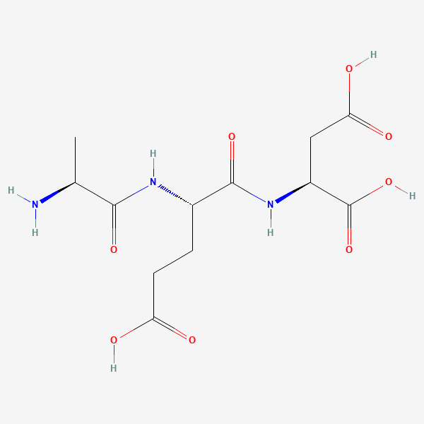
Cartalax is a programme trade name for the synthetic tripeptide L-alanyl-L-glutamyl-L-aspartic acid (Ala-Glu-Asp, AED), C12H19N3O8, 333.29 g/mol as the free peptide, a small, strongly acidic molecule with no modifications. It belongs to the short-peptide series developed by the Khavinson group at a St Petersburg institute, which paired it with cartilage, and differs from Epitalon (AEDG) by one glycine. No experimental structure or measured binding constant exists; its proposed DNA and transporter interactions are docking calculations. The experimental literature is small, mostly marker expression in cell cultures, and comes almost entirely from that one institute. The cartilage label is the least evidenced part of its identity.[1,2,3,4,5,6]
Cartalax regulatory status
No FDA, EMA or Health Canada marketing authorisation for cartalax (Ala-Glu-Asp) was found in Drugs@FDA, the European Medicines Agency medicines database or the Health Canada Drug Product Database, and a ClinicalTrials.gov search on the name returned no study of it (all searched November 2026). It is not named on the World Anti-Doping Agency 2026 Prohibited List, whose class S0 covers pharmacological substances with no current approval for human therapeutic use that are not addressed elsewhere on the list, and it does not appear on the FDA's list of bulk drug substances that may present significant safety risks (current as of 22 April 2026).[7,8,9,10,11,12]
Cartalax at a glance
- A synthetic tripeptide, L-alanyl-L-glutamyl-L-aspartic acid, three residues and 333.29 g/mol as the free peptide, with two free acidic side chains in addition to the C-terminal carboxylate.[1,2,3]
- The name Cartalax is a programme label rather than a chemical one. In the peer-reviewed literature the molecule is called AED, Ala-Glu-Asp or the internal code T-31, and the trade name appears mainly in tables and in two Russian-language papers; the registry number 85806-95-7 predates the cartilage patent, which claims a use rather than a new substance.[13,14,15,3]
- The chemistry of an actual sample has been published once, in the 2007 patent: solution-phase synthesis, isolation as the acetate salt, 98.75% by reversed-phase HPLC at 220 nm, a single thin-layer spot, and a specific optical rotation of minus 20 degrees. No mass spectrum is reported there; the only mass-spectrometric work found is a 2020 targeted search by the originating group, which reported the tripeptide in a cartilage tissue complex at 0.2 mg/g.[3,16]
- No experimental structure exists, and neither does any measured binding constant. The DNA minor-groove preference and the transporter interactions attributed to this peptide are docking calculations; in the largest of them AED ranks mid-table among 26 related peptides on LAT1, LAT2 and PEPT1.[17,4,18]
- The experimental literature is small, almost entirely from one St Petersburg institute and its collaborators, and reports marker expression in cultures rather than molecular interactions. The most on-subject experiment found raised SOX9, aggrecan, type II collagen and COMP in ageing human mesenchymal stem cells at an assay concentration of 200 ng/mL.[6,19,20,21]
What is Cartalax?
Cartalax is one of a series of two- to four-residue synthetic peptides developed from the 1990s onward by the group of V. Kh. Khavinson at the St Petersburg Institute of Bioregulation and Gerontology. The series pairs each short sequence with a tissue label and a trade name: AEDG with the pineal gland (Epitalon), AEDL with the lung (Bronchogen), AEDR with the heart (Cardiogen), EDL with the kidney and liver (Ovagen), and this tripeptide, AED, with cartilage, as Cartalax. The labels come from the group's own reading of its experiments; they are not independent classifications, and the same tables in which they appear also carry the sequences, which is how a reader can check which molecule is meant.[13,5,22]
The molecule itself is older than the programme's claim on it. The 2007 patent, filed by Khavinson, Grigoriev, Malinin and Ryzhak, cites the registry number 85806-95-7 as prior art and claims a previously unknown activity in bone and cartilage metabolism rather than a new compound, and its examples are an explant preparation from rat femoral-head cartilage and morphometry of thyroid C-cells in epiphysectomised rats. The document is also the only place found where the material a reader might encounter is chemically described rather than merely named.[3,1]
Two neighbouring kinds of material are easy to confuse with it. The first is the tetrapeptide AEDG, which differs by one glycine and has its own much larger literature; the two are tested in the same experiments and separate on the endpoints measured. The second is the family of cartilage and bone tissue extracts from the same institute, Sigumir and Chondrolux among them, which are mixtures with no formula or mass. One review from the group states that the cartilage polypeptide complex contains short peptides of about 75 to 846 Da, this tripeptide included, and cites a 2020 Russian-language paper for it. That paper describes the order of events plainly: the short sequences were designed and synthesised from the amino-acid composition of the extracts, and only afterwards were the extracts searched for them. Using liquid chromatography with mass-spectrometric detection and synthetic AED as the reference standard, it reported the tripeptide in the cartilage complex at 0.2 mg per gram (0.02%), a minor component. So AED is a reported constituent of such a preparation, found by a search aimed at it, rather than a synonym for it.[23,16,24,14]
What is worth knowing about the supporting literature is how narrow it is, and that none of it is in people. No human study of this tripeptide was found: PubMed searches on the sequence, on all three spellings of the trade name and on the internal code, each combined with human, patients, trial and clinical, return only the cell and animal reports cited here, and ClinicalTrials.gov holds no study of it. Clinical wording does appear twice in this literature and neither instance is a study of the molecule that can be read: the 2007 patent's clinical passage is internally inconsistent on units and is not relied on here, and the 2020 identification paper asserts clinical usefulness for the tissue complexes in its introduction without reporting a trial. What the searches do return is a few dozen papers, nearly all from one institute, in three journals, mostly short reports with abstracts rather than full methods. The one clearly external laboratory found, in Chieti, co-authors with the originating group, and in its experiment AED was not the peptide carrying the reported effect.[21,6,20,10,3,16]
Not to be confused with
- Epitalon / epithalon (AEDG, Ala-Glu-Asp-Gly)
- A tetrapeptide, this sequence plus a C-terminal glycine: C14H22N4O9, 390.35 g/mol, its own CAS number and its own library entry. The two are routinely tested side by side in the same papers and do not behave alike: in old rats AED lowered urinary protein and raised distal sodium transport while AEDG lowered glomerular filtration rate, and in skin fibroblast cultures the reported marker changes differ. Almost all of the measured DNA-binding and histone-binding work in this literature was done on AEDG, not on AED, so evidence is frequently transferred from the tetrapeptide to the tripeptide by inference.[24,26,5]
- AEDL (bronchogen), AEDR (cardiogen), AEDP, KED (vesugen), EDL (ovagen)
- Other members of the same synthetic series, each one or two residues different and each assigned a different tissue label by the same group. One-letter codes of three or four characters are easy to transpose, and the differences are chemically real: in the transporter docking study AED, AEDL, AEDR and AEDP rank differently on every carrier tested, and in the cisplatin kidney model the authors name EDL rather than AED as the agent that moved their panel.[4,34,13]
- Sigumir, Chondrolux and the cartilage polypeptide complex (PCC)
- Tissue preparations extracted from calf cartilage and bone, not single molecules, and they have no formula or mass. One review states that the cartilage polypeptide complex contains short peptides of roughly 75 to 846 Da including this tripeptide, and the analytical paper it cites reports AED in that complex at 0.2 mg/g, so AED is a reported minor constituent of a preparation rather than a synonym for it. Where the two have been compared the authors report the complex and the single peptide behaving differently, in one bone study explicitly ranking the extract above the tripeptide.[23,16,14,29]
- The abbreviation AED in unrelated literatures
- The same three letters are an established abbreviation in unrelated fields, so a search on them alone returns papers concerning neither this molecule nor peptides of this class. The reliable search handles are the sequence spelled out, the CAS number and the internal code T-31, which the kidney papers print as T-31 (AED) beside T-35 (EDL).[15,28]
- The PubChem synonym 'H-Asp-Glu-Asp-OH'
- That string appears in the synonym list of CID 87815447 and contradicts the record's own structure, formula and InChIKey, all of which describe an N-terminal alanine. It is a defect in an aggregated synonym list rather than an alternative identity, and a reader who takes the synonym rather than the structure gets a different molecule.[1,2]
Cartalax structure and sequence
The graph is as simple as a peptide gets: alanine, then glutamate, then aspartate, joined head to tail through alpha-carboxyl groups, with a free N-terminal amine on Ala1 and the C-terminal carboxylate on Asp3. PubChem and ChEBI agree on C12H19N3O8, an average mass of 333.29, a monoisotopic mass of 333.11721 and the InChIKey KXEVYGKATAMXJJ-ACZMJKKPSA-N, and the patent writes the same molecule as H-Ala-Glu-Asp-OH. There are three stereocentres, one per residue, all L, so eight stereoisomers of the sequence exist on paper and only the all-L form is this compound; the one measured stereochemical datum on a real sample is the patent's optical rotation.[1,2,3]
The chemically interesting feature is charge density. Glu2 contributes a free gamma-carboxyl and Asp3 a free beta-carboxyl, so beside the terminal carboxylate the molecule carries three acidic groups against one basic amine, and at neutral pH it is a small, strongly anionic zwitterion. That is the property the group's models work with: their DNA docking places the peptide in the minor groove and their transporter docking treats it as a substrate or inhibitor candidate for carriers that move acidic amino acids and small peptides. Nothing has measured a charge state, a pKa or an affinity for this sequence.[18,4,17]
The 3D view here is a calculated conformer from PubChem, and it should be read as connectivity and stereochemistry only. No experimental structure of the molecule exists, and the Protein Data Bank entries that surface on a search for the string Ala-Glu-Asp are designed amyloid-beta macrocycles containing those three residues inside a longer chain, which are a different subject entirely. For a free tripeptide in water there is in any case no single shape to show, and no NMR or circular dichroism study of this sequence was found that would describe its conformational preferences.[1,4]
- Ala1
- Glu2
- Asp3
Three stereocentres, one per residue, all L. PubChem and ChEBI express that as the all-(S) configuration in the systematic name and agree on the standard InChIKey KXEVYGKATAMXJJ-ACZMJKKPSA-N, so eight stereoisomers of this sequence are possible and only the all-L form is the molecule described here. There is no cysteine and therefore no disulfide, and no cyclisation, lipidation, PEG, glycosylation or metal coordination. The only measured piece of stereochemical evidence found for a real sample is the specific optical rotation of minus 20 degrees reported in the patent for its synthesised material. At neutral pH the molecule is a zwitterion carrying one ammonium group and three carboxylates, two of them free side chains, which is an unusually acidic molecule for its size and is the feature the docking work leans on.[1,2,3]
What studies on Cartalax show
Each card names the experimental system and what was measured. A result in one system is not evidence about another.
Peptide synthesis with physico-chemical characterisation, plus a cartilage explant assay · Analytical chemistry on the synthesised material; explant fragments of femoral-head cartilage from Wistar rats
The material was reported at 98.75% by HPLC with a single thin-layer spot at Rf 0.75 and a specific optical rotation of minus 20 degrees, and the explant area index was about 26% above control at day three at the most effective assay concentration of 100 ng/mL.[3]
- Material
- H-Ala-Glu-Asp-OH prepared by solution-phase synthesis and isolated as the acetate salt
- Measured
- HPLC purity at 220 nm on a C18 column, thin-layer mobility, moisture, pH of a dilute solution, specific optical rotation; explant area index over three days across a range of assay concentrations
- Compared with
- Untreated explant fragments
- Limitation
- A patent is an interested document that was not peer reviewed, no mass spectrum is reported, and the explant readout is a morphometric area index on a small number of fragments. The experiment did not establish a molecular target, and the clinical material in the same specification is internally inconsistent on units and is not relied on here.
Khavinson VK et al., 2007, Russian patent RU2299741C1 (applicant Sia Peptides; PCT WO2007139433A1)
Chondrogenic differentiation of stem cells · Human mesenchymal stem cells carried through replicative ageing in culture
All four chondrogenic markers were reported higher with the tripeptide at an assay concentration of 200 ng/mL, and with the tissue complex at 2000 ng/mL.[6]
- Material
- AED tripeptide, with the cartilage polypeptide complex tested alongside
- Measured
- Gene expression and protein synthesis of SOX9, aggrecan, type II collagen and COMP
- Compared with
- The tissue complex at a tenfold higher assay concentration, against untreated ageing cultures
- Limitation
- Read as abstract only, from the originating institute, with one stated concentration per agent so no dose-response is visible and no sequence-scrambled control described. Marker expression in a stem-cell culture is not cartilage, and the experiment did not establish how the peptide reaches those genes.
Myakisheva SN et al., 2023, Advances in Gerontology (in Russian)
Immunocytochemistry with confocal microscopy · Human skin fibroblasts under replicative ageing
The two peptides separated on the panel: AED was reported to raise sirtuin-1, sirtuin-6 and collagen I staining, while KE lowered IL-1, NF-kB and TGF-beta staining and raised sirtuin-6.[20]
- Material
- AED tripeptide
- Measured
- Staining for sirtuin-1, sirtuin-6, collagen I, IL-1, TGF-beta and NF-kB
- Compared with
- The dipeptide KE on the same endpoints in the same experiment
- Limitation
- Read as abstract only; staining intensity as the endpoint, no stated concentrations or molar matching between the two peptides, and one laboratory. A difference between two peptides on a marker panel does not identify what either one binds.
Fridman NV et al., 2020, Bulletin of Experimental Biology and Medicine
Transcript measurement in two ageing models · Human embryonic bone-marrow mesenchymal stem cells (FetMSC line), aged by passaging and by stationary culture
IGF1 transcript rose 3.5 to 5.6 fold with the peptides in both models and all three raised NFkB transcript; the peptide-specific effects the authors highlight, on FOXO1 and the reversal of the TNKS2 response between models, belong to KED and KE rather than to AED.[25]
- Material
- AED, with KED and KE tested alongside at nanomolar assay concentrations
- Measured
- IGF1, FOXO1, TERT, TNKS2 and NFkB transcript levels
- Compared with
- The other two peptides and the two ageing models against each other
- Limitation
- Read as abstract only. Transcripts without protein, one immortalised line, and no scrambled-sequence control stated. The experiment did not establish that any of these genes is a direct target.
Ashapkin V et al., 2020, Molecular Biology Reports
Immunofluorescence and western blot in primary human stem cells · Periodontal ligament stem cells from ten donors, in basal and neuroinductive medium (Chieti laboratory, with the originating group)
All four peptides and the mixture raised GAP43 staining against untreated cells, but the increases the authors single out, for GAP43 in differentiated cells and for nestin, were those of KED and of the four-peptide mixture rather than of AED alone.[21]
- Material
- AED, KED, KE and AEDG individually and as a mixture of all four
- Measured
- GAP43 and nestin protein by staining and by blot
- Compared with
- Untreated cells in each medium, and the peptides against one another
- Limitation
- Full text read. Staining quantified from ten randomly collected images, no scrambled-peptide control, and no purity or characterisation reported for the peptides used. This is the nearest thing to an outside replication found, and in it this tripeptide is not the agent carrying the effect.
Caputi S et al., 2019, International Journal of Immunopathology and Pharmacology
Molecular docking against transporter structures · Published structures of the amino-acid carriers LAT1 and LAT2 and the peptide carriers PEPT1 and PEPT2, in silico
AED scored mid-table: 14th of 26 on LAT1 (ICM-Score minus 28.14), 12th on LAT2 (minus 23.64), 13th on PEPT1 (minus 18.95) and 7th on PEPT2 (minus 7.97). It is one of the eight peptides the authors name as likely LAT1 inhibitors, and they state that a good score is as consistent with the peptide blocking a carrier as with its being carried.[4]
- Material
- AED with free N- and C-termini, among 26 named peptides and a comparison library of 8,400 di- and tripeptides
- Measured
- ICM-Pro docking scores
- Compared with
- Known substrates and inhibitors of each carrier, and the other 25 peptides
- Limitation
- Full text read. Docking scores are not transport: no uptake assay, no competition experiment and no mutagenesis was done, and the ranking depends on which receptor conformation was chosen. The authors further report that the single human PEPT2 structure in the Protein Data Bank is not representative of the conformation in which di- and tripeptides bind, and call every PEPT2 result preliminary, so the PEPT2 column carries no weight: only the dipeptides DG and DA were said to bind PEPT2 better than its known inhibitors. The study did not establish that this peptide enters any cell.
Khavinson VK et al., 2023, Biomolecules
Immunofluorescent confocal microscopy in an ageing cell culture · Human skin fibroblasts aged in culture
All four peptides were reported to lower MMP-9 staining, which rises as these cultures age, and to raise Ki-67 and CD98hc staining, which fall. On caspase-3 the four separated: AED and AEDG were reported to suppress caspase-dependent apoptosis and the other two were not. This is the proliferation and apoptosis result that most secondary pages about this peptide lead with.[26]
- Material
- AED, with KE, KED and AEDG tested on the same endpoints in the same experiment
- Measured
- Staining for Ki-67 (proliferation), CD98hc, caspase-3 (apoptosis) and MMP-9 (matrix remodelling)
- Compared with
- The three other peptides, and the ageing cultures untreated
- Limitation
- Read as abstract only, from the originating institute. Staining intensity is the endpoint, no concentrations or molar matching between the four peptides are given in the abstract, and no scrambled-sequence control is described. Four peptides moving three markers in the same direction is as consistent with a shared non-specific effect as with four separate mechanisms, and the experiment did not establish what any of them binds.
Lin'kova NS et al., 2016, Bulletin of Experimental Biology and Medicine
Aged cell-culture comparison of three agents · Thymocyte culture at passage 8
Of the three agents only AB-9 was reported to show the combined effect across the three endpoints. This is the clearest reported non-result for this molecule, and it is the paper PubChem cross-references from the compound record.[27]
- Material
- T-31 (this tripeptide), with the peptoids AB-17 and AB-9
- Measured
- Proliferative activity, differentiation and apoptosis of the cultured cells
- Compared with
- The other two agents in the same experiment
- Limitation
- Read as abstract only, and the abstract does not quantify what T-31 did, so this is a reported absence rather than a measured null. The experiment did not establish why the three agents differed.
Lin'kova NS et al., 2011, Bulletin of Experimental Biology and Medicine
What is proposed for Ala-Glu-Asp at the molecular level, and what has actually been measured
The programme proposes one mechanism for the whole series: a peptide small enough to cross the plasma and nuclear membranes enters the nucleus, binds a short DNA sequence or a histone tail, and changes the transcription of nearby genes. It is a hypothesis about a class, stated for short peptides generally rather than demonstrated for this sequence.[5,22]
For this tripeptide the whole account is docking, and the two calculations disagree. One paper models its most favourable complexes against an AT-rich decamer in the minor groove and, in the same abstract, asserts that this interaction causes the expression changes seen in the accompanying cell cultures, which the experiments reported there do not test. A later survey docks nineteen peptides of the series against DNA sites and assigns this one the sequence acct, shared with KEDW, while placing KE and EDP at agat and AEDL and EDL at ctcc; its authors describe nuclear entry followed by DNA binding as a supposition. A third study docks twenty-six of these peptides against the amino-acid carriers LAT1 and LAT2 and the peptide carriers PEPT1 and PEPT2, where this one ranks mid-table, and notes that a good score is as consistent with blocking a carrier as with being carried by it. No uptake measurement, competition assay, labelled tracer or affinity measurement for this sequence was found.[18,17,4]
What has been measured was measured on other sequences, and the attribution is worth getting right. In the systematic review where the DNA and histone work is set out, the fluorescence quenching against labelled wheat histones and the eukaryotic endonuclease assays were done on AEDG, EDR, AEDL, KEDG, AEDR and KEDW, the ultraviolet and circular dichroism work on KEDW, the histone modelling on AEDG, EDR and DS, and the duplex melting measurements on free amino acids and a tetrapeptide. This tripeptide appears in that review's gene-expression sections and in its summary table, not in its binding sections. So the molecular mechanism usually attached to the Cartalax name is inherited from its neighbours.[5,13]

What is still unknown about Cartalax
Identity is settled; provenance and material are not. The sequence, formula, masses, stereochemistry and registry number agree across PubChem, ChEBI and the patent, and the patent adds purity and optical rotation for a real synthesised sample, isolated as the acetate salt. What no public document states is which form or counterion a given non-pharmaceutical preparation contains. The one analytical report placing the peptide in a tissue material is a targeted search by the originating group, identifying it by retention time and mass number only. The CAS number is quoted by PubChem and the patent but could not be confirmed at the registry, which refuses lookups without a subscription. One synonym in the PubChem record, H-Asp-Glu-Asp-OH, contradicts the record's own structure and is a registry defect rather than an alternative reading.[1,2,3,16]
The structural file is empty. There is no deposited structure, no NMR, no circular dichroism, no calorimetry and no melting measurement for this sequence, so every statement about its shape or its interactions is a calculation, and the two calculations of its DNA preference from the same group do not agree with each other. For a molecule whose proposed mechanism is sequence-specific binding, that is the central gap: an affinity measurement against a defined oligonucleotide would be the obvious experiment and has not been published.[17,18,5]
Replication is the second gap. Almost everything found comes from one institute and its named collaborators, much of it in short-report format where concentrations, controls and statistics are not visible in what is publicly readable, and several of the papers are abstract-only in English. In the one experiment run largely outside that institute, the reported effects belonged to a different peptide in the same set. Where this tripeptide has been tested beside a tissue extract or beside its neighbours, the authors themselves have twice ranked it below the comparator, once in rat kidney explants and once in a rat bone study.[21,28,29]
Finally, the cartilage label is the least evidenced part of the identity, and the commonest claim made in support of it is false. AED is widely described as a fragment of type XI collagen. The sequence does not occur in that protein: UniProt P12107, collagen alpha-1(XI), 1,806 residues, and P13942, collagen alpha-2(XI), 1,736 residues, were each searched through and neither contains the run AED at any position, and nor does P02458, collagen alpha-1(II), which supplies the third chain of the type XI heterotrimer. The originating programme never made that claim either: its own account is that the sequence was composed from the amino-acid COMPOSITION of a cartilage extract, which is a different thing from a position in a protein, and the peptide was detected in that extract only afterwards, at 0.2 mg/g. A second question, which vendor pages raise and this literature does not answer, is how a small acidic peptide would reach chondrocytes at all, since cartilage is avascular: nothing found measures this peptide's distribution in any tissue, and the only treatment of its transport is a docking calculation against carrier structures. Meanwhile the measured work on the molecule is spread across skin fibroblasts, kidney cultures and explants, thymocytes and mesenchymal stem cells, with two 2023 reports on chondrogenic markers and chondrocyte secretory markers from the originating institute as the only cartilage-directed cell experiments found. The review that places this peptide inside a cartilage tissue complex spends most of its length on growth-factor-derived and matrix-derived peptides that have a named receptor mechanism, B2A and CK2.1 at bone morphogenetic protein receptors, GFOGER and CMP at collagen, RGD at fibronectin, and names this tripeptide in its conclusions with no mechanism of that kind attached to it.[30,31,32,16,4,6,19,23]
Key papers on Cartalax
- Peptide normalizing metabolism in osseous and cartilage tissue, pharmaceutical composition based thereon, and method of use.
Khavinson VK, Grigoriev EI, Malinin VV, Ryzhak GA (2007), Russian patent RU2299741C1 (applicant Sia Peptides; PCT WO2007139433A1) · Patent with synthesis, characterisation and animal and explant examples
The only document that describes a real sample of this peptide rather than a name: the synthetic route, the acetate salt, the HPLC purity, the thin-layer spot and the optical rotation, plus the cartilage explant assay the whole cartilage claim rests on. Read it knowing it is a patent and an interested document.
- PubChem Compound Summary for CID 87815447, Alanyl-glutamyl-aspartic acid.
National Center for Biotechnology Information, PubChem · Database record
The fastest check that the name in front of you and the molecule are the same thing: formula, both masses, all-L stereochemistry, the CAS number, and the computed conformer. Also the place to see the one contradictory synonym for yourself.
- Идентификация коротких пептидов в составе полипептидных комплексов, выделенных из органов животных [Identification of short peptides as part of polypeptide complexes isolated from animal organs].
Zhurkovich IK, Kovrov NG, Ryzhak GA, Mironova ES, Khavinson VKh (2020), Uspekhi Sovremennoi Biologii (in Russian) · Targeted liquid chromatography with mass-spectrometric detection of tissue complexes
The only analytical report placing this tripeptide inside a cartilage tissue complex, and the source of the order of events: sequences designed first, extracts searched for them afterwards. Read the methods for what the identification rests on, retention time and mass number against a synthetic standard. Russian-language, open on the publisher platform.
- Feasibility of Transport of 26 Biologically Active Ultrashort Peptides via LAT and PEPT Family Transporters.
Khavinson VK, Linkova NS, Rudskoy AI, Petukhov MG (2023), Biomolecules · Molecular docking against transporter structures
The most quantitative treatment of this peptide anywhere, and a lesson in what it is: 26 peptides docked against four carriers, with this one mid-table and the authors noting that a good score may mean inhibition rather than transport. Open access.
- Peptide Regulation of Gene Expression: A Systematic Review.
Khavinson VK, Popovich IG, Linkova NS, Mironova ES, Ilina AR (2021), Molecules · Systematic review of peptide-DNA and peptide-histone work
Read it to separate this peptide's evidence from its neighbours'. The binding experiments it describes were done on other sequences; this one appears in the gene-expression sections and in the summary table that assigns it the Cartalax name. Open access.
- The influence of peptides on the chondrogenic differentiation of human mesenchymal stem cells during replicative aging.
Myakisheva SN, Linkova NS, Diatlova AS, Polyakova VO, Ryzhak GA (2023), Advances in Gerontology (in Russian) · Stem-cell differentiation study
The most on-subject experiment in existence: chondrogenic markers in ageing human mesenchymal stem cells, with the tissue complex beside it. Abstract-only in English, which is itself informative about the state of this literature.
- Effect of short peptides on neuronal differentiation of stem cells.
Caputi S, Trubiani O, Sinjari B, Trofimova S, Diomede F, Linkova N, Diatlova A, Khavinson V (2019), International Journal of Immunopathology and Pharmacology · Primary human stem-cell study with immunofluorescence and western blot
The closest thing to an outside laboratory, and the clearest illustration of how effects get attributed in this series: four peptides and a mixture tested together, with the highlighted results belonging to KED and the mixture. Full text open access.
- Peptidergic regulation of thymocyte differentiation, proliferation, and apoptosis during aging of the thymus.
Lin'kova NS, Polyakova VO, Trofimov AV, Kvetnoy IM, Khavinson VKh (2011), Bulletin of Experimental Biology and Medicine · Aged cell-culture comparison
A short report where this peptide was tested and was not the agent that worked, which is rare in this literature and worth reading for that reason. It is also the paper PubChem links from the compound record.
References
- 1.National Center for Biotechnology Information. PubChem Compound Summary for CID 87815447, Alanyl-glutamyl-aspartic acid. PubChem. source · database record
- 2.ChEBI, European Bioinformatics Institute. Ala-Glu-Asp, CHEBI:158137. ChEBI. source · database record
- 3.Khavinson VK, Grigoriev EI, Malinin VV, Ryzhak GA. (2007). Peptide normalizing metabolism in osseous and cartilage tissue, pharmaceutical composition based thereon, and method of use. Russian patent RU2299741C1 (applicant Sia Peptides; PCT WO2007139433A1). source · full text read
- 4.Khavinson VK, Linkova NS, Rudskoy AI, Petukhov MG. (2023). Feasibility of Transport of 26 Biologically Active Ultrashort Peptides via LAT and PEPT Family Transporters. Biomolecules. 13:552. doi:10.3390/biom13030552 · full text read
- 5.Khavinson VK, Popovich IG, Linkova NS, Mironova ES, Ilina AR. (2021). Peptide Regulation of Gene Expression: A Systematic Review. Molecules. 26:7053. doi:10.3390/molecules26227053 · full text read
- 6.Myakisheva SN, Linkova NS, Diatlova AS, Polyakova VO, Ryzhak GA. (2023). The influence of peptides on the chondrogenic differentiation of human mesenchymal stem cells during replicative aging. Advances in Gerontology (in Russian). 36:383-390. PMID 37782646 · abstract only
- 7.U.S. Food and Drug Administration. (2026). Drugs@FDA: FDA-Approved Drugs. U.S. Food and Drug Administration. source · database record
- 8.European Medicines Agency. (2026). Medicines. European Medicines Agency. source · database record
- 9.Health Canada. (2026). Drug Product Database online query. Health Canada. source · database record
- 10.U.S. National Library of Medicine. (2026). ClinicalTrials.gov search results for "cartalax". ClinicalTrials.gov. source · database record
- 11.World Anti-Doping Agency. (2026). The 2026 Prohibited List: International Standard. World Anti-Doping Agency. source · full text read
- 12.U.S. Food and Drug Administration. (2026). Certain Bulk Drug Substances for Use in Compounding that May Present Significant Safety Risks. U.S. Food and Drug Administration. source · full text read
- 13.Khavinson V, Linkova N, Kozhevnikova E, Dyatlova A, Petukhov M. (2022). Transport of Biologically Active Ultrashort Peptides Using POT and LAT Carriers. International Journal of Molecular Sciences. 23:7733. doi:10.3390/ijms23147733 · full text read
- 14.Myakisheva SN, Linkova NS, Kozhevnikova EO, Ryzhak GA. (2023). Chondrocytes secretory phenotype associated with aging: role in the pathogenesis of osteoarthritis and prospects for peptide bioregulation. Advances in Gerontology (in Russian). 36:313-323. PMID 37782637 · abstract only
- 15.Khavinson VKh, Lin'kova NS, Polyakova VO, Durnova AO, Nichik TE, Kvetnoi IM. (2014). Peptides regulate expression of signaling molecules in kidney cell cultures during in vitro aging. Bulletin of Experimental Biology and Medicine. 157:261-264. doi:10.1007/s10517-014-2540-y · abstract only
- 16.Zhurkovich IK, Kovrov NG, Ryzhak GA, Mironova ES, Khavinson VKh. (2020). Идентификация коротких пептидов в составе полипептидных комплексов, выделенных из органов животных [Identification of short peptides as part of polypeptide complexes isolated from animal organs]. Uspekhi Sovremennoi Biologii (in Russian). 140:140-148. doi:10.31857/S004213242002012X · full text read
- 17.Khavinson VK, Lin'kova NS, Tarnovskaya SI. (2016). Short Peptides Regulate Gene Expression. Bulletin of Experimental Biology and Medicine. 162:288-292. doi:10.1007/s10517-016-3596-7 · abstract only
- 18.Khavinson VKh, Tarnovskaia SI, Lin'kova NS, Poliakova VO, Durnova AO, Nichik TE, Kvetnoi IM, D'iakonov MM, Iakutseni PP. (2014). Tripeptides slow down aging process in renal cell culture. Advances in Gerontology (in Russian). 27:651-656. PMID 25946838 · abstract only
- 19.Myakisheva SN, Linkova NS, Kozhevnikova EO, Polyakova VO, Ryzhak GA. (2023). Peptides prevent the forming of secretory phenotype of chondrocytes associated with the aging. Advances in Gerontology (in Russian). 36:234-238. PMID 37356100 · abstract only
- 20.Fridman NV, Linkova NS, Kozhevnikova EO, Gutop EO, Khavinson VK. (2020). Comparison of the Effects of KE and AED Peptides on Functional Activity of Human Skin Fibroblasts during Their Replicative Aging. Bulletin of Experimental Biology and Medicine. 170:154-157. doi:10.1007/s10517-020-05022-1 · abstract only
- 21.Caputi S, Trubiani O, Sinjari B, Trofimova S, Diomede F, Linkova N, Diatlova A, Khavinson V. (2019). Effect of short peptides on neuronal differentiation of stem cells. International Journal of Immunopathology and Pharmacology. 33:2058738419828613. doi:10.1177/2058738419828613 · full text read
- 22.Khavinson V, Linkova N, Diatlova A, Trofimova S. (2020). Peptide Regulation of Cell Differentiation. Stem Cell Reviews and Reports. 16:118-125. doi:10.1007/s12015-019-09938-8 · abstract only
- 23.Linkova N, Khavinson V, Diatlova A, Myakisheva S, Ryzhak G. (2023). Peptide Regulation of Chondrogenic Stem Cell Differentiation. International Journal of Molecular Sciences. 24:8415. doi:10.3390/ijms24098415 · full text read
- 24.Zamorskii II, Shchudrova TS, Zeleniuk VG, Linkova NS, Nichik TE, Khavinson VK. (2018). The influence of peptides on the morphofunctional state of old rats kidneys. Advances in Gerontology (in Russian). 31:498-504. PMID 30607912 · abstract only
- 25.Ashapkin V, Khavinson V, Shilovsky G, Linkova N, Vanuyshin B. (2020). Gene expression in human mesenchymal stem cell aging cultures: modulation by short peptides. Molecular Biology Reports. 47:4323-4329. doi:10.1007/s11033-020-05506-3 · abstract only
- 26.Lin'kova NS, Drobintseva AO, Orlova OA, Kuznetsova EP, Polyakova VO, Kvetnoy IM, Khavinson VKh. (2016). Peptide Regulation of Skin Fibroblast Functions during Their Aging In Vitro. Bulletin of Experimental Biology and Medicine. 161:175-178. doi:10.1007/s10517-016-3370-x · abstract only
- 27.Lin'kova NS, Polyakova VO, Trofimov AV, Kvetnoy IM, Khavinson VKh. (2011). Peptidergic regulation of thymocyte differentiation, proliferation, and apoptosis during aging of the thymus. Bulletin of Experimental Biology and Medicine. 151:239-242. doi:10.1007/s10517-011-1298-8 · abstract only
- 28.Chalisova NI, Lin'kova NS, Nichik TE, Ryzhak AP, Dudkov AV, Ryzhak GA. (2015). Peptide Regulation of Cells Renewal Processes in Kidney Tissue Cultures from Young and Old Animals. Bulletin of Experimental Biology and Medicine. 159:124-127. doi:10.1007/s10517-015-2906-9 · abstract only
- 29.Povorozniuk VV, Khavinson VKh, Makogonchuk AV, Ryzhak GA, Kreslov EA, Gopkalova IV. (2007). Effect of peptide regulators on the structural and functional status of bone tissue in ageing rats. Advances in Gerontology (in Russian). 20:134-137. PMID 18306703 · abstract only
- 30.UniProt Consortium. UniProtKB P12107, Collagen alpha-1(XI) chain, Homo sapiens. UniProt. source · database record
- 31.UniProt Consortium. UniProtKB P13942, Collagen alpha-2(XI) chain, Homo sapiens. UniProt. source · database record
- 32.UniProt Consortium. UniProtKB P02458, Collagen alpha-1(II) chain, Homo sapiens. UniProt. source · database record
- 33.Slugina AS, Iordanishvili AK, Serikov AA, Samsonov VV, Ryzhak GA. (2016). Optimization of reparative osteogenesis jaws on aging (preclinical studies). Advances in Gerontology (in Russian). 29:128-133. PMID 28423258 · abstract only
- 34.Zamorskii II, Shchudrova TS, Lin'kova NS, Nichik TE, Khavinson VKh. (2015). Peptides Restore Functional State of the Kidneys During Cisplatin-Induced Acute Renal Failure. Bulletin of Experimental Biology and Medicine. 159:736-739. doi:10.1007/s10517-015-3062-y · abstract only
- 35.ChemSpider (Royal Society of Chemistry). ChemSpider record 16568512: ala-glu-asp. ChemSpider. source · database record
- 36.National Center for Biotechnology Information. PubChem Compound Summary for CID 7010502, Lysylglutamic acid. PubChem. source · database record
Related entries
- Epithalon (epitalon)Epithalon is a four-residue synthetic peptide (Ala-Glu-Asp-Gly) modelled on the amino-acid composition of a bovine pineal extract. It has been studied mainly for its effect on telomere length in cell culture.
- PinealonPinealon is the trade name one Russian research programme uses for the synthetic tripeptide L-glutamyl-L-aspartyl-L-arginine (Glu-Asp-Arg, one-letter EDR). It is one of the Khavinson short peptides proposed to act on gene expression.
